2662
Motion and B0 correction for multiparametric mapping with jointly acquired FID and spherical navigators1Western University, London, ON, Canada, 2Siemens Healthcare Limited, Oakville, ON, Canada, 3Robarts Research Institute, London, ON, Canada
Synopsis
Keywords: Motion Correction, Brain
Motivation: In cases of motion, multi-echo GRE sequences are susceptible to motion artifacts as well as additional phase errors caused by motion-induced B0 inhomogeneity.
Goal(s): Implement jointly acquired FID and spherical navigators for prospective correction of motion and retrospective correction of field offsets in a multi-echo GRE protocol for multiparametric mapping.
Approach: One volunteer was scanned with/without prospective motion correction. Motion was guided by visual prompt to produce repeatable and clinically relevant trajectories.
Results: Prospective motion correction alone provided a notable improvement in image quality, as well as quantitative fat-fraction, R2*, and susceptibility maps. Additional retrospective correction of field offsets reduced residual artifacts.
Impact: Combining spherical navigators with an additional FID readout is a promising approach to simultaneous motion and B0 correction with multiple applications, including quantitative mapping techniques.
Introduction
Multi-echo GRE has many widely used quantitative brain imaging applications including Dixon MRI,1 R2* mapping,2 and susceptibility mapping (QSM).3 As with most MRI applications, such approaches are sensitive to motion. Prospective motion correction (PMC) is a promising solution in such cases;4 however, data acquired at longer echo times is also sensitive to additional phase errors caused by motion-induced spatiotemporal variations in B0 as subject orientation diverges from that for which shimming was optimized.5Navigator-based approaches to simultaneous motion and B0 correction6-10 are preferable to external monitoring systems11 in that they require no additional hardware and impose minimal changes to the clinical workflow. In particular, spherical navigators12 (SNAVs) have shown promise for PMC with relatively short acquisition and processing times.13 Incorporating an additional free induction decay (FID) readout enables simultaneous field monitoring.14 Previous work has used these FID-SNAVs for combined PMC and retrospective field correction of a susceptibility weighted imaging protocol.15 In this study, we extend their application from single- to multi-echo GRE for multiparametric mapping.16
Methods
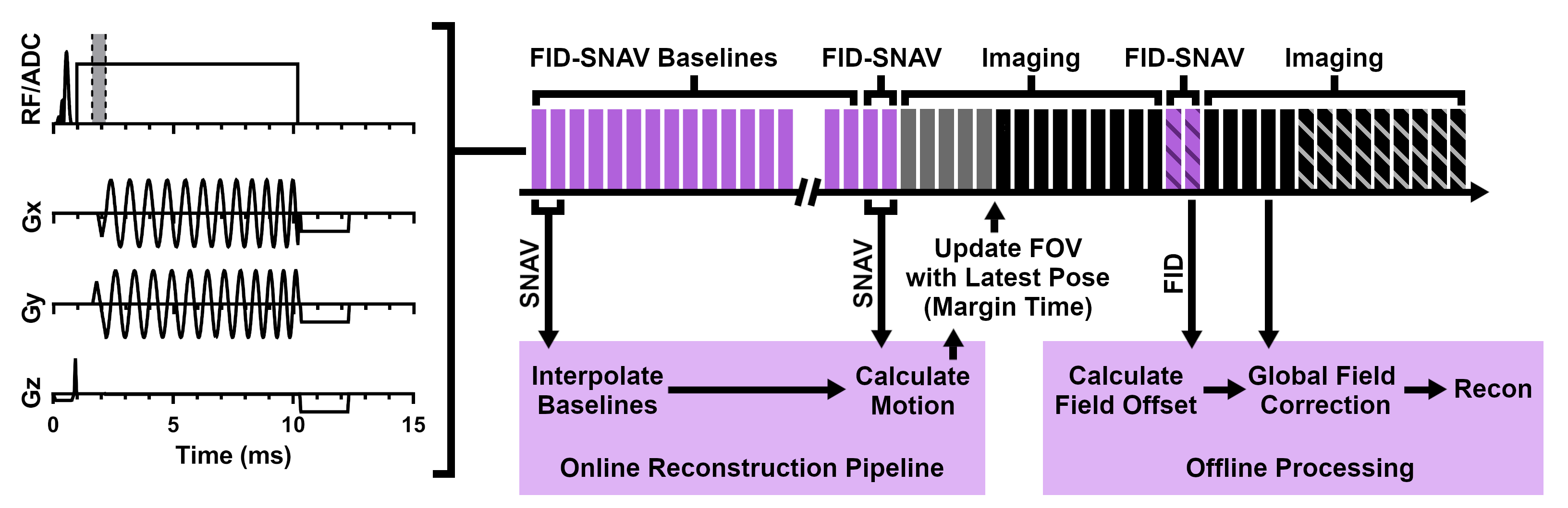
All experiments were performed on a 3T scanner (MAGNETOM PrismaFit, Siemens Healthcare GmbH, Erlangen, Germany) using the product 32-channel head coil. Imaging parameters (FOV 25.6x25.6x19.2 cm, resolution 1.14x1.14x2.00 mm, TR 51 ms, TEs 3.28/4.72/6.22/7.72/9.49/16.75/23.90/31.10/38.16/45.40 ms, FA 15°, BW 1015 Hz/Pixel, GRAPPA R = 2, scan time 10:07) and processing pipeline for mapping fat fraction, R2*, and susceptibility (using MEDI,17 λ = 2e4, 2 iterations) were based on a previously developed protocol.16 FID-SNAVs (acquired as two hemispheres) were interleaved with imaging acquisition at a frequency of 1.96 Hz (scan time increase 2:32); an additional baseline scan (0:17) was also acquired to accelerate rotation estimation.18 SNAV data were processed online to provide motion estimates for PMC (using real-time sequence processing, margin time 10 ms).13 FID data were processed offline to perform zeroth order field correction of imaging data on a channel-wise basis prior to GRAPPA reconstruction.15 An overview is given in Figure 1.Motion measurement accuracy was evaluated using a pineapple phantom; rotations were simulated via gradient update while translation was controlled using a linear motion stage.19 A separate QSM phantom20 was also imaged to ensure the navigators had no impact on image contrast or the resulting parametric maps. One volunteer was scanned with approval from the local Research Ethics Board. Following an initial reference acquisition (no motion), two images were acquired with motion: one uncorrected and one with PMC (processed with and without field correction). To produce similar trajectories for scans with and without correction, motion was guided using a visual indicator projected into the scanner,21 with the prompt itself derived from a true undeliberate motion trajectory obtained from an open-source database.22 Lastly, one additional scan was acquired with no motion to provide a measure of interscan variation. All scans were processed to produce quantitative maps before registering the results to the initial reference scan.
Results
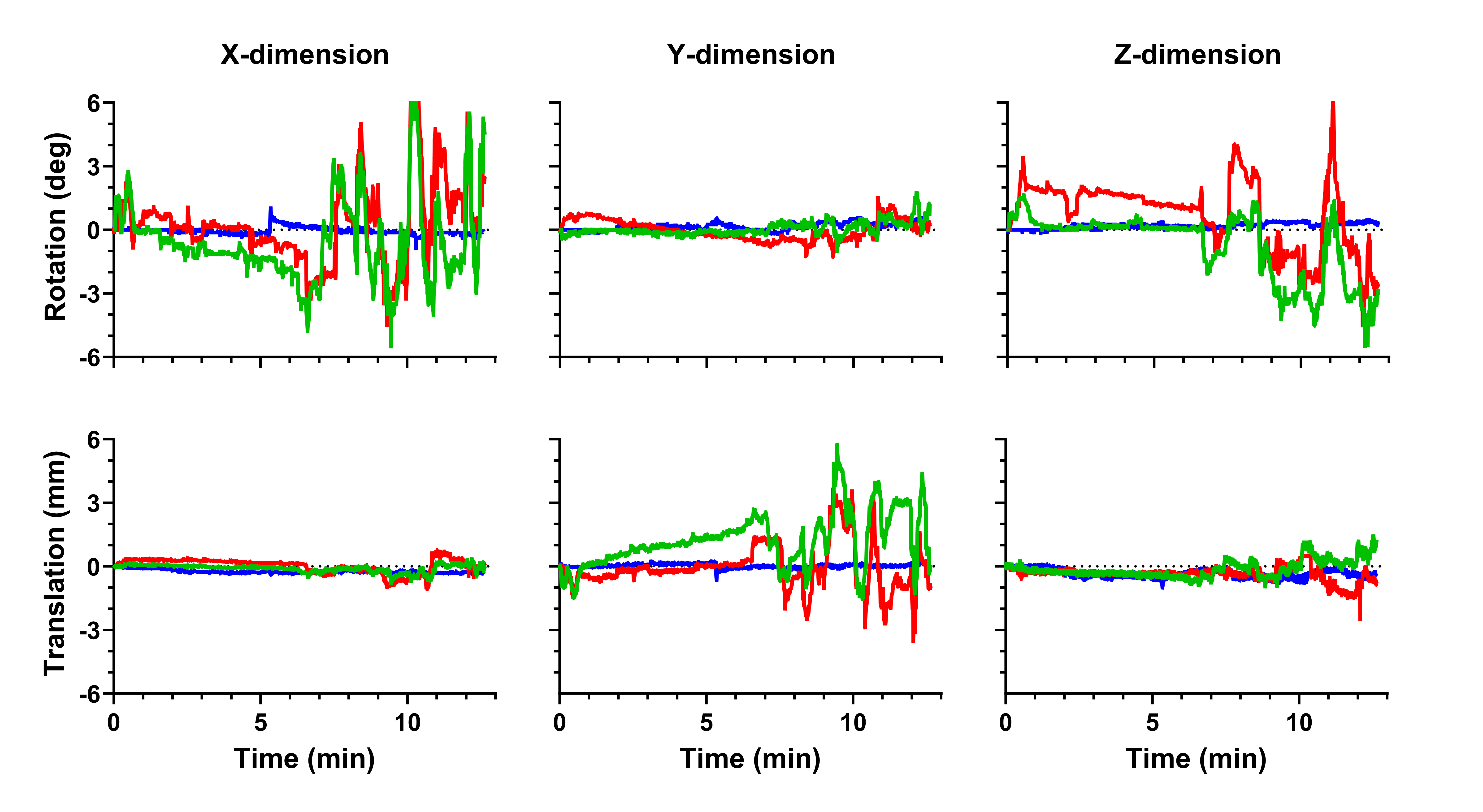
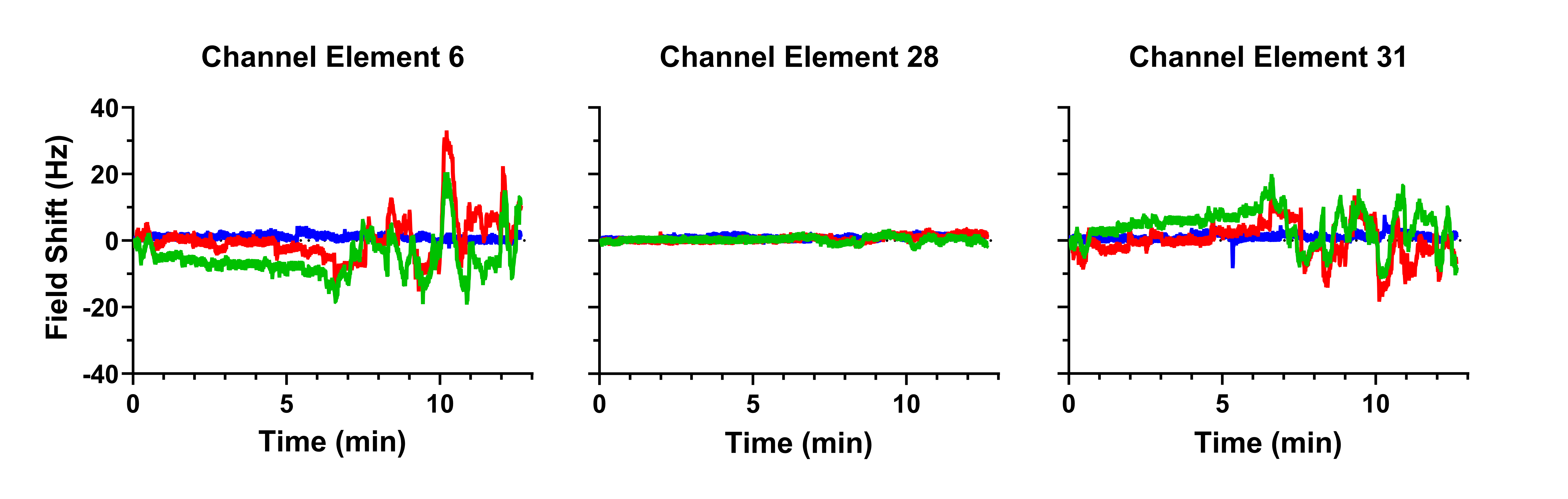
Phantom evaluation of motion estimation demonstrated sub-degree, millimeter accuracy (mean absolute error 0.04 ± 0.06 deg, 0.21 ± 0.14 mm). Comparison of scans with and without navigator acquisition showed no effect on image contrast or the resulting quantitative maps.Measured motion traces for the volunteer study are given in Figure 2. Motion estimates were computed in 26.1 ± 0.4 ms (total latency < 50 ms when considering margin time). Field offsets (shown for three sample channels) are given in Figure 3. Similar trajectories, offsets, were observed for scans with and without PMC.
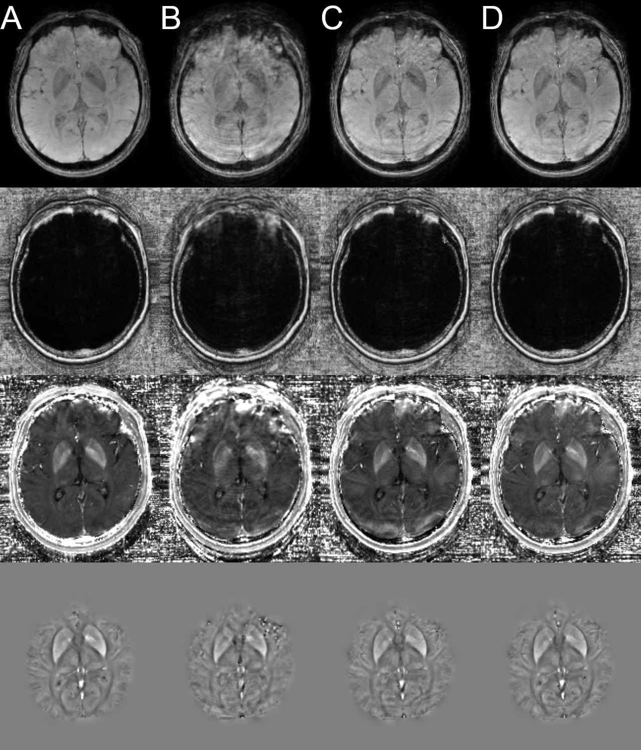
Clear improvement in image quality was seen with PMC alone for early echoes; later echoes also showed improvement but suffered from additional artifacts caused by motion induced B0 inhomogeneity. Additional retrospective correction of field offsets further improved image quality. Sample images and corresponding quantitative maps are displayed in Figure 4.
Discussion
In this work, combined FID-SNAVs were implemented for PMC and retrospective correction of resulting field offsets in a multi-echo GRE protocol for R2* mapping and QSM. Motion correction alone provided a notable improvement in image quality, though retrospective correction of field offsets did reduce the impact of residual artifacts. High field applications would likely experience an even greater benefit.5Remaining artifacts, most apparent in the anterior frontal lobe, will likely benefit from prospective correction of higher order field offsets.23,24 Future work will also investigate FID-based triggering of navigator acquisition25 to optimize the tradeoff with scan time, as well as prospective update of navigator acquisition to reduce the time required for baseline acquisition.
Conclusion
FID-SNAVs are a promising approach to combined motion and B0 correction for quantitative mapping techniques with no requirement for additional hardware or modifications to clinical workflow.Acknowledgements
This work was completed with support from the Natural Sciences and Engineering Council of Canada, the Brain Canada Foundation, and the Center for Metabolic Mapping at Robarts Research Institute.References
1. Reeder SB, McKenzie CA, Pineda AR, Yu H, Shimakawa A, Brau AC, Hargreaves BA, Gold GE, Brittain JH. Water–Fat Separation With IDEAL Gradient-Echo Imaging. J Magn Reson Imaging 2007;25:644-652.2. Fernández-Seara MA, Wehrli FW. Postprocessing Technique to Correct for Background Gradients in Image-Based R*2 Measurements. Magn Reson Med 2000;44:358-366.
3. Liu J, Liu T, Rochefort Ld, Ledoux J, Khalidov I, Chen W, Tsiouris AJ, Wisnieff C, Spincemaille P, Prince MR, Wang Y. Morphology enabled dipole inversion for quantitative susceptibility mapping using structural consistency between the magnitude image and the susceptibility map. NeuroImage 2012;59:2560-2568.
4. Mattern H, Sciarra A, Lüsebrink F, Acosta‐Cabronero J, Speck O. Prospective motion correction improves high‐resolution quantitative susceptibility mapping at 7T. Magn Reson Med 2019;81:1605-1619.
5. Liu J, Zwart JAd, Gelderen Pv, Murphy-Boesch J, Duyn JH. Effect of head motion on MRI B0 field distribution. Magn Reson Med 2018;80:2538-2548.
6. Hess AT, Tisdall MD, Andronesi OC, Meintjes EM, van der Kouwe AJW. Real-Time Motion and B0 Corrected Single Voxel Spectroscopy Using Volumetric Navigators. Magn Reson Med 2011;66:314-323.
7. Alhamud A, Taylor PA, van der Kouwe AJW, Meintjes EM. Real-Time Measurement and Correction of Both B0 Changes and Subject Motion in Diffusion Tensor Imaging Using a Double Volumetric Navigated (DvNav) Sequence. NeuroImage 2016;126:60-71.
8. Simegn GL, Van der Kouwe AJW, Robertson FC, Meintjes EM, Alhamud A. Real-time simultaneous shim and motion measurement and correction in glycoCEST MRI using double volumetric navigators (DvNavs). Magn Reson Med 2019;81(4):2600-2613.
9. Liu J, van Gelderen P, Zwart JAd, Duyn JH. Reducing motion sensitivity in 3D high-resolution T2*-weighted MRI by navigator-based motion and nonlinear magnetic field correction. NeuroImage 2020;206:116332.
10. van der Kouwe AJW, Benner T, Dale AM. Real-Time Rigid Body Motion Correction and Shimming Using Cloverleaf Navigators. Magn Reson Med 2006;56:1019-1032.
11. Jorge J, Gretsch F, Gallichan D, Marques JP. Tracking Discrete Off-Resonance Markers With Three Spokes (trackDOTS) for Compensation of Head Motion and B0 Perturbations: Accuracy and Performance in Anatomical Imaging. Magn Reson Med 2018;79:160-171.
12. Welch EB, Manduca A, Grimm RC, Ward HA, Jack CR. Spherical navigator echoes for full 3D rigid body motion measurement in MRI. Magn Reson Med 2002;47:32-41.
13. Hewlett M, Oran O, Liu J, Drangova M. First application of spherical navigators for prospective correction of intra-scan motion. In Proceedings of the ISMRM Workshop on Motion Detection & Correction, Oxford, UK, 2022.
14. Johnson PM, Liu J, Drangova M. Simultaneous motion and B0 correction using FID-SNAVs. In Proceedings of the Annual Meeting of the ISMRM, Honolulu, USA, 2017. Program Number 3948.
15. Hewlett M, Liu J, Drangova M. Combined motion and B0 correction of susceptibility weighted imaging with jointly acquired FID and spherical navigators. In Proceedings of the Annual Meeting of the ISMRM, Toronto, CA, 2023. Program Number 1822.
16. Liu J, Christiansen SD, Drangova M. Single multi-echo GRE acquisition with short and long echo spacing for simultaneous quantitative mapping of fat fraction, B0 inhomogeneity, and susceptibility. NeuroImage 2018;172:703-717.
17. Liu T, Wisnieff C, Lou M, Chen W, Spincemaille P, Wang Y. Nonlinear Formulation of the Magnetic Field to Source Relationship for Robust Quantitative Susceptibility Mapping. Magn Reson Med 2013;69:467-476.
18. Liu J, Drangova M. Rapid six-degree-of-freedom motion detection using prerotated baseline spherical navigator echoes. Magn Reson Med 2011;65:506-514.
19. Tavallaei MA, Johnson PM, Liu J, Drangova M. Design and evaluation of an MRI-compatible linear motion stage. Med Phys 2016;43:62-71.
20. Hong G, Khazaee T, Cobos SF, Christiansen SD, Liu J, Drangova M, Holdsworth DW. Quantitative MRI to characterize diffusion-controlled release of gadolinium from a calcium sulphate matrix. In Proceedings of the Annual Meeting of the ISMRM, London, UK, 2022. Program Number 1976.
21. Berglund J. MotionParadigm. gitlab.com/johan.berglund/MotionParadigm. Accessed February 3, 2022.
22. Skare S. Brain MRI Motion Data Base. brainmrimotion.org. Accessed June 12, 2023.
23. Wallace TE, Afacan O, Kober T, Warfield SK. Rapid measurement and correction of spatiotemporal B0 field changes using FID navigators and a multi‐channel reference image. Magn Reson Med 2020;82:575-589.
24. Wallace TE, Kober T, Stockmann JP, Polimeni JR, K.Warfield S, Afacan O. Real-time shimming with FID navigators. Magn Reson Med 2022;88:2548-2563.
25. Waszak M, Falkovskiy P, Hilbert T, Bonnier G, Marechal B, Meuli R, Gruetter R, Kober T, Krueger G. Prospective Head Motion Correction Using FID-Guided On-Demand Image Navigators. Magn Reson Med 2017;78:193-203.
Figures