2659
Real-time Multislice-to-volume Motion Correction with B1+ Shimming for Task-based Functional MRI at 7T1Imaging Centre of Excellence, University of Glasgow, Glasgow, Scotland, 2Fraunhofer Institute for Digital Medicine MEVIS, Bremen, Germany, 3Siemens Healthineers UK, Frimley, United Kingdom, 4MR CoilTech Ltd, Glasgow, Scotland
Synopsis
Keywords: Motion Correction, Motion Correction, Brain, neuroscience, parallel transmit imaging, pTx, b1+ shimming, slice-by-slice, Siemens ICE, Terra, BrainVoyager
Motivation: 7T MRI has the capacity for higher resolution imaging, but is also more sensitive to motion artefacts and B1+ field inhomogeneity. A motion-robust, B1+ homogeneous technique will enable routine 7T usage for motion-sensitive protocols such as in fMRI.
Goal(s): Our goal was to develop integrated real-time motion correction and parallel transmission technique that improves the data quality in 7T fMRI.
Approach: The multislice-to-volume motion correction technique and the capability to execute slice-specific B1+ shims were integrated into an in-house-developed EPI sequence. A cohort was scanned in a task fMRI study.
Results: The combined technique demonstrated reduced motion, increased brain activation and improved tSNR.
Impact: With improved data quality, the integrated real-time motion correction and B1+ shimming parallel transmission technique provides an option for better statistical parametric mapping in neuroscience studies with 7T fMRI, and potentially benefiting other future 7T applications.
Introduction
MRI at 7T has higher inherent signal-to-noise ratio than typical clinical field strengths (3T and below). This enables a higher spatial resolution, but also features an associated increase in sensitivity to motion-induced artefacts. Functional MRI (fMRI) protocols are particularly affected due to the long acquisition, in which involuntary motion effects are more evident1. These are commonly corrected with retrospective motion correction2. Prospective motion correction can be performed to reduce these effects further3. The restricted environment in 7T scanners, which have narrower bores and tighter head coils, limits the use of marker-based optical tracking methods.Previous work4,5 demonstrated the use of markerless, image-based, real-time Multislice Prospective Acquisition Correction (MS-PACE6,7) to reduce the effects of motion in 7T fMRI. In-plane and through-plane motion are estimated by registering a small number of equidistant echo-planar imaging (EPI) slices to a reference volume (registration subset), differing from PACE which uses whole volumes8. By using multislice-to-volume registration, sub-TR motion detection and thus a faster rate of real-time measurement-system updates can be achieved.
B1+ inhomogeneity is an issue at this field strength as radiofrequency wavelength becomes shorter9. This study explores the integration of MS-PACE with slice-specific B1+ shimming parallel transmission (pTx) method to provide the combined benefits of real-time motion correction and improved B1+ homogeneity respectively.
Methods
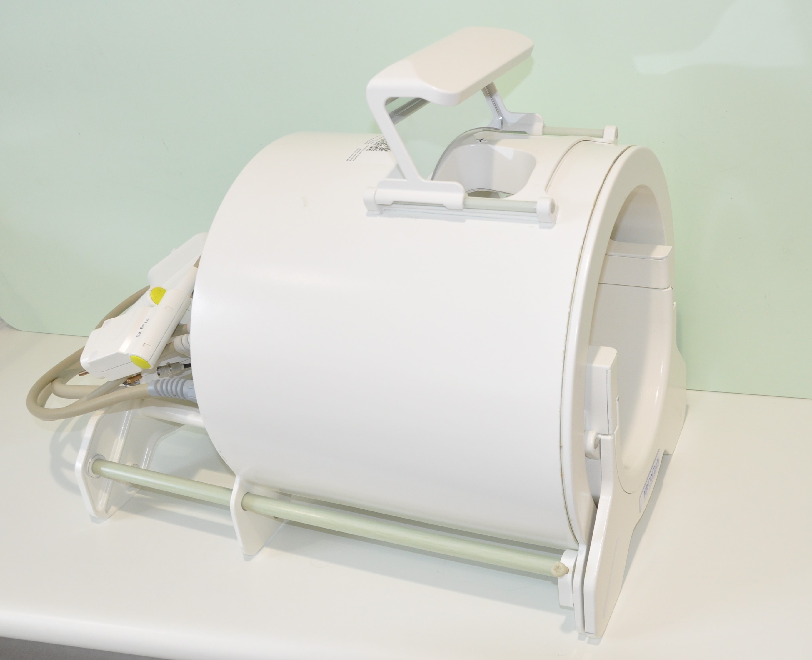
6 healthy subjects (age 45±15) were scanned in a MAGNETOM Terra 7T scanner (Siemens Healthineers, Erlangen, Germany) with an 8Tx64Rx head coil (Figure 1) (MR CoilTech Ltd, Glasgow, Scotland)10,11 using a GRE-EPI sequence developed in-house. A right-handed finger-tapping task fMRI study was performed. A block design paradigm with interleaving resting and finger-tapping blocks (shown in Figure 1) was instructed to the subject using the PsychoPy software12.Following localisation and B0 shimming, B1+ and B0 field mapping was performed at the slice positions used by the EPI protocol, and the results were used in MATLAB (MathWorks, Natick, MA, USA) offline to generate slice-specific B1+ shim weights13. T1-weighted data were also acquired using a 3D FLASH sequence. Following the structural scan, 4 EPI datasets were acquired in: circularly-polarised (CP) mode (equivalent to single transmission) without and with MS-PACE; B1+ shimming pTx mode.without and with MS-PACE. The scan parameters were otherwise identical: voxel size 2×2×2mm3, resolution 96×96, GRAPPA factor 3, 51 slices, 110 volumes, echo spacing 580ms, TR 4s, TE 18ms and acquisition time 7m32s. A 3-slice registration subset was used for real-time motion correction. Estimated motion parameters were used to update the scanner’s imaging gradients.
The correction robustness was evaluated post-acquisition by estimating residual rigid-body motion parameters. Image processing was done within the Image Calculation Environment (Siemens Healthineers, Erlangen, Germany) using ITK open-source libraries. The functional analysis maps were calculated using BrainVoyager (v.22.4, Brain Innovation BV, Maastricht, The Netherlands).
Results
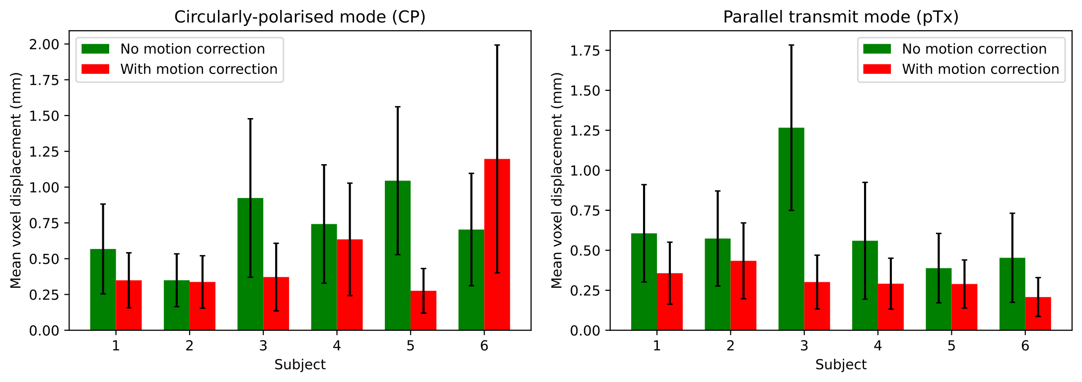
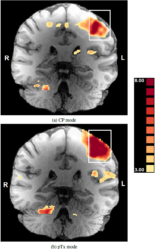
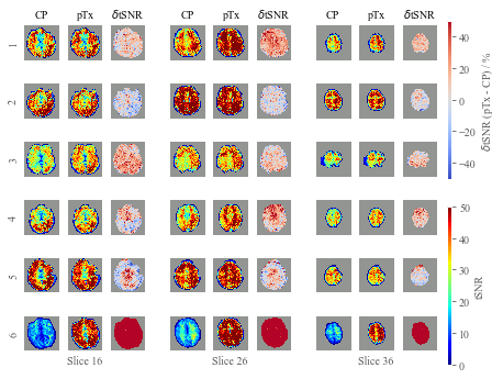
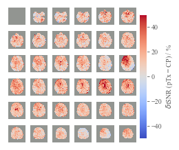
Figure 2 compares the mean voxel displacement for the acquisitions without (green) and with (red) MS-PACE in the CP and pTx scans. Figure 3 visualises the all-subject integrated general linear model (GLM) activation maps, comparing MS-PACE-corrected scans acquired in CP and pTx modes. Figure 4 displays the temporal SNR (tSNR) maps and percentage differences in tSNR between CP and pTx, δtSNR(pTx-CP) mode scans with MS-PACE in each subject. The average δtSNR(pTx-CP) of all subjects is then shown on Figure 5.Discussion
The technique was able to reduce overall motion in both CP and pTx modes. For subjects with both high and low levels of motion, a consistent reduction in residual motion is observed.The integration of slice-specific B1+ shimming with MS-PACE demonstrates an overall improvement in data quality. Comparing the group functional maps of the CP-mode and pTx-mode scans, the primary motor cortex activation is observed to be increased in the latter. There is also stronger activation in the ipsilateral cerebellum, which suffers from low B1+ at 7T14. Subject-wise tSNR comparison also shows positive changes when slice-specific B1+ shimming was used, although there are exceptions in some slices or subjects. Improvements are the most prominent in slices where the equivalent CP-acquired slices have low tSNR. Averaging the δtSNR across all subjects also demonstrates overall increases in tSNR with slice-specific B1+ shimming.
Conclusion
This study has implemented and validated an integration of real-time multislice-to-volume motion correction and pTx slice-specific B1+ shimming for task-based fMRI at 7T. It has been demonstrated that this combined approach harnessed both techniques’ advantages, providing a high spatial resolution and motion-agnostic option for 7T fMRI.Acknowledgements
The authors are grateful for the support of Kristian Stefanov at the University of Glasgow, Patrick Liebig, Jürgen Herrler and Robin Heidemann at Siemens Healthineers, Erlangen, Germany, and Iulius Dragonu and Radhouene Neji from Siemens Healthineers UK.References
[1] Herbst M et al. Reproduction of motion artifacts for performance analysis of prospective motion correction in MRI. Magn Reson Med. 2014;71:182-90.
[2] Maknojia S et al. Resting state fMRI: Going through the motions. Front Neurosci. 2019;13:1-13.
[3] Zaitsev M et al. Prospective motion correction in functional MRI. Neuroimage. 2017;154.
[4] Winata S et al. Multislice-to-volume Prospective Motion Correction for Functional MRI protocols at 7T. Proceedings of the 2023 Annual Meeting of the ISMRM (Toronto, ON, Canada, 3-8 June 2023).
[5] Winata S et al. Real-time multislice-to-volume motion correction for task-based functional MRI at 7T. 39th Annual Scientific Meeting of the ESMRMB (Basel, Switzerland, 4-7 October 2023).
[6] Hoinkiss DC, Porter DA. Prospective motion correction in 2D multishot MRI using EPI navigators and multislice-to-volume image registration. Magn Reson Med. 2017;78:2127-35.
[7] Hoinkiss DC et al. Prospective motion correction in functional MRI using simultaneous multislice imaging and multislice-to-volume image registration. Neuroimage. 2019;200:159-73.
[8] Thesen S et al. Prospective Acquisition Correction for head motion with image-based tracking for real-time fMRI. Magn Reson Med. 2000;44:457-65.
[9] Webb AG, Collins CM. Parallel transmit and receive technology in high-field magnetic resonance neuroimaging. Intl J Imaging Syst Technol. 2010; 20.1: 2–13.
[10] Williams SN et al. A Nested Eight-Channel Transmit Array with Open-Face Concept for Human Brain Imaging at 7 Tesla”. Front Phys. 2021;9.
[11] Shajan G, Feinberg D. An 8-channel transmit 64-channel receive compact head coil for Next Gen 7T scanner with head gradient insert”. Proceedings of the 2022 Annual Meeting of the ISMRM (London, England, UK, 7–12 May 2022).
[12] Peirce J et al. PsychoPy2: Experiments in behavior made easy. Behav Res Methods. 2019;51:195–203.
[13] Williams SN et al. Multi-Slice 2D pTx Readout-Segmented Diffusion-Weighted Imaging Using Slice-by-Slice B1+ Shimming. Proceedings of the 2021 Annual Meeting of the ISRM (Online, 15–20 May 2021).
[14] Padormo F et al. Parallel transmission for ultrahigh-field imaging. NMR Biomed. 2016;29.9:1145–1161.
Figures