2654
Gibbs Ringing Correction for non-Cartesian Acquisitions1Bernard and Irene Schwartz Center for Biomedical Imaging, Department of Radiology, New York University Grossman School of Medicine, New York, NY, United States, 2Center for Advanced Imaging Innovation and Research (CAI2R), Department of Radiology, New York University Grossman School of Medicine, New York, NY, United States, 3Department of Biomedical Engineering, New York University, Brooklyn, NY, United States, 4Athinoula A. Martinos Center for Biomedical Imaging, Charlestown, MA, United States, 5Department of Radiology, Harvard Medical School, Boston, MA, United States
Synopsis
Keywords: Artifacts, Artifacts
Motivation: Truncation in k-space leads to Gibbs ringing. Removal of Gibbs artifact for non-Cartesian isotropic sampling remains unaddressed.
Goal(s): To develop Gibbs ringing correction method for non-Cartesian isotropic k-space readouts.
Approach: We generalize the subvoxel-shift Gibbs ringing correction to isotropic sampling schemes.
Results: The developed correction removes Gibbs ringing for isotropic sampling schemes.
Impact: Gibbs ringing leads to artifacts and biases in parametric maps, especially in diffusion MRI. We generalize the subvoxel-shift Gibbs ringing correction, previously developed for cartesian EPI acquisitions, to non-Cartesian sampling. The method will increase the reproducibility of MRI processing pipelines.
Introduction
Gibbs ringing is a well-known MRI artifact that occurs near sharp boundaries due to truncation of the k-space1,2. In 1 dimension (1d), it can be understood as a result of convolving the image with the sinc function, which is the point-spread function (PSF)3,4 of a rectangular low-pass filter. This intuition is easily generalized to Cartesian sampling1 via a product of 1d PSFs in the x and y directions. For such cases, the de-facto standard Gibbs artifact correction is the subvoxel-shift method1, based on resampling images at the zeros of a sinc function (where Gibbs effect is minimized). The method1 was recently generalized for partial Fourier cartesian EPI acquisitions5.Here, we further generalize the subvoxel-shift method1 to non-Cartesian sampling by noticing that subvoxel-shifting sharp walls also minimizes the Gibbs effect.
Theory
For isotropic sampling schemes, with no preferential angle from the center of k-space such as spiral or radial acquisitions, with $$$k<k_{max}=\pi/a$$$, where a is the voxel size (henceforth set to 1 without the loss of generality), the Gibbs pattern is a result of the convolution with the corresponding PSF6.$$\mathrm{PSF}_{2d}(r)=\int_{k<k_\mathrm{max}}{d^2k\over(2\pi)^2}e^{ikr}={\pi\over2r}J_1(\pi{r})\quad(1)$$
where $$$J_1$$$ is the first-order Bessel function. Fig.1 shows the result of such convolution,
$$f_G(r)=R\int_0^{k_\mathrm{max}}\,dk\,J_0(kr)J_1(kR)$$
for a disk $$$f = \theta(R-r)$$$ of radius R, where $$$\theta(x)$$$ is a unit step function. We notice the following:
- Gibbs pattern for a disk in the radial direction is nearly identical numerically to that of the 1d case, where a rectangle $$$\theta(R-|x|)$$$is convolved with 1d $$$PSF_{1d}(x)=sinc(\pi x)$$$.
- The Gibbs pattern is notably minimized if disk radius R is close to half-voxel for large R>>1, much like in 1d. This behavior for small-curvature walls is explained by the connection to the 1d case in the zero-curvature limit: A strip with a straight wall sets the tangential projection of k to zero, which results in integrating the 2d $$$PSF_{2d}(r)$$$ along a tangential dimension, yielding $$$PSF_1d(x)$$$ and the exact 1d Gibbs pattern normal to the wall.
- If the disk shrinks, R~1, the optimal sub-voxel shift tends to a quarter of a voxel, which can be deduced from the asymptotic behavior of the Bessel function $$$J_1(z) \simeq \sqrt{2\over \pi z} \sin(z-\pi/4)$$$ entering Eq.(1), which means that the pattern of PSF zeros is shifted by a quarter-voxel relative to that of 1d sinc function, shown in Fig. 2. Importantly, the intervals between successive zeros are asymptotically equal to those for the 1d case (the overall shift is not important as the algorithm1 would find it automatically). Such asymptotic behavior already practically occurs for z~1.
Methods
The above considerations justify applying the subvoxel-shift method1 radially. As it is implemented for x and y directions1,3, we suggest applying the subvoxel-shift method also for a rotated k-space, e.g., by $$$\pi/4$$$, such that the Gibbs pattern in every direction is effectively eliminated, as shown in Fig.3.A phantom was created via the Michigan Image Reconstruction Toolbox (MIRT)7, and a spiral trajectory was simulated via vdspiral8. No $T_2^*$ decay was considered for this simulation. Reconstruction was performed offline via Matlab (Mathworks, USA) with NUFFT and Conjugate Gradient.
Results
Figure 1: Gibbs ringing patterns in 1d (blue), for a rectangle of length 2R, and 2d (red), for a disk of radius R, are very similar and strongly depend on the subvoxel shift of the object boundary. Top: zero-shift (boundary coincides with a voxel). Middle: half-voxel shift (minimal ringing). Bottom: L2-norm of deviation from an ideal object as a function of the subvoxel shift for different radius R=1,2,5,10.Figure 3 and 5: Comparing the undersampled to the Gibbs corrected image 1x and Gibbs corrected image 2x, the Gibbs artifact was reduced by ~49% for correction only in the x and y direction and reduced by ~58% for correction in the x and y along with the rotated x and y. The image is rotated in k-space by $$$\pi/4$$$, and Gibbs ringing correction was applied before rotating back to proper orientation. The 1D case is shown in Fig.5.
Discussion and Conclusion
We demonstrate the feasibility of Gibbs ringing artifact correction based on the radial local subvoxel shifts for spiral MRI, and by extension, non-Cartesian isotropic sampling schemes. The local subvoxel shifts are applied in only the x and y directions for cartesian EPI imaging. In this work, we applied the local subvoxel shifts by rotating the k-space by $$$\pi/4$$$ to eliminate the artifact approximately isotropically. Further work will focus on improving the isotropic correction by local subvoxel shifts in different directions.Acknowledgements
This work was performed under the rubric of the Center for Advanced Imaging Innovation and Research (CAI2R, www.cai2r.net), an NIBIB National Center for Biomedical Imaging and Bioengineering (NIH P41 EB017183).References
1. Kellner E, Dhital B, Kiselev VG, Reisert M. Gibbs-ringing artifact removal based on local subvoxel-shifts. Magn Reson Med. 2016 Nov;76(5):1574-1581. doi: 10.1002/mrm.26054. Epub 2015 Nov 24. PMID: 26745823.
2. Veraart J, Fieremans E, Jelescu IO, Knoll F, Novikov DS. Gibbs ringing in diffusion MRI. Magn Reson Med. 2016 Jul;76(1):301-14. doi: 10.1002/mrm.25866. Epub 2015 Aug 10. PMID: 26257388; PMCID: PMC4915073.
3. Feizollah S, Tardif CL. High-resolution diffusion-weighted imaging at 7 Tesla: Single-shot readout trajectories and their impact on signal-to-noise ratio, spatial resolution and accuracy. Neuroimage. 2023 Jul 1;274:120159. doi: 10.1016/j.neuroimage.2023.120159. Epub 2023 May 5. PMID: 37150332.
4. Tsai CM, Nishimura DG. Reduced aliasing artifacts using variable-density k-space sampling trajectories. Magn Reson Med. 2000 Mar;43(3):452-8. doi: 10.1002/(sici)1522-2594(200003)43:3<452::aid-mrm18>3.0.co;2-b. PMID: 10725889.
5. Lee HH, Novikov DS, Fieremans E. Removal of partial Fourier-induced Gibbs (RPG) ringing artifacts in MRI. Magn Reson Med. 2021 Nov;86(5):2733-2750. doi: 10.1002/mrm.28830. Epub 2021 Jul 5. PMID: 34227142; PMCID: PMC9212190.
6. Qin Q. Point spread functions of the T2 decay in k-space trajectories with long echo train. Magn Reson Imaging. 2012 Oct;30(8):1134-42. doi: 10.1016/j.mri.2012.04.017. Epub 2012 Jul 18. PMID: 22817958; PMCID: PMC3443331.
7. Fessler JA, “Michigan Image Reconstruction Toolbox” available at https://web.eecs.umich.edu/~fessler/code/
8. Lee JH, Hargreaves BA, Hu BS, Nishimura DG. Fast 3D imaging using variable-density spiral trajectories with applications to limb perfusion. Magn Reson Med. 2003 Dec;50(6):1276-85. doi: 10.1002/mrm.10644. PMID: 14648576.
Figures
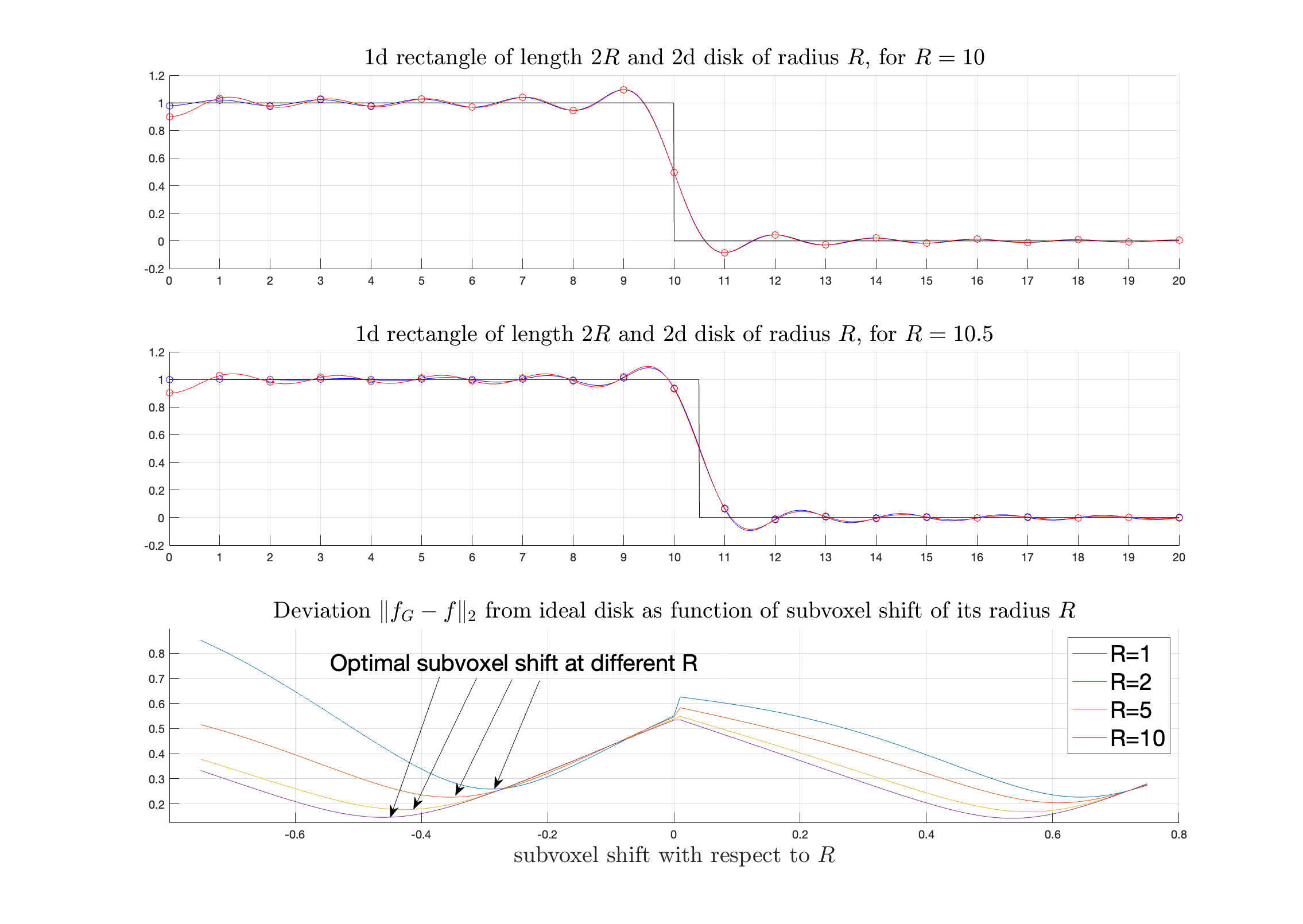
Figure 1: Comparison of Gibbs ringing oscillations between Cartesian and non-Cartesian isotropic acquisitions. Gibbs ringing pattern are shown for the 1d Cartesian case in blue and 2d radial case in red for R = 10 and R = 10.5. These Gibbs ringing patterns are similar to one another. The middle figure demonstrates the effect of a half voxel shift. The bottom figure shows the optimal subvoxel shift relative to radius R.
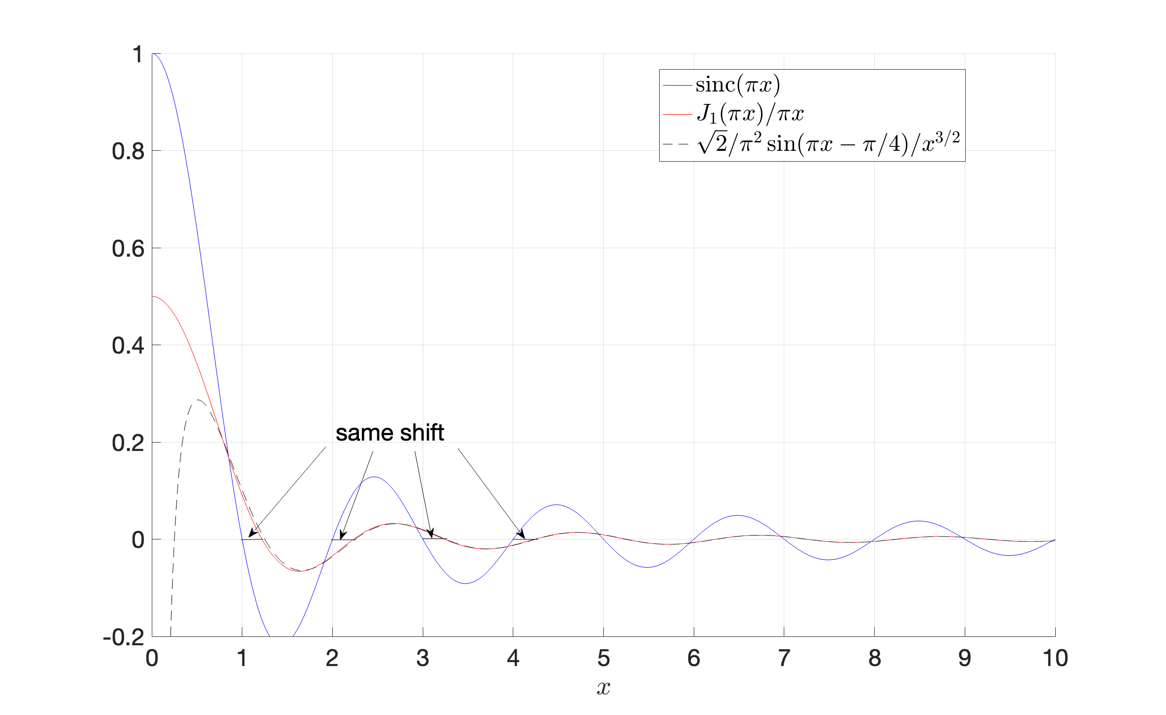
Figure 2: Point spread function (PSF) for Cartesian and non-Cartesian isotropic acquisition. Point spread function (PSF) of a disk-like k-space pattern (red) versus PSF for Cartesian 1d sampling (blue). The zeros of $$$J_1(\pi x)$$$ (PSF of a disk), are asymptotically given by that of a shifted sinc function and, hence, are nearly similar (up to an overall shift) to the zeros of a Cartesian PSF. This suggests that the subvoxel-shift method, based on resampling in the PSF zeros, will work well for disk-like sampled k-space.
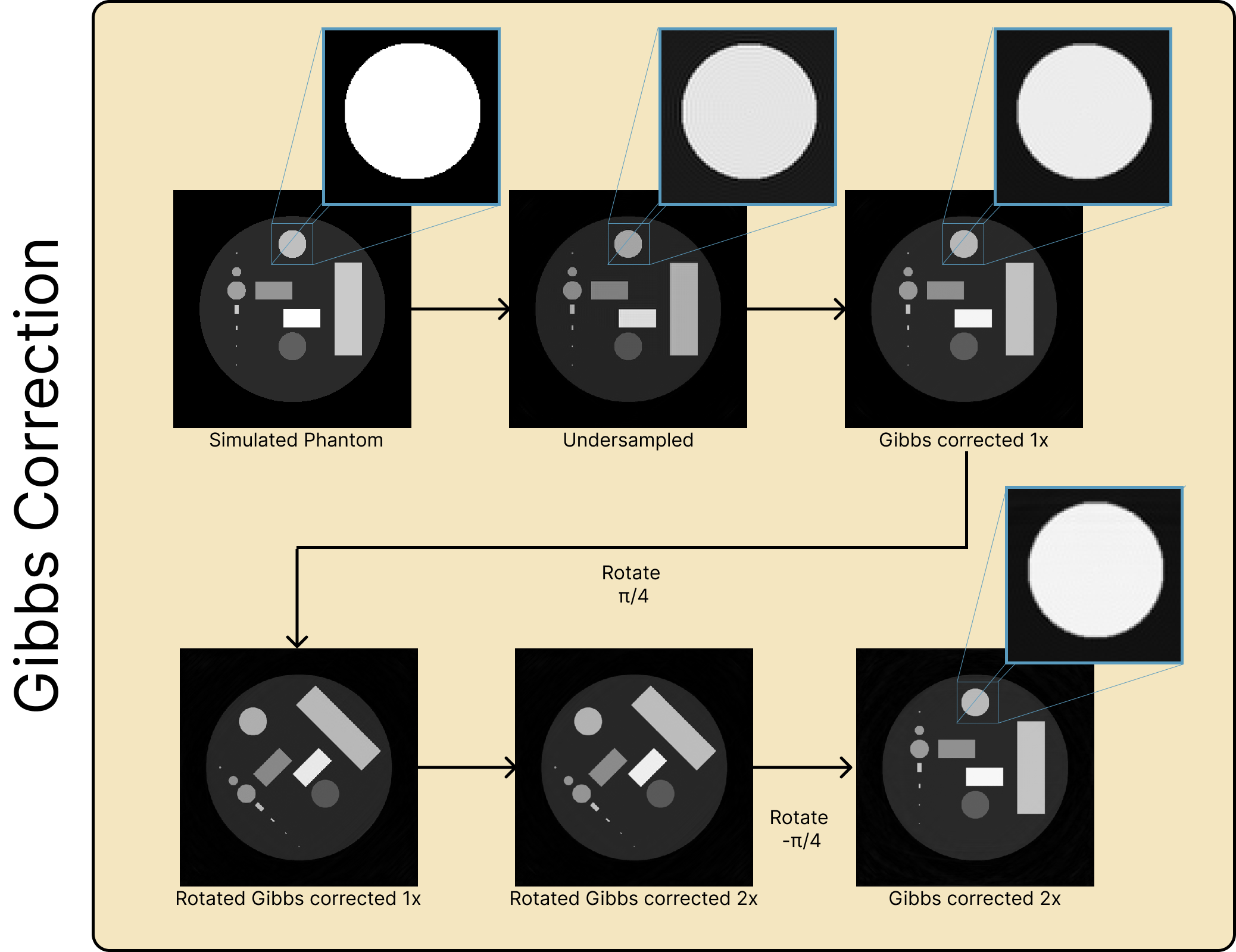
Figure 3: DeGibbs-ing pipeline for an image with a spiral trajectory. The simulated phantom is undersampled to create Gibbs artifacts. The phantom is then processed once (Gibbs corrected 1x) for artifacts in the x and y direction. The image is then rotated about $$$\pi/4$$$ to correct for the diagonal directions. Lastly, the image is rotated back (Gibbs corrected 2x).
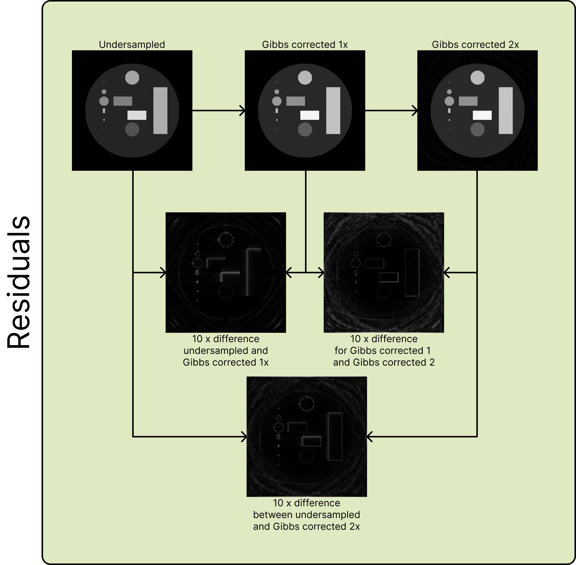
Figure 4: Residuals for the deGibbs-ing pipeline. Residuals are shown between the undersampled image (top left), Gibbs corrected 1x (top center), and Gibbs corrected 2x (top right). The residuals are magnified by 10x.
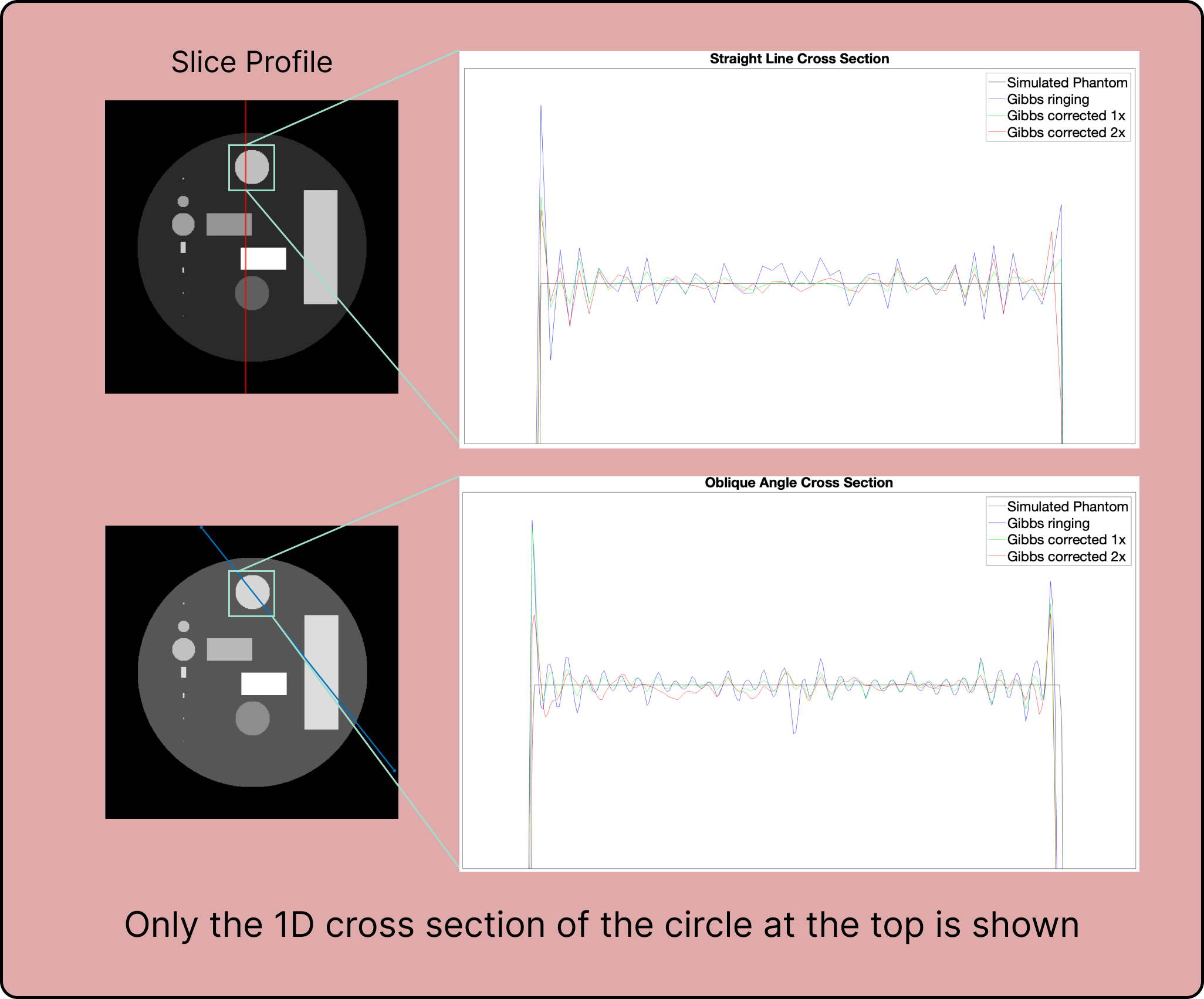
Figure 5: 1D cross sections. The 1d cross section is shown for the simulated phantom across the circle at the top for a straight line and at an oblique slice. For each image, the simulated phantom (black), undersampled (blue), Gibbs corrected 1x (green), and Gibbs corrected 2x (red) are compared to one another.