2653
Self-supervised Contrastive Learning for Automatic Image Quality Assessment in Whole-body MRI: Preliminary results in UK Biobank1University Hospital of Tübingen, Tübingen, Germany, 2University of Stuttgart, Stuttgart, Germany
Synopsis
Keywords: Artifacts, Artifacts, Image Quality Assessment, Motion Correction, Self-supervised Contrastive Learning
Motivation: MRI is vital for many medical decisions, yet susceptible to motion artifacts. Impairment by motion artifacts can reduce the reliability of diagnoses and a motion‐free reacquisition can become time-/cost‐intensive. Moreover, in large-scale cohorts, manual inspection is impractical. An automated quality assessment is desirable, but collection of motion-free references is challenging or even impractical.
Goal(s): We aim for automatic image quality assessment without extensive labeled training data.
Approach: We present a self-supervised quality classification framework based on SimCLR operating as zero-shot learning.
Results: The framework achieves promising results for binary quality classification, while showcasing its potential for future work as continuous quality score.
Impact: By automating MRI quality assessment, our approach helps in preventing artifact propagation into downstream tasks without additional efforts for manual inspection or data labeling.
Introduction
MR imaging provides valuable information for shaping medical decisions. However, patient motion such as breathing or rigid movements are still one of the main extrinsic sources of image quality degradation. To mitigate the risk of misdiagnosis due to artifact-affected images, the image quality needs to be evaluated. Manual inspection is time- and cost-intensive and may not be feasible for large-scale cohort data like the UK Biobank (UKB)1 or German National Cohort (NAKO)2. Therefore, an automated approach for assessing the image quality is desirable. Obtained quality scores can facilitate data classification or initiate retrospective motion correction3-8. Deep-learning-based approaches for automatic quality assessment have been previously proposed9-15. However, they often rely on quality labels as ground truth for training, which require extensive manual annotation. Moreover, the definition or collection of motion-free reference data is very demanding if not even impractical. Previously, we introduced a label-efficient approach using a ViT-UNet architecture, which is trained in a self-supervised manner with a supervised fine-tuning step, hence requiring only few labeled images16. In this work, we present an image quality classification framework based on the SimCLR17, employing self-supervised contrastive learning and entirely eliminating the dependence on labeled data.Methods
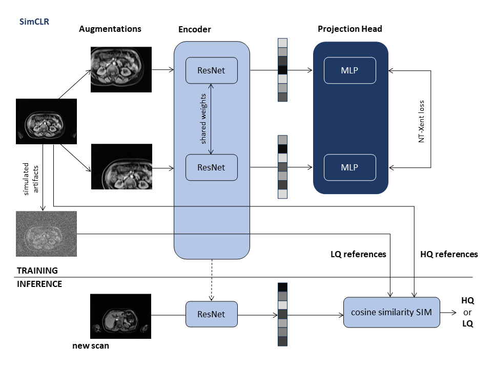
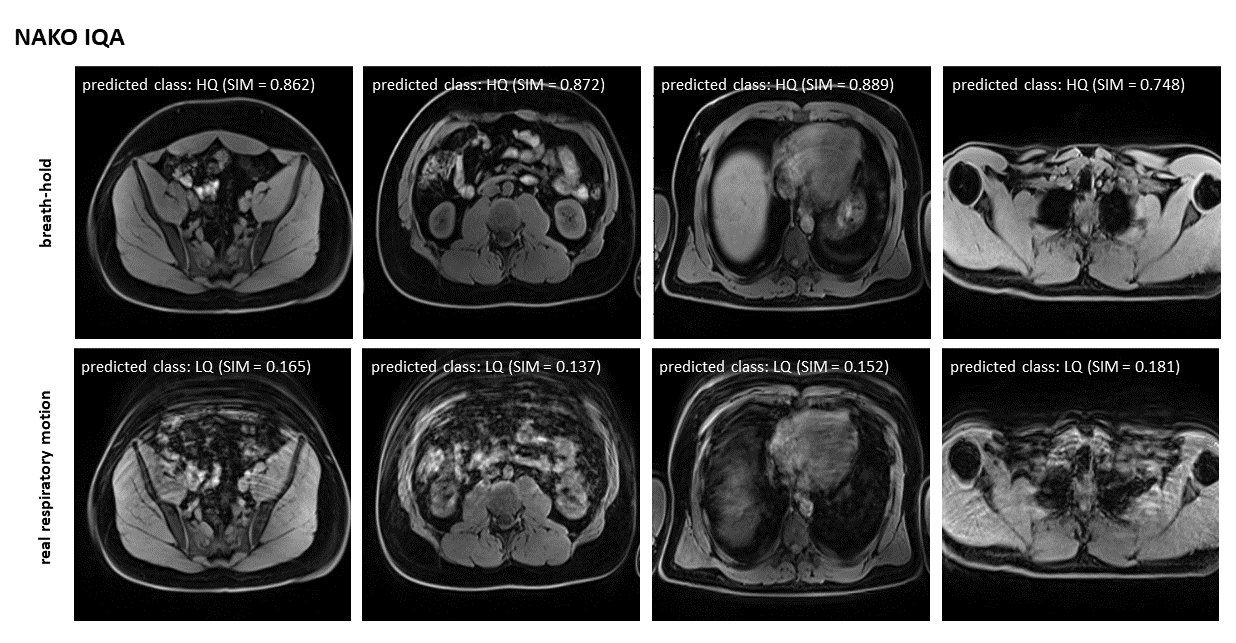
Data: The training data consists of 40,000 abdominal MRIs from the UK Biobank, acquired with a Dixon-3D dual-echo GRE sequence (1.5T Siemens Aera, resolution: 2.23×2.23×4mm3, TE/TR: 2.39 ms, 4.77 ms/6.69 ms, α: 10°). The water contrast images were used for training. For testing, 20 healthy subjects were scanned with a similar protocol (based on the NAKO cohort) which consists of breath-held and free-breathing abdominal MRIs (3T Siemens Skyra, resolution: 1.4×1.4×3mm3, TE/TR: 1.23ms, 2.46ms/ 4.36ms, α: 9°) (NAKO-IQA).Network: The network is based on the SimCLR17, which is trained to find a suitable feature embedding of the input image using self-supervised contrastive learning. During training, the network receives a pair of data augmented versions of the same 2D image slice as input. Augmentation methods include resizing and cropping. The image pairs are then passed through feature encoders (ResNet18) with shared weights and projection heads (two-layer perceptron). The contrastive loss (Normalized Temperature-Scaled Cross-Entropy) is designed to maximize the agreement between feature representations of similar pairs and maximize the distance between dissimilar pairs. During inference, the feature embeddings computed by the pre-trained encoder are used to determine the quality class by comparing a distance metric (cosine similarity) between reference images of high quality (HQ) and low quality (LQ) (Fig. 1). Manually inspected input scans of the UK Biobank serve as HQ reference data, while LQ data is generated by introducing simulated artifacts. The mean similarity is then computed to 20 LQ and HQ scans, considering individuals of the same sex, and similar height and weight. The quality class is assigned based on the higher mean value. The SIM metric, reflecting the quality level, is determined by the similarity value (predicted class: HQ) or by 1-similarity (predicted class: LQ).
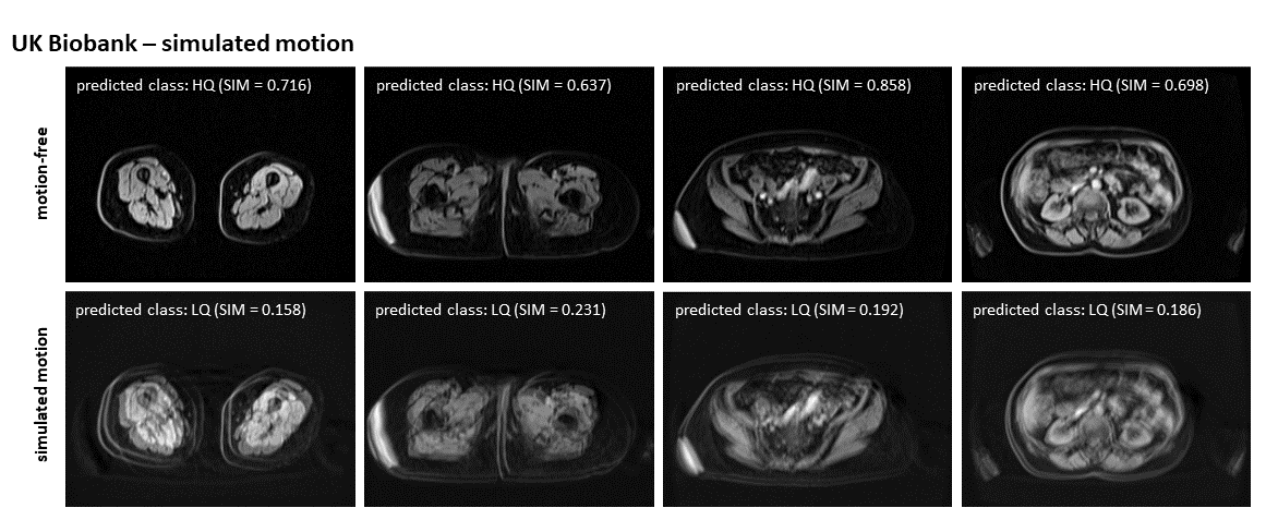
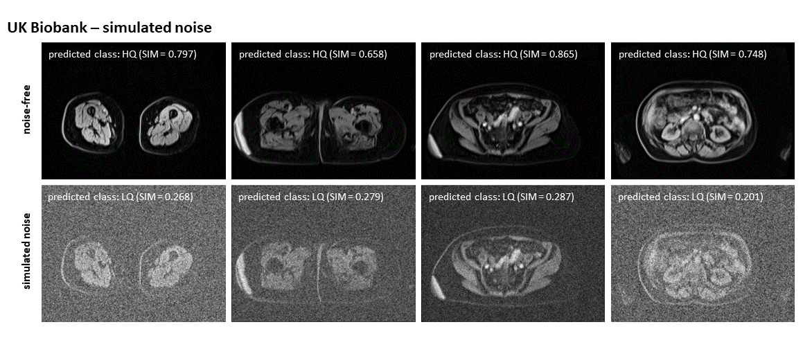
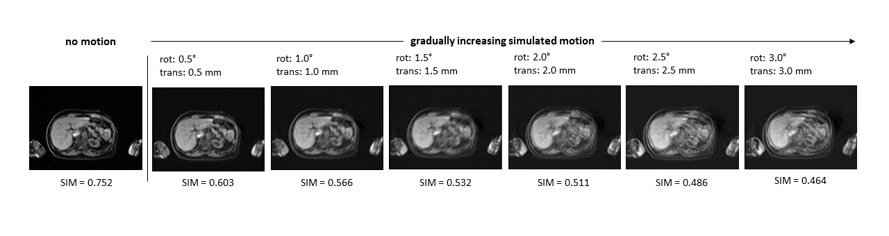
Training: Training was performed for 200 epochs using ADAM optimizer with a linear warmup scheduler on eight NVIDIA A100 GPUs. The framework was tested on 4174 simulated motion (translational: 1.0°, rotational: 1.0mm) and noise ($$$\mathcal{N}(0,0.25)$$$) cases of the UKB and 20 cases of motion-affected (free-breathing) and motion-free (breath-hold) images (NAKO-IQA data). To test the framework's potential in producing a continuous quality score, we conducted another experiment using gradually increasing rotational and translational motion on the UKB data.
Results and Discussion
We found that the framework is able to correctly classify HQ and LQ images for simulated motion (Fig. 2) and noise (Fig. 3) in the UKB. The framework also performs well for the NAKO-IQA data (Fig. 4), which the network has not seen during training and which contains real respiratory motion artifacts, indicating a robust performance across domains. While a binary classification is already beneficial, we aim to develop a continuous quality score. Preliminary results are promising, as the mean similarity to LQ images continuously increases while the image quality deteriorates (Fig. 5).We acknowledge several limitations. A more refined evaluation of image quality is desirable, necessitating further experiments to establish a reliable and conclusive numerical quality score. External validation methods for this score should be addressed. Additionally, testing image quality assessment during acquisition and under various imaging conditions is essential to progress toward our goal and will be implemented in the future.
Conclusion
Our self-supervised image quality assessment framework is trained without any labeled data and was tested in a binary classification setting on two different datasets. The framework performs well in the classification tasks and the results demonstrate potential for establishing a numerical quality score.Acknowledgements
Marcel Früh and Veronika Ecker contributed equally.
This work was funded by the Deutsche Forschungsgemeinschaft (DFG, German Research Foundation) project #428219130 and supported under Germany’s Excellence Strategy EXC 2064/1 #390727645. This work was carried out under UK Biobank Application 60520. We thank all participants who took part in the UKBB study and the staff in this research program.
References
1. Sudlow et al. UK biobank: An open access resource for identifying the causes of a wide range of complex diseases of middle and old age. vol. 12, no. 3, p. e1001779, 2015. Publisher: Public Library of Science.
2. Schlett et al. Population-Based Imaging and Radiomics: Rationale and Perspective of the German National Cohort MRI Study. RoFo Fortschr Gebiet Rontgenstrahlen Bildgebenden Verfahren. 2016;188(7):652–61
3. Küstner et al. Retrospective correction of motion-affected MR images using deep learning frameworks. Magnetic Resonance in Medicine 2019;82(4):1527-1540.
4. Lee et al. MC2-Net: motion correction network for multi-contrast brain MRI. Magnetic Resonance in Medicine 2021;86(2):1077-1092.
5. Levac et al. Accelerated Motion Correction for MRI Using Score-Based Generative Models. 2023 IEEE 20th International Symposium on Biomedical Imaging (ISBI), Cartagena, Colombia, 2023, pp. 1-5
6. Hossbach et al. Deep learning-based motion quantification from k-space for fast model-based magnetic resonance imaging motion correction. Med Phys. 2023 Apr;50(4):2148-2161.
7. Rizzuti et al. Joint Retrospective Motion Correction and Reconstruction for Brain MRI With a Reference Contrast. in IEEE Transactions on Computational Imaging, vol. 8, pp. 490-504, 2022.
8. Lee et al. MC2-Net: motion correction network for multi-contrast brain MRI. Magnetic Resonance in Medicine 2021;86(2):1077-1092.
9. Küstner et al. Automated reference-free detection of motion artifacts in magnetic resonance images. Magnetic Resonance Materials in Physics,Biology and Medicine 2018;31(2):243-256.:
10. Iglesias et al. Retrospective Head Motion Estimation in Structural Brain MRI with 3D CNNs. Springer, Cham; 2017. p 314-322.
11. Kapsner et al. Image quality assessment using deep learning in high b-value diffusion-weighted breast MRI. Sci Rep. 2023 Jun 29;13(1):10549.
12. Lei et al. Med Image Anal. 2022 Apr;77:102344.
13. Sujit et al. Automated image quality evaluation of structural brain MRI using an ensemble of deep learning networks. J Magn Reson Imaging. 2019 Oct;50(4):1260-1267.
14. Oksuz et al. Automatic CNN-based detection of cardiac MR motion artefacts using k-space data augmentation and curriculum learning. Medical Image Analysis 2019;55:136-147.
15. Kastryulin et al. Image quality assessment for magnetic resonance imaging. IEEE Access, 11:14154–14168, 2023.
16. Küstner et al. Self-supervised contrastive learning for motion artiact detection in whole-body MRI. Quality assessment across multiple cohorts. ISMRM. 2023.
17. Chen et al. A simple framework for contrastive learning of visual representations, 2020.
Figures