2652
Evaluation of Accelerated SEMAC at 0.55T using Hexagonal Sampling and Parallel Imaging1Electrical and Computer Engineering, University of Sourthern California, Los Angeles, CA, United States, 2Biomedical Engineering, University of Southern California, Los Angeles, CA, United States, 3Radiology, Stanford University, Stanford, CA, United States, 4Electrical Engineering, Stanford University, Stanford, CA, United States, 5Bioengineering, Stanford University, Stanford, CA, United States
Synopsis
Keywords: Artifacts, Low-Field MRI
Motivation: Low-field and mid-field MRI systems have tremendous potential for imaging near metal implants with reduced artifacts, but reduced ability to do parallel imaging limits encoding options.
Goal(s): To achieve 50% scan time reduction in addition to conventional parallel imaging at 0.55T.
Approach: Hexagonal sampling (in ky-kf space) combined with GRAPPA-2 in SEMAC acquisitions was evaluated qualitatively and quantitatively via phantom and in vivo experiments at 0.55T.
Results: GRAPPA-2 hexagonal sampling achieved comparable image quality to conventional GRAPPA-2 SEMAC with slight SNR reduction and a modest increase in artifact area, while allowing a 50% decrease in scan time.
Impact: We evaluate the performance of hexagonal sampling combined with GRAPPA-2 at 0.55T where high parallel imaging factors are challenging via phantom and in vivo experiments. 50% additional scan time reduction is achieved with a modest increase in artifact area.
INTRODUCTION
Metallic implants cause significant distortion in the static magnetic field1,2. The resulting artifacts can be mitigated using multi-spectral imaging (MSI) techniques, namely, MAVRIC, MAVRIC-SL, and SEMAC3,4. MSI requires longer scan times that are partially compensated using parallel imaging, partial Fourier, and/or compressed-sensing5,6.Imaging near metallic implants at lower-field strengths7,8 is promising due to substantially reduced off-resonance9,10. However, the parallel imaging factor is limited at low-to-mid field (<1.5T) because achieving body noise dominance limits the ability to use high channel-count arrays with small coils11.
In this work, we evaluate the use of hexagonal sampling combined with GRAPPA-2 to halve the scan time of conventional GRAPPA-2 SEMAC acquisitions at 0.55T. The technique was shown to achieve a 50% scan time reduction without introducing additional artifacts at 1.5T12. Performance is evaluated using phantom and in-vivo experiments.
METHODS
Experiments: Experiments were performed using a whole-body 0.55T MRI (prototype MAGNETOM Aera, Siemens Healthineers, Erlangen, Germany) with high-performance shielded gradients (45mT/m amplitude, 200T/m/s slew rate). The 6-channel body coil (anterior) and 6-elements of the table-integrated spine array were used for signal reception. Phantom and in-vivo experiments were performed with GRAPPA-2 SEMAC acquisitions in coronal orientation.Phantom: A hip implant with a cobalt-chromium head and titanium stem was scanned with parameters: 300x244mm2 FOV, 320x260 matrix size, 20 slices, 4mm slice thickness, SEMAC factor 12.
In-Vivo: One volunteer with a hip implant consisting of a titanium stem and a high susceptibility head was scanned after providing written informed consent and under a protocol approved by our IRB. Scan parameters: 280x228mm2 FOV, 320x260 matrix size, 75% phase oversampling (to prevent aliasing from the other hip), 20 slices, 4mm slice thickness, SEMAC factor 8.
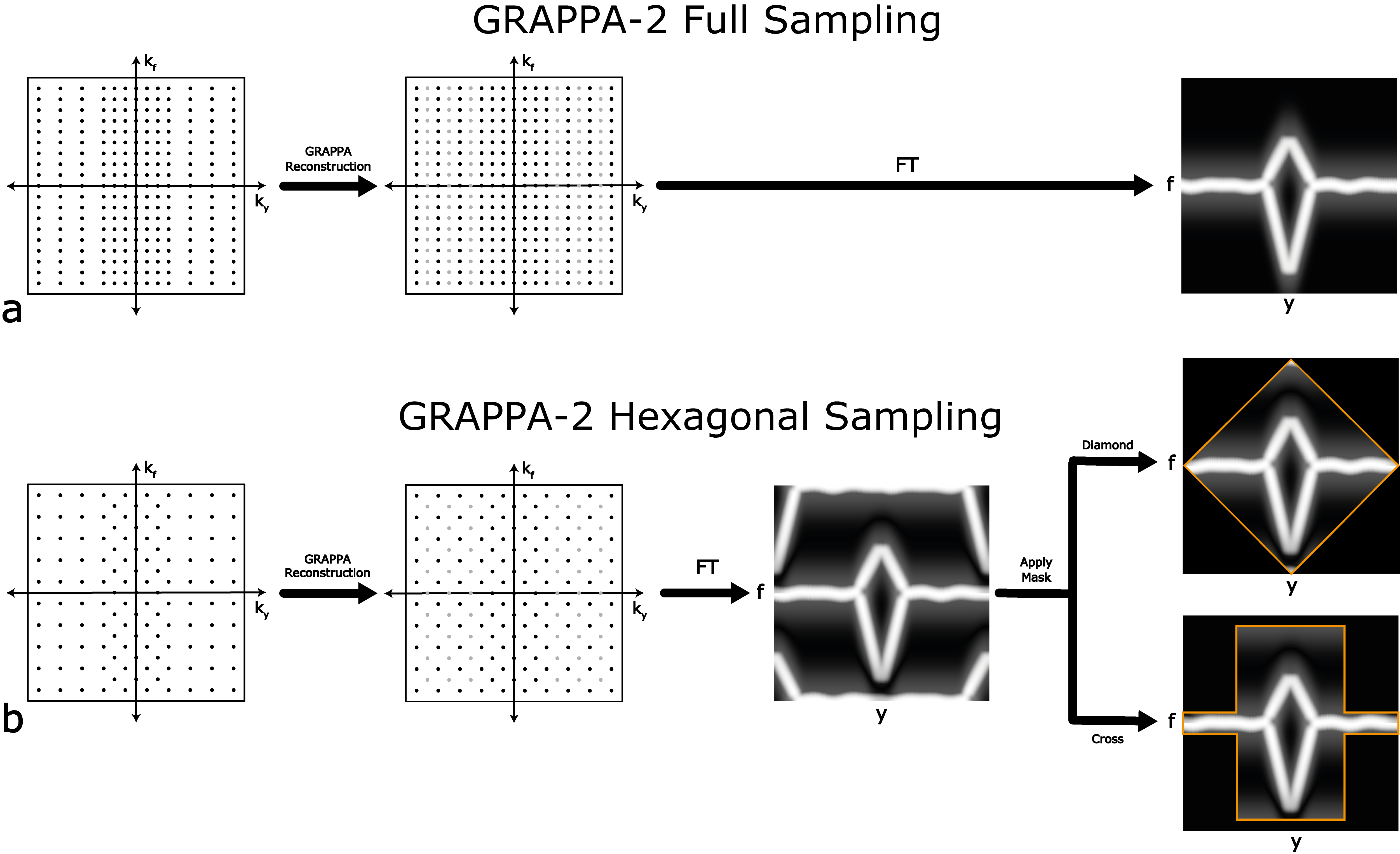
Image Reconstruction & Analysis: Figure 1 illustrates conventional GRAPPA-accelerated SEMAC and GRAPPA+hexagonal sampling. For the hexagonal case, 3D-GRAPPA is performed with a kernel size [5,5,3] along x-,y- and f- (bin) directions, respectively, to fill k-space in a checkerboard fashion (gray-dots reconstructed). The reconstructed k-space had the full hexagonal pattern, leading to aliasing replicas centered at the corners in y-f space. “Diamond” or “Cross” masks were applied to remove replicas while preserving the target y-f signal. The diamond and cross masks were designed to prioritize robustness against residual artifacts and sensitivity against implant position along y-direction. The performance of two masks with GRAPPA reconstruction is quantitatively compared by measuring the signal void artifact area and computing the noise levels as a function of y-position. After reconstruction, images were obtained by adaptive coil combination13 followed by the sum-of-squares (SOS) reconstruction of SEMAC images14.
RESULTS
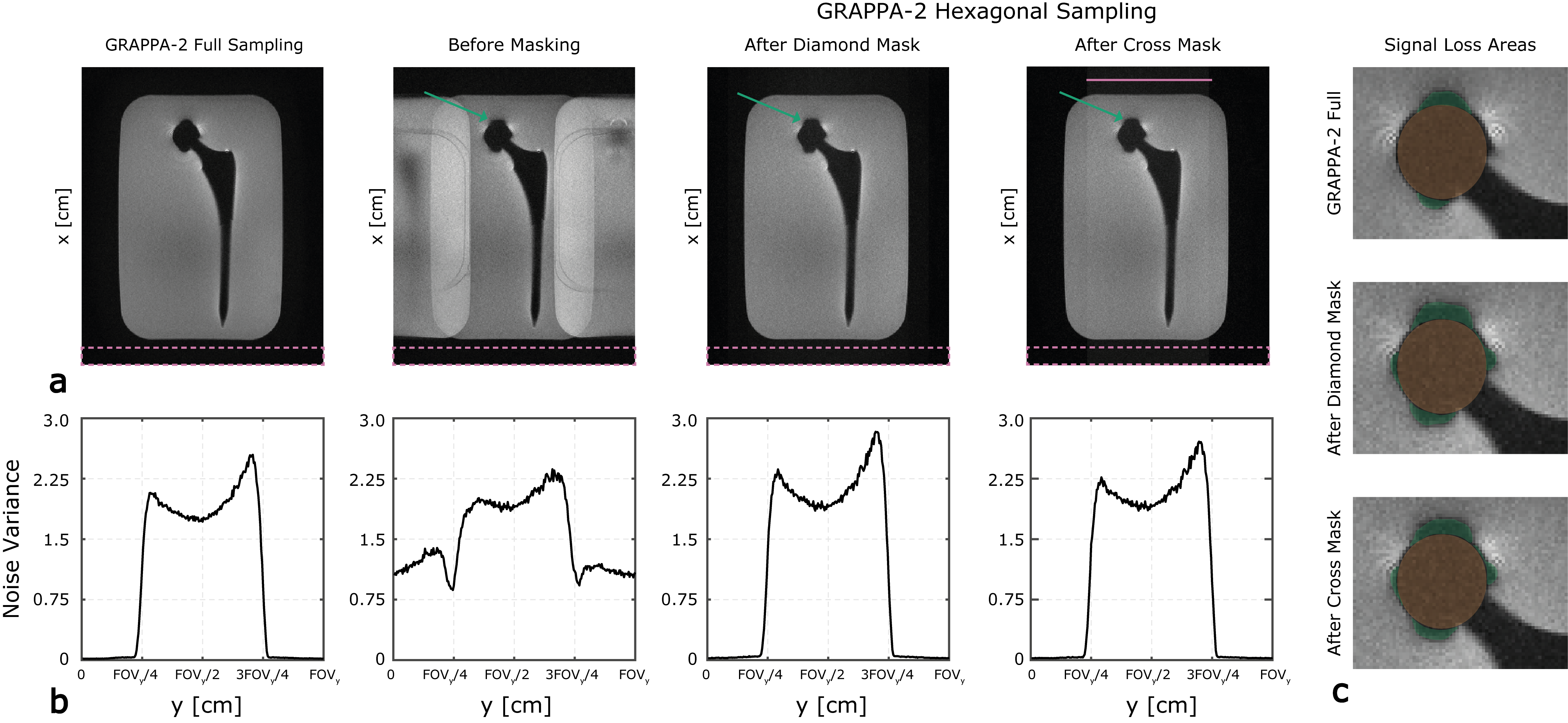
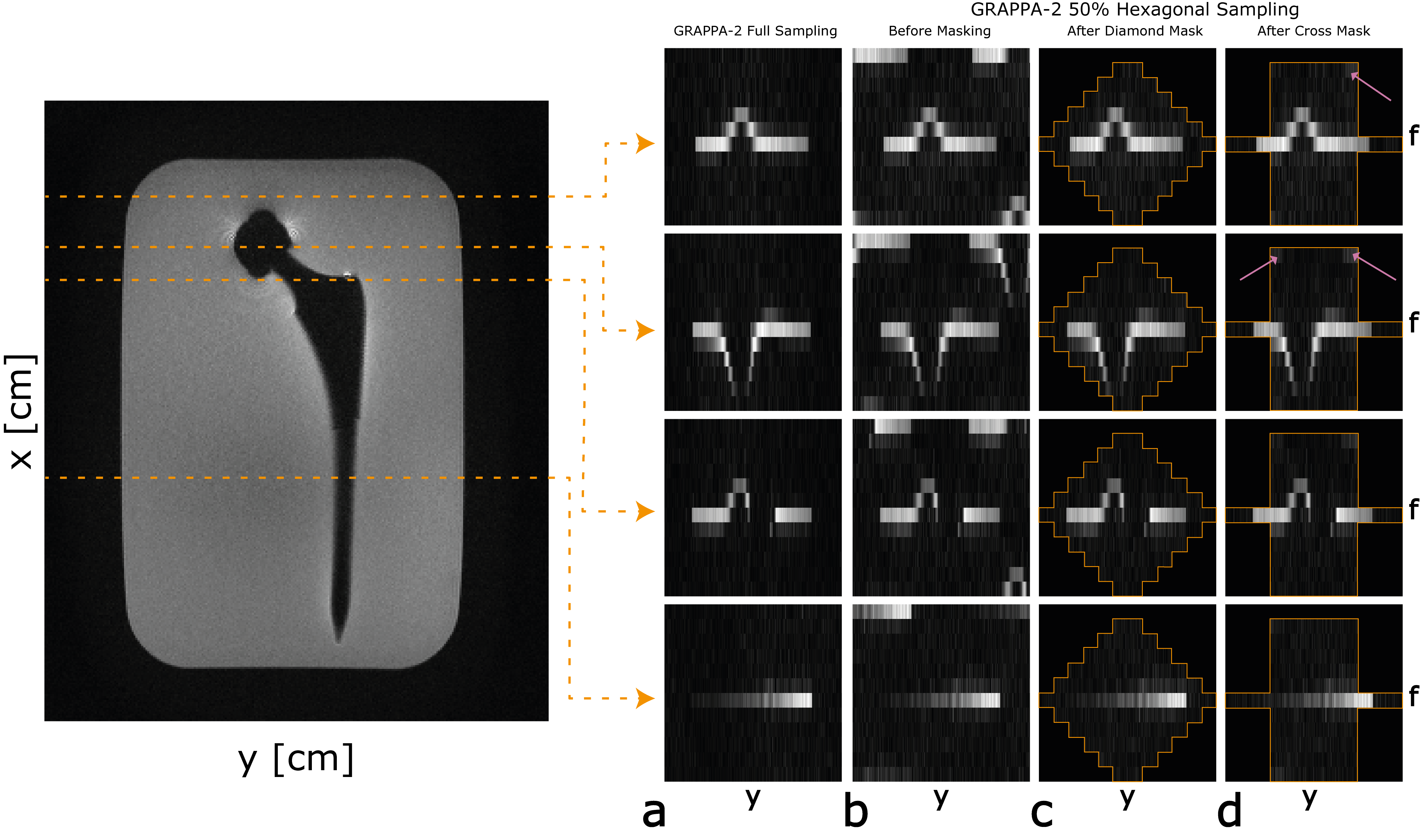
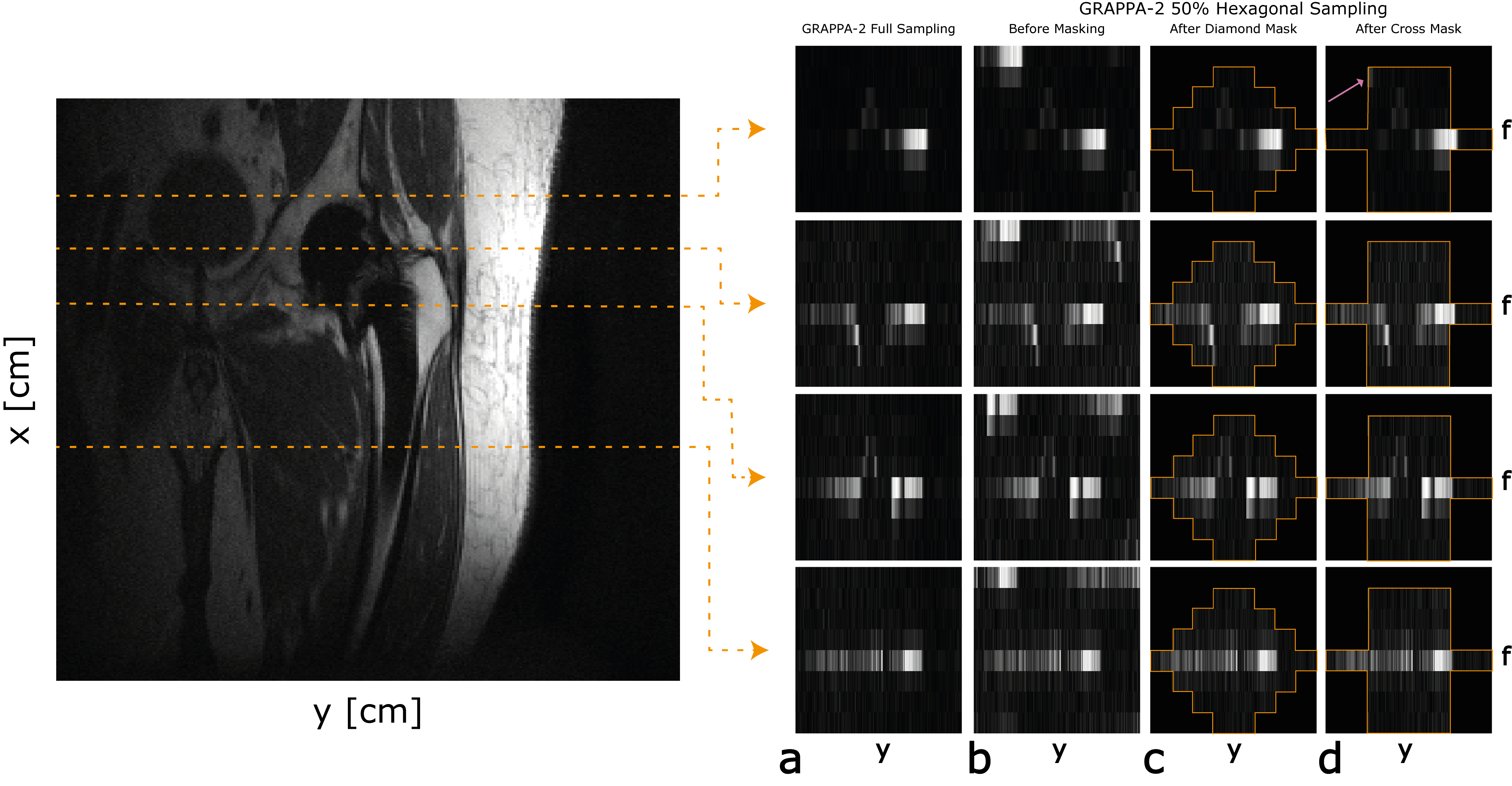
Figure 2 shows reconstructed SEMAC images acquired from the central slice of the hip implant phantom. Qualitatively, GRAPPA+hexagonal with diamond and cross masks appear comparable to the GRAPPA only. Masks suffer from slight signal loss around the head (green arrows). Small residual artifacts are observed for the cross mask (pink solid line). While slight SNR reduction is observed for diamond and cross cases, the shape of the masks has minimal effect on SNR along y-direction.Figure 3 illustrates y-f profiles from the phantom (from yellow-dashed lines). The replicas are centered at the diagonal corners, and SNR is slightly reduced for hexagonal sampling. Leakage from replicas is apparent (pink arrow) for “cross” mask due to its mask shape. “Diamond” mask eliminated the replicas while preserving the target profile.
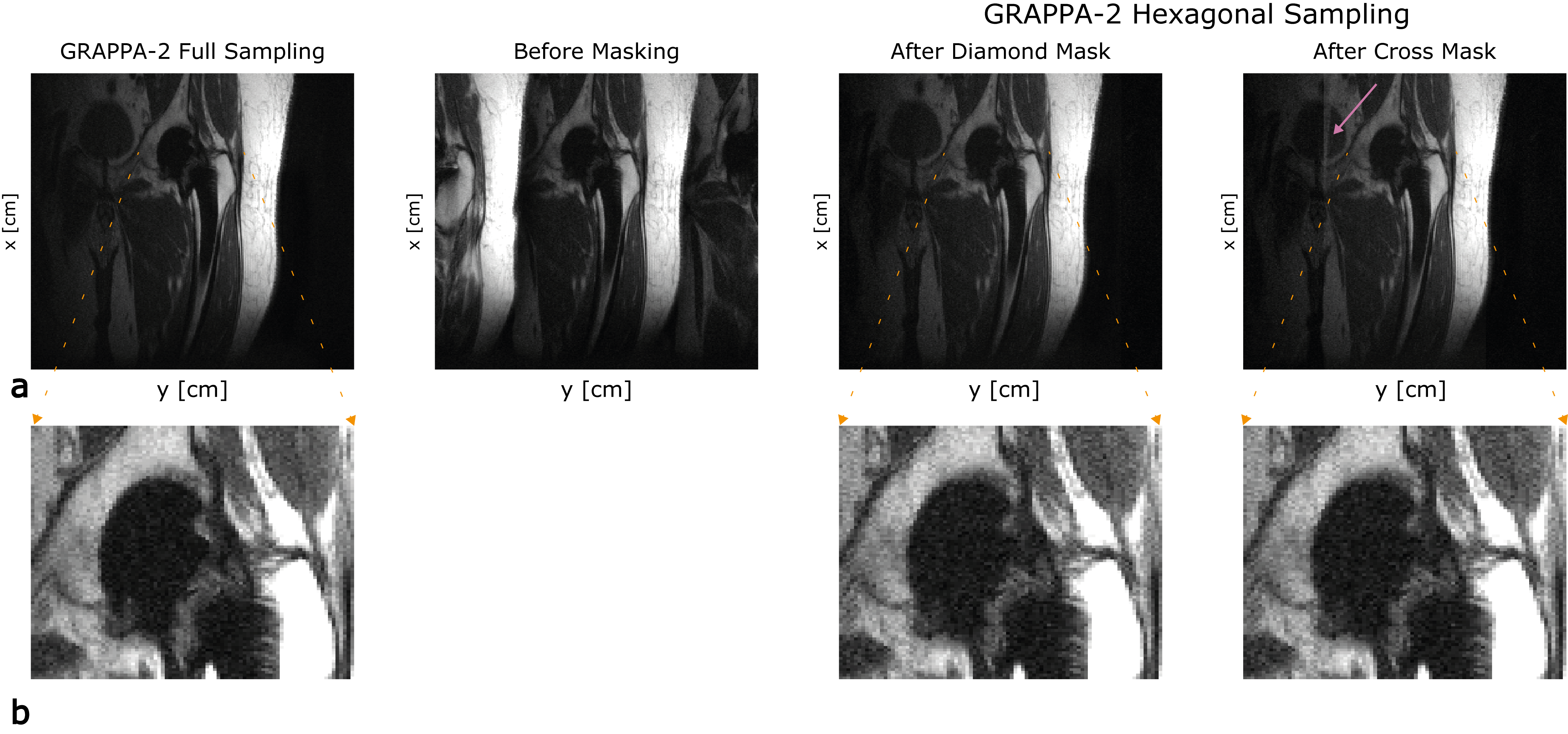
Figure 4 shows the SEMAC images acquired from a volunteer. Both masks preserved the off-resonance profile (2% signal loss for both masks). “Cross” mask significantly suffered from residual artifact (pink arrow) due to the unsuppressed fat signal from the replicas. “Diamond” mask achieved comparable image quality to the GRAPPA-2 full sampling case.
Figure 5 illustrates y-f profiles for the in-vivo experiment. The targeted y-f profile is maintained for both masks, verifying in-vivo SEMAC reconstructed images. The residual artifact is shown by the pink arrow for “cross” mask case.
DISCUSSION
We demonstrate that hexagonal sampling can be combined with GRAPPA-2 for accelerated SEMAC at 0.55T, where high parallel imaging factors are challenging. Masks can be optimized to improve robustness to implant placement and residual leakage. We suspect that mask centralization (along the y-direction) will improve the performance of “diamond” mask. Automatic mask centralization15 will be investigated as a future work. The technique will benefit from testing in a larger number of THA subjects with different material compositions, and in subjects with different orthopedic implants, such as spinal-fixation or knee replacement.CONCLUSION
Hexagonal sampling combined with GRAPPA in SEMAC achieves an additional 50% scan time reduction. The necessary tradeoff is SNR, based on reduced scan time and a small increase in artifact area.Acknowledgements
We acknowledge grant support from the National Institutes of Health (R01-AR078912) and National Science Foundation (Award #1828736), and research support from Siemens Healthineers.References
- Hargreaves B. A., Worters P. W., Pauly K. B., Pauly J. M., Koch K. M., & Gold G. E. (2011). Metal-Induced Artifacts in MRI. American Journal of Roentgenology, 197(3), 547–555. doi: 10.2214/AJR.11.7364Koch K.
- Dillenseger J. P., Molière S., Choquet P., Goetz C., Ehlinger M., & Bierry G. (2016). An illustrative review to understand and manage metal-induced artifacts in musculoskeletal MRI: a primer and updates. Skeletal Radiology, 45(5), 677–688. doi: 10.1007/s00256-016-2338-2
- Koch K. M., Hargreaves B. A., Pauly K. B., Chen W., Gold G. E., & King K. F. (2010). Magnetic resonance imaging near metal implants. Journal of Magnetic Resonance Imaging, 32(4), 773–787. doi: 10.1002/jmri.22313
- Jungmann P. M., Agten C. A., Pfirrmann C. W., & Sutter R. (2017). Advances in MRI around metal. Journal of Magnetic Resonance Imaging,46(4), 972–991. doi: 10.1002/jmri.25708
- Hargreaves B. A., Chen W., Lu W., Alley M. T., Gold G. E., Brau A. C. S., Pauly J. M., & Pauly K. B. (2010). Accelerated slice encoding for metal artifact correction. Journal of Magnetic Resonance Imaging, 31(4), 987–996. doi: 10.1002/jmri.22112
- Fritz J., Ahlawat S., Demehri S., Thawait G. K., Raithel E., Gilson W. D & Nittka M. (2016) Compressed Sensing SEMAC: 8-fold Accelerated High-Resolution Metal Artifact Reduction MRI of Cobalt-Chromium Knee Arthroplasty Implants. Invest Radiol. 51(10), 666-76. doi: 10.1097/RLI.0000000000000317. PMID: 27518214.
- Khodarahmi I., Brinkmann I. M., Lin D. J., Bruno M., Johnson P. M., Knoll F., Keerthivasan M. B., Chandarana H., & Fritz J. (2022). New-Generation Low-Field Magnetic Resonance Imaging of Hip Arthroplasty Implants Using Slice Encoding for Metal Artifact Correction. Investigative Radiology, 57(8), 517–526. doi. 10.1097/RLI.0000000000000866
- Khodarahmi I., Keerthivasan M.B., Brinkmann I.M., Grodzki D. and Fritz J., (2023). Modern Low-Field MRI of the Musculoskeletal System: Practice Considerations, Opportunities, and Challenges. Invest Radiol. 58(1), 76-87. doi: 10.1097/RLI.0000000000000912. Epub 2022 Sep 13. PMID: 36165841.
- Marques J. P., Simonis F. F. J., & Webb A. G. (2019). Low‐field MRI: An MR physics perspective. Journal of Magnetic Resonance Imaging,49(6), 1528–1542. doi: 10.1002/jmri.26637
- Campbell-Washburn A. E., Ramasawmy R., Restivo M. C., Bhattacharya I., Basar B., Herzka D. A., Hansen M. S., Rogers T., Bandettini W.P., McGuirt D. R., Mancini C., Grodzki D., Schneider R., Majeed W., Bhat H., Xue H., Moss J., Malayeri A. A., Jones E. C., … Balaban R. S. (2019). Opportunities in Interventional and Diagnostic Imaging by Using High-Performance Low-Field-Strength MRI. Radiology, 293(2), 384–393. doi: 10.1148/radiol.2019190452
- Gruber B., Froeling M., Leiner T., & Klomp D. W. J. (2018). RF coils: A practical guide for nonphysicists. Journal of Magnetic Resonance Imaging, 48(3), 590–604. doi: 10.1002/jmri.26187
- Sveinsson B., Worters P. W., Gold G. E., & Hargreaves B. A. (2015). Hexagonal undersampling for faster MRI near metallic implants. Magn Reson Med. 73(2), 662-8. doi: 10.1002/mrm.25132. Epub 2014 Feb 18. PMID: 24549782; PMCID: PMC4156560.
- Walsh D.O., Gmitro A.F., Marcellin M.W. (2000). Adaptive reconstruction of phased array MR imagery. Magn Reson Med.;43(5), 682-690. doi:10.1002/(SICI)1522-2594(200005)43:5<682::AID-MRM10>3.0.CO;2-G
- Lu W., Pauly K.B., Gold G.E., Pauly J.M., Hargreaves B.A. (2009) SEMAC: slice encoding for metal artifact correction in MRI. Magn Reson Med. 62(1), 66-76. doi:10.1002/mrm.21967
- Sveinsson B., Taviani V., Gold G., & Hargreaves B. (2015). Automatic Detection of Metal Implant Location in Hexagonally Sampled MAVRIC-SL. In Proc. Intl. Soc. Mag. Reson. Med (Vol. 23, p. 2508).
Figures