2650
Imaging Near Metallic Implants: Initial 0.55T vs. 3T MRI Comparison in Hip Implant Patients1Electrical and Computer Engineering, University of Southern California, Los Angeles, CA, United States, 2Siemens Healthineers, Los Angeles, CA, United States, 3Biomedical Engineering, University of Southern California, Los Angeles, CA, United States, 4Diagnostic Radiology, University of California Los Angeles, Los Angeles, CA, United States, 5Gerling Institute, New York, NY, United States, 6Orthopaedic Surgery, University of Southern California, Los Angeles, CA, United States, 7Radiology, Stanford University, Stanford, CA, United States, 8Electrical Engineering, Stanford University, Stanford, CA, United States, 9Bioengineering, Stanford University, Stanford, CA, United States
Synopsis
Keywords: Artifacts, Low-Field MRI, Artifacts, MSK, Metallic Implants, Susceptibility, Multi-Spectral Imaging, SEMAC, Metal artifacts
Motivation: Patients with orthopedic metallic implants often require diagnostic imaging to evaluate adjacent tissues. MRI performance, including artifact levels and SNR, varies with field strength.
Goal(s): To compare 0.55T and 3T MRI for imaging patients with total hip arthroplasty (THA).
Approach: Patients with THA were scanned with similar protocols at 0.55T and 3T, including multi-spectral imaging (MSI). We qualitatively compared the images from both scanners.
Results: Metal artifact severity was reduced at 0.55T compared to 3T at the expense of SNR. Diagnostic imaging of patients with titanium hip implants at 0.55T is possible without MSI.
Impact: We qualitatively compared 0.55T and 3T image quality in patients with hip replacements. Our findings indicate that 0.55T MRI offers substantially reduced metal artifacts and advanced multi-spectral techniques may not be required in many cases.
Introduction
Orthopedic metallic implants improve quality of life, increasing mobility and possibly longevity. MRI offers an essential non-invasive imaging tool to provide soft tissue contrast to evaluate the tissues near the metallic implants, satisfying a critical need in patients with metal implants. However, artifacts occur due to magnetic susceptibility differences between tissues and implants1,2. Multi-spectral imaging (MSI) techniques are commonly used to reduce the artifacts3,4,5. Recent work with implant phantoms has demonstrated that metal artifacts are significantly reduced at lower field strengths, with fewer spectral encodings required for effective artifact mitigation6. Residual artifacts depend on the material composition, with titanium alloys producing the smallest artifacts compared to cobalt-chromium and stainless steel7. Titanium implants may be imaged using optimized turbo spin echo (TSE) sequences at 0.55T6. In this study, we scanned patients with total hip arthroplasty (THA) implants at both 0.55T and 3T field strengths using similar protocols, comparing SNR and severity of metal artifacts.Methods
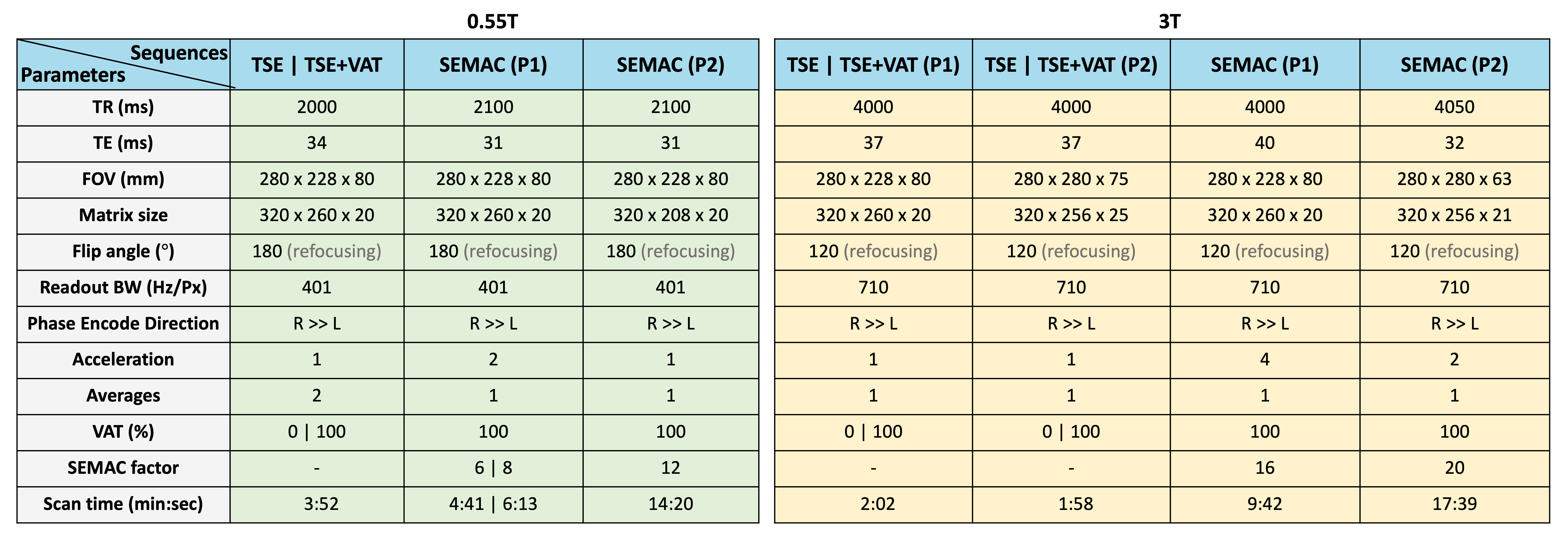
0.55T experiments were performed on a whole-body 0.55T scanner (prototype MAGNETOM Aera, Siemens Healthineers, Erlangen, Germany) equipped with shielded gradients (45 mT/m amplitude, 200 T/m/s slew rate). Data were collected using 6 elements of a table-integrated spine array (posterior) and a 6-channel body coil (anterior). 3T experiments were performed on a whole-body 3T scanner (MAGNETOM Prisma Fit, Siemens Healthineers, Erlangen, Germany) equipped with gradients (80 mT/m amplitude, 200 T/m/s slew rate). Data were collected using 4 elements of a table-integrated spine array (posterior) and an 18-channel body coil (anterior).We employed standard clinical sequences including Turbo Spin Echo (TSE), TSE with View-Angle Tilting (TSE-VAT), Slice Encoding for Metal Artifact Correction (SEMAC), and Short Tau Inversion Recovery with SEMAC (STIR-SEMAC) at both field strengths. Imaging was performed with a coronal prescription with respect to the alignment of the implant stem. Detailed scan parameters are listed in Table 1. The diagnostic quality of the images was evaluated by a board-certified MSK radiologist with 10 years of experience. All patients were scanned under a protocol approved by our institutional review board after providing written informed consent.
Results
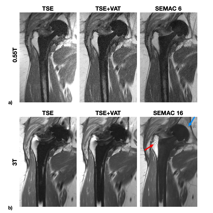
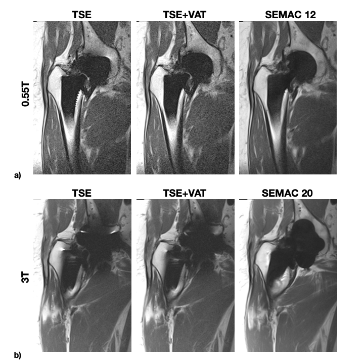
Figure 1 illustrates a comparison of TSE, TSE with VAT, and SEMAC sequences at a) 0.55T and b) 3T for a 58-year-old woman with a titanium THA. Imaging of the titanium implant at 0.55T produces diagnostic quality images for all the sequences, versus imaging at 3T which still requires MSI techniques to reduce artifact.Figure 2 illustrates a comparison of TSE, TSE with VAT, and SEMAC sequences at a) 0.55T and b) 3T for a 79-year-old man with a higher magnetic susceptibility THA. Tissues in close proximity of the implant are visible at 0.55T and are obscured by the metal artifacts at 3T even with a SEMAC factor of 20.
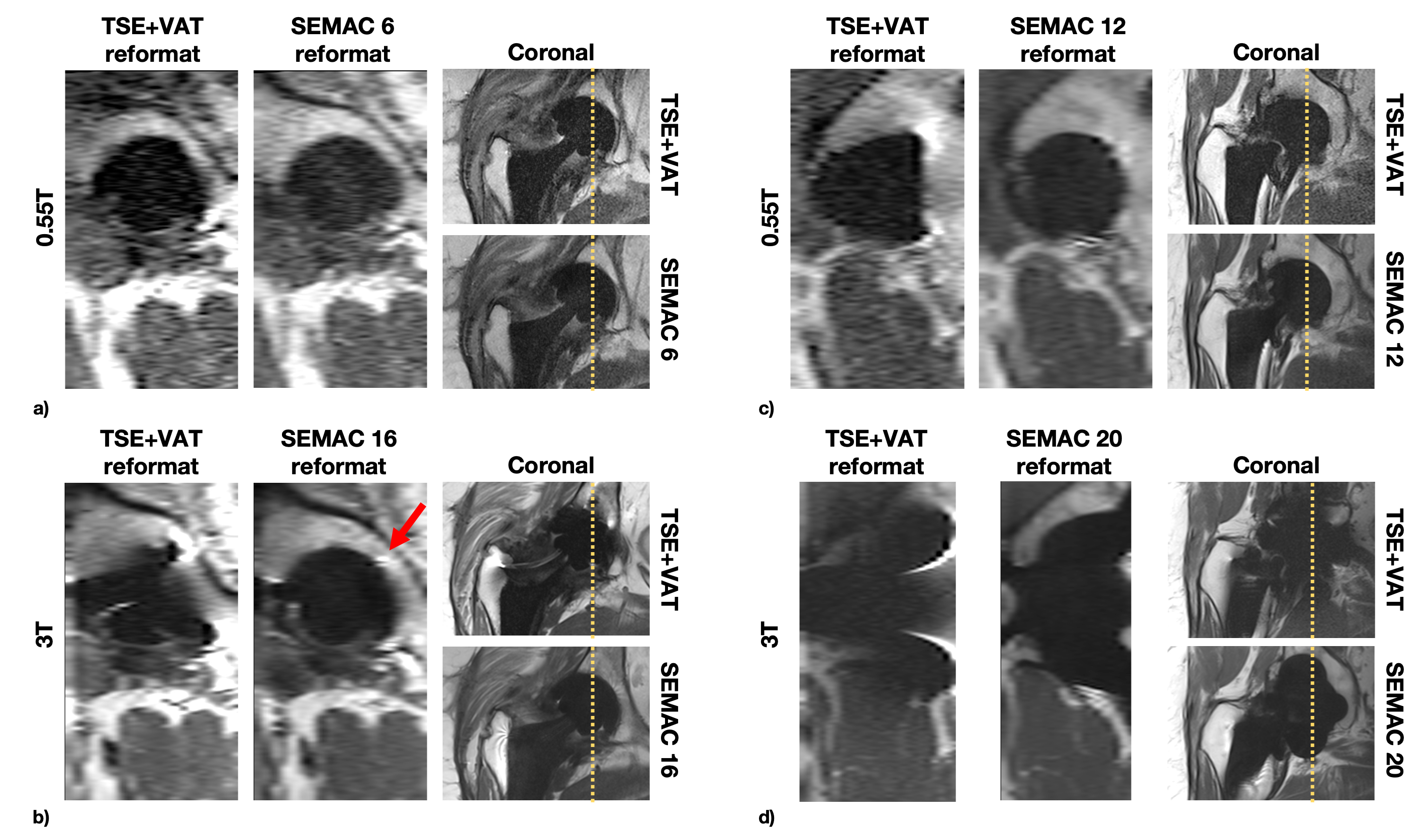
Figure 3 illustrates reformatted images along the slice dimension and compares TSE with VAT and SEMAC at a) 0.55T and b) 3T for a 58-year-old woman, and at c) 0.55T and d) 3T for a 79-year-old man. Titanium implant at 0.55T has very low in-plane and through-plane distortions, and conventional TSE provides diagnostic quality.
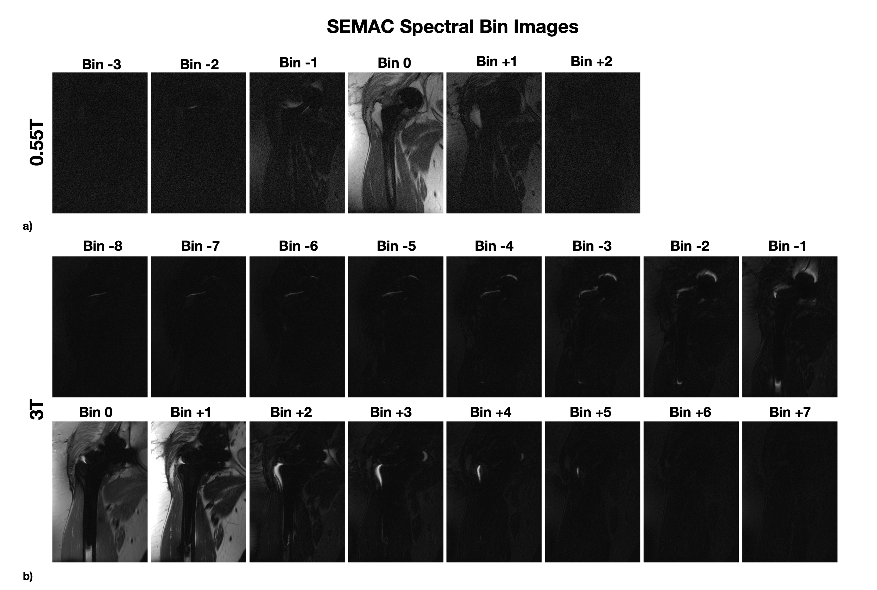
Figure 4 shows SEMAC spectral bin images for a 58-year-old woman with THA at a) 0.55T and b) 3T. Titanium implant has a low spectral range at 0.55T so the main contribution of signal comes from very few spectral bins.
Discussion
We observed a significant reduction in the severity of metal artifacts at 0.55T compared to 3T. This presents a promising advancement in imaging for patients with orthopedic metallic implants. However, there is a trade-off between metal artifact severity and SNR, with 0.55T exhibiting a lower SNR compared to 3T as expected.For titanium THA at 0.55T, simple sequences like TSE and TSE+VAT provided diagnostic quality. This outcome is not surprising due to the relatively low magnetic susceptibility of titanium compared to other materials used in orthopedic implants. The range of off-resonance frequencies caused by the titanium implant can mostly fall under one spectral bin, avoiding the need for advanced multi-spectral imaging techniques used at conventional MRI field strengths6. This might simplify the diagnostic process, reduce the duration of image acquisition, and shorten the scan protocols, leading to more efficient patient care. It might also allow other sequences to be applied without metal artifact concerns (e.g., quantitative relaxometry, diffusion).
Conclusion
We demonstrate an initial field strength comparison study for patients with hip replacement at 0.55T and 3T. The substantial reduction in metal artifact severity at 0.55T, particularly in the case of titanium hip implants, might enhance patient care and the diagnostic process.Acknowledgements
We acknowledge grant support from the National Institutes of Health (R01-AR078912), National Science Foundation (Award #1828736), research support from Siemens Healthineers, and USC Annenberg Graduate Fellowship (to K.K.). We thank Mary Yung for the research coordination.References
1. Koch KM, Hargreaves BA, Pauly KB, et al. Magnetic Resonance Imaging Near Metal Implants. J Magn Reson Imaging. 2010;32(4):773-787.
2. Hargreaves B, Worters PW, Pauly KB, et al. Metal-induced artifacts in MRI. AJR Am. J. Roentgenol. 2011;197(3):547-555.
3. Lu W, Pauly KB, Gold GE, et al. SEMAC: slice encoding for metal artifact correction in MRI. Magn Reson Med. 2009;62(1):66-76.
4. Koch KM, Lorbiecki JE, Hinks RS, et al. A multispectral three-dimensional acquisition technique for imaging near metal implants. Magn Reson Med. 2009;61(2):381-390.
5. Koch KM, Brau AC, Chen W, et al. Imaging near metal with a MAVRIC-SEMAC hybrid. Magn Reson Med. 2010;65(1):71-82.
6. Khodarahmi I, Brinkmann IM, Lin D, et al. New-generation low-field magnetic resonance imaging of hip arthroplasty implants using slice encoding for metal artifact correction. Invest Radiol. 2022;57(8):517-526.
7. Breit HC, Vosshenrich J, Clauss M, et al. Visual and quantitative assessment of hip implant-related metal artifacts at low field MRI: a phantom study comparing a 0.55-T system with 1.5-T and 3-T systems. Eur Radiol Exp. 2023.
Figures