2647
Quantification of breathing-induced B0 field variations along the human spinal cord at 3T1Department of Physics, Norwegian University of Science and Technology, Trondheim, Norway, 2Spinal Cord Injury Center, Balgrist University Hospital, University of Zurich, Zurich, Switzerland, 3Department of Neuro-Urology, Balgrist University Hospital, University of Zurich, Zurich, Switzerland, 4Department of Physics, University of British Columbia, Vancouver, BC, Canada
Synopsis
Keywords: Artifacts, Spinal Cord, Breathing
Motivation: Breathing-induced B0 field variations cause artefacts in spinal cord MRI. Characterizing the variations is useful for optimizing correction strategies, but has not yet been done below the thoracic region.
Goal(s): To quantify breathing-induced fields in the cervical, thoracic, and lumbar spinal cord, and to investigate their effect on image quality in multi-echo GRE.
Approach: Field measurements were obtained from navigator projection lines in 2D slices, and were quantified by taking their standard deviation over time.
Results: Breathing-induced field variations were detected in every subject, with a similar profile across vertebral levels. Levels with more intense field variations showed higher artifact load.
Impact: Breathing-induced B0 fields impact image quality and exhibit a consistent pattern of variation along the spinal cord that is highly similar between subjects. Characterizing these variations lays the foundation for future optimized corrections to achieve consistently high image quality.
Introduction
Breathing induces periodic changes in the air volume within the lungs, causing B0 field variations in the surrounding area1,2. Time-varying B0 fields interfere with the signal encoding, leading to ghosting, blurring, and signal loss in T2*-weighted acquisitions, apparent motion in EPI scans, and line broadening in spectroscopy.Characterizing and modelling field variations helps understanding their impact on the data and optimizing prospective (e.g. dynamic shimming) or retrospective correction techniques. Two studies have reported measurements of breathing-induced fields in the cervical and upper thoracic spinal cord, based on field maps during breath-holds1,2. Only one of them presented a free-breathing investigation2, which was limited to the cervical region.
In this study, we use navigator-based field measurements3 during free breathing to confirm the published results, and to extend the characterization to the lumbar region.
Methods
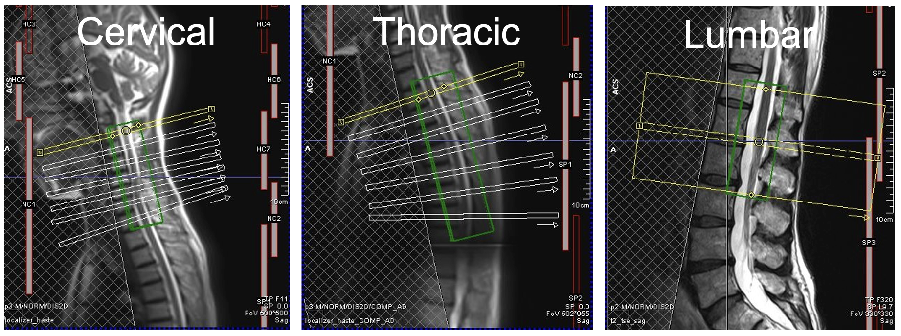
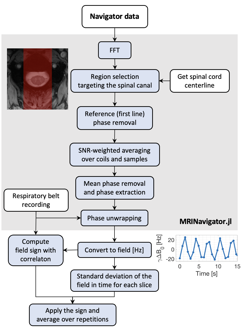
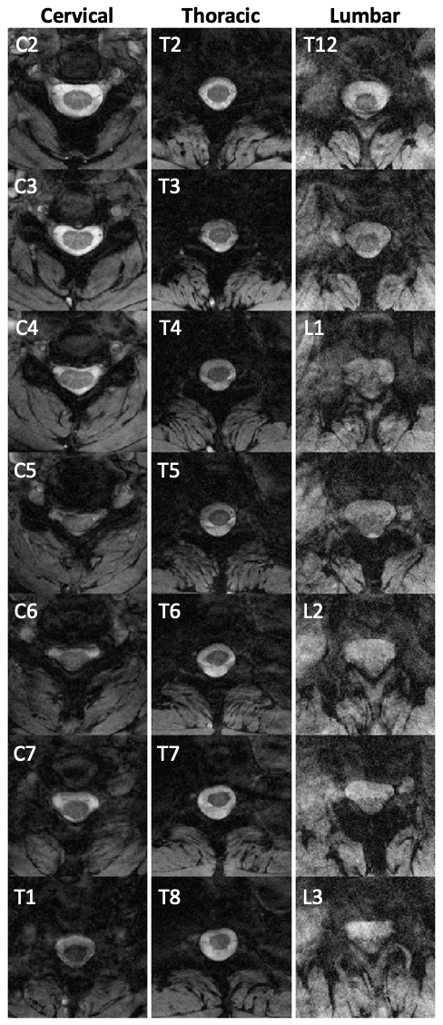
Data were acquired during free breathing in six healthy volunteers on a 3T Siemens Prisma system, using a 32-channel receive spine coil. 2D multi-echo gradient-echo (ME-GRE) sequences targeting the cervical, thoracic, and lumbar spinal cord were acquired (Fig. 1). The acquisition parameters were: slice thickness=5mm, FOV=192x192mm2, resolution=0.5x0.5mm2, TE=7/11/15/19ms, TR=700ms (cervical, thoracic) or 899ms (lumbar), flip angle=38° (cervical, thoracic) or 44° (lumbar), GRAPPA=2x, 3 repetitions. After the last echo in each TR, a navigator3 was acquired reading out a single line through the centre of k-space. A trace from the Siemens respiratory belt was simultaneously recorded. Field measurements for each TR were computed using the FFT_unwrap algorithm from MRINavigator.jl4. The main algorithm steps are shown in Fig. 2.The standard deviation of the navigator field estimates over time was computed for each slice and repetition as a measure of the magnitude of the field variations. The sign was determined by the sign of the correlation between the navigator estimates and the belt recording (positive when in phase). Then, the mean value across repetitions was computed, giving one estimate of the field variations per slice and subject. To model the profile of the field variations across subjects, an Eilers smoothing5 with 95% confidence interval was applied. Images were reconstructed offline4,6 with an iterative SENSE7,8 algorithm (10 iterations) making use of a low resolution (2x2mm2) fully sampled reference scan.
Results
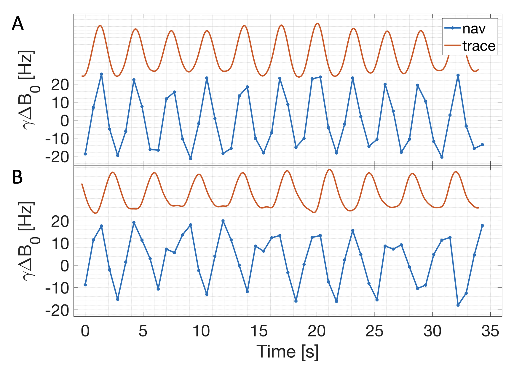
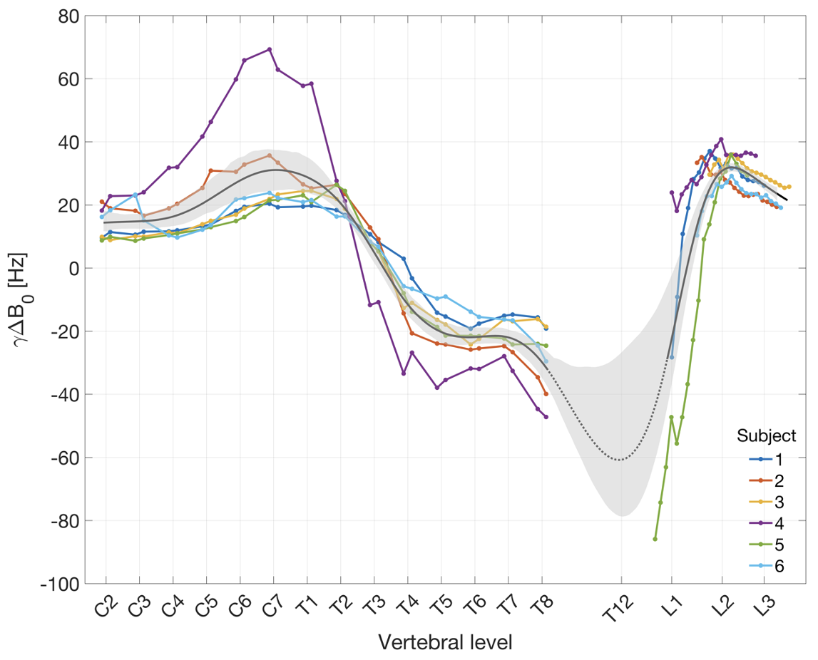
The navigator-based field measurements showed the same periodic pattern as the belt recordings in all subjects (Fig. 3), indicating the presence of breathing-induced B0 field variations. The profile of the field variations across vertebral levels was highly similar between subjects (Fig. 4). In the upper cervical cord, the magnitude of field variations was low, with a positive correlation sign. The magnitude increased towards lower vertebral levels, up to a peak around C7 (31±17 Hz). Then, the magnitude decreased steadily reaching a sign inversion point around T3/T4. Below that, the correlation sign was negative in the thoracic region, with a mostly flat profile between T5-T7, then increasing slightly in magnitude towards T8 (-28±11 Hz). Data were not acquired between T8-T12, but the Eilers smoothing indicates the likely presence of a negative peak with another sign inversion point around T12-L1. In the lumbar cord, the sign was positive, reaching a peak around L2 (32±4 Hz). Vertebral levels with larger field variations also showed higher artefact load, especially at later echoes (Fig. 5).Discussion and conclusion
The pattern of field variations across vertebral levels is consistent with a model of the lungs as air-filled spheres (positive magnetic susceptibility relative to water), which expand upon inspiration. The strongest image artifacts were observed close to the upper and lower borders of the lungs, in correspondence with peaks in the local field variations. The field gradient was largest near the sign inversion points, which likely contributes to artifacts.The left-right projection-line navigator does not allow for spatial selection in the anterior-posterior direction. The field estimate is therefore not specific to the spinal canal, unlike the field map approach used in previous studies1,2. Moreover, because of the standard deviation calculation, the measured values are smaller than the maximum peak-to-peak variation. Nevertheless, the obtained results in the cervical and upper thoracic areas are in the expected range according to the literature and follow a very similar pattern to previously reported values1,2. This serves as a confirmation of the validity of the approach for the extended measurements in the lumbar region.
One advantage of using the navigator approach is that the field variations can be computed from a standard ME-GRE anatomical acquisition and can be directly related to artifact load in the images. In future work, data should be collected in the region between T8 and L1, to investigate the inferred local peak.
Acknowledgements
This work was financially supported by the Swiss National Science Foundation (SNSF) (33IC30_179644). Imaging was performed with support of the Swiss Center for Musculoskeletal Imaging, SCMI, Balgrist Campus AG, Zürich.References
- Verma, T., & Cohen-Adad, J. (2014). Effect of respiration on the B0 field in the human spinal cord at 3T. Magnetic Resonance in Medicine, 72(6), 1629–1636. https://doi.org/10.1002/mrm.25075
- Vannesjo, S. J., Miller, K. L., Clare, S., & Tracey, I. (2018). Spatiotemporal characterization of breathing-induced B0 field fluctuations in the cervical spinal cord at 7T. NeuroImage, 167, 191–202. https://doi.org/10.1016/j.neuroimage.2017.11.031
- Versluis, M. J., Peeters, J. M., van Rooden, S., van der Grond, J., van Buchem, M. A., Webb, A. G., & van Osch, M. J. P. (2010). Origin and reduction of motion and f0 artifacts in high resolution T2*-weighted magnetic resonance imaging: Application in Alzheimer’s disease patients. NeuroImage, 51(3), 1082–1088. https://doi.org/10.1016/j.neuroimage.2010.03.048
- Beghini L., David G., Liechti D. M., Büeler S., Vannesjo S. J. (2023). Optimised navigator correction of physiological field fluctuations in multi-echo GRE of the lumbar spinal cord at 3T. Proceedings of the International Society for Magnetic Resonance in Medicine (ISMRM). https://github.com/NordicMRspine/MRINavigator.jl
- Eilers PH. A perfect smoother. Anal Chem. 2003 Jul 15;75(14):3631-6. doi: 10.1021/ac034173t. PMID: 14570219.
- Knopp, T., & Grosser, M. (2021). MRIReco.jl: An MRI reconstruction framework written in Julia. Magnetic Resonance in Medicine, 86(3), 1633–1646. https://doi.org/10.1002/mrm.28792
- Pruessmann, K. P., Weiger, M., Scheidegger, M. B., & Boesiger, P. (1999). SENSE: Sensitivity encoding for fast MRI. Magnetic Resonance in Medicine, 42(5), 952–962. https://doi.org/10.1002/(SICI)1522-2594(199911)42:5<952::AID-MRM16>3.0.CO;2-S
- Pruessmann, K. P., Weiger, M., Börnert, P., & Boesiger, P. (2001). Advances in sensitivity encoding with arbitrary k -space trajectories: SENSE With Arbitrary k -Space Trajectories. Magnetic Resonance in Medicine, 46(4), 638–651. https://doi.org/10.1002/mrm.1241
- De Leener B, Lévy S, Dupont SM, et al. (2017) SCT: Spinal Cord Toolbox, an open-source software for processing spinal cord MRI data. NeuroImage. 145, 24-43. https://doi.org/10.1016/j.neuroimage.2016.10.009
Figures