2645
Strain tensor imaging using single-shot multi-slice DENSE in a pediatric population at 7T1University Medical Center Utrecht, Utrecht, Netherlands, 2Princess Máxima Center for Pediatric Oncology, Utrecht, Netherlands
Synopsis
Keywords: Artifacts, Artifacts, pediatric, neuro
Motivation: DENSE can provide information about the brain pulsations that likely reflect the condition of blood vessels, which may deteriorate following treatment in pediatric brain tumors.
Goal(s): The goal of this study is to perform an initial analysis of this single-shot multi-slice DENSE data in a pediatric population to study the robustness of this sequence during motion.
Approach: By using a single-shot multi-slice DENSE sequence, brain motion maps were acquired from which strain maps could be derived on a voxel-wise level.
Results: Even though the pediatric participants moved during the MR-acquisition, good quality strain maps were obtained with the expected patterns (as in adults).
Impact: Single-shot strain tensor imaging allows evaluation of cardiac-related brain tissue strain, in a pediatric cohort of posterior fossa tumor, despite the presence of unwanted head motion. This enables investigating strain as potential new biomarker of neurovascular integrity in patients.
Introduction
Many children who have been treated for a posterior fossa tumor experience neurocognitive problems after treatment[1,2]. Recently, increasing evidence has shown that early neurobiological changes that occur after brain tumor treatment are predictive of neurocognitive functioning[3-5]. Displacement encoding with stimulated echoes (DENSE) sequence can provide information about the pulsations that are induced by the microvascular bed embedded in the tissue[6,7]. These pulsations likely reflect the condition of these vessels, which may deteriorate following treatment. However, when applied to a pediatric population, robustness against unwanted motion is crucial. Different approaches have been investigated to reduce these movements without sedation, however, these approaches cannot eliminate movement[8]. Recently, strain tensor imaging, which uses a single-shot multi-slice DENSE sequence, was proposed to obtain voxel-wise strain tensor maps of the brain tissue strain as induced by the heartbeat[9]. The strength of the single-shot approach lies in its inherent robustness against motion artefacts, which is an improvement over earlier multi-shot 3D acquisitions[6,7]. Head motion and respiration can induce inter-shot phase variability in multi-shot 3D acquisition that propagates to artifacts in the DENSE images, particularly towards the end of the cardiac cycle[6,7]. The goal of this study is to perform an initial analysis of this single-shot multi-slice DENSE data in a pediatric population to study the robustness of this sequence during motion.Methods
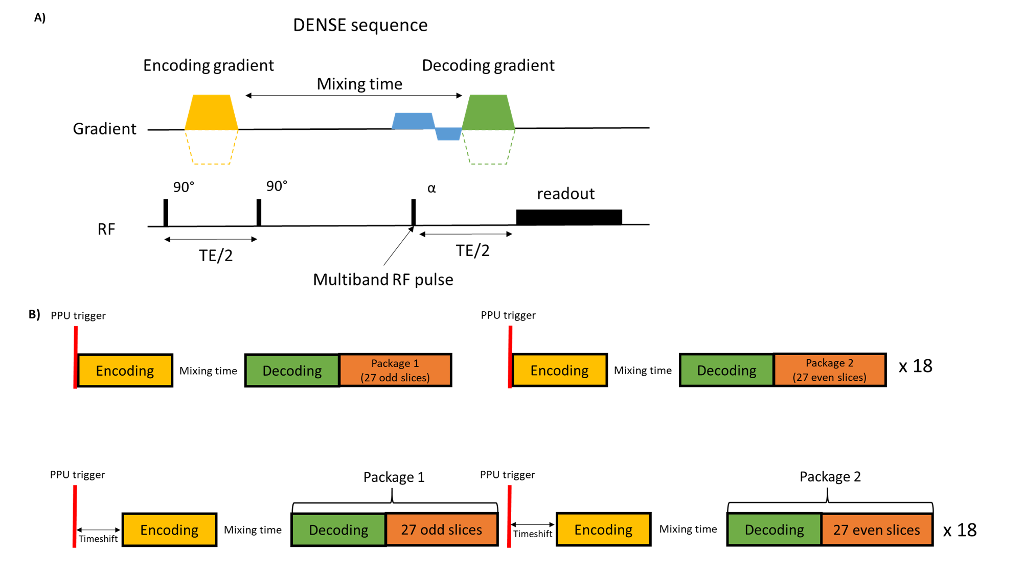
A cardiac triggered single-shot DENSE sequence was combined with a multi-band acquisition to be able to obtain whole brain coverage and was implemented as described previously[9] (Figure 1). DENSE measurements were repeated 36 times during which 54 slices were acquired using a motion encoding strength of 80 µm. These 54 slices were divided over 2 packages and acquired with slice order permutations. Two time shifts after triggering at 0 ms and 7x slice time interval, respectively, were used to cover the entire cardiac cycle (resolution: 3x3x3mm3, SENSE 2.6, MB factor 3). During the acquisition physiological data was simultaneously recorded, using a Peripheral Pulse Unit (PPU) for triggering and a respiration belt to trace abdominal breathing. Six DENSE datasets were acquired with different motion encoding directions and slice orientations(TRA:RL&AP, COR:FH&RL, SAG:FH&AP). Analysis of the data was performed as described previously[9]. This scan was acquired as part of the SIMBA study (a prospective study with posterior fossa brain tumor patients: https://www.isrctn.com/ISRCTN15453405) at 7T MRI (Philips healthcare) using an 8-channel transmit and 32-channel receive head coil (Nova Medical). This study received medical ethical approval, and written informed consent was obtained from the participants and/or legal guardians. 19 participants have been scanned for this study, and 6 DENSE datasets were used for this initial analysis (6 girls, age: 12-18 y.o., brain tumor survivors). Nine DENSE datasets could not yet be analyzed due to the quality of the PPU signal, three datasets were exported in the wrong format and the DENSE sequence was not acquired in one participant. Motion of the participants was estimated during the acquisition of a separate BOLD sequence within the same scan session using MCflirt[10] during the analysis. The standard deviation was calculated for both rotation and translation parameters.Results
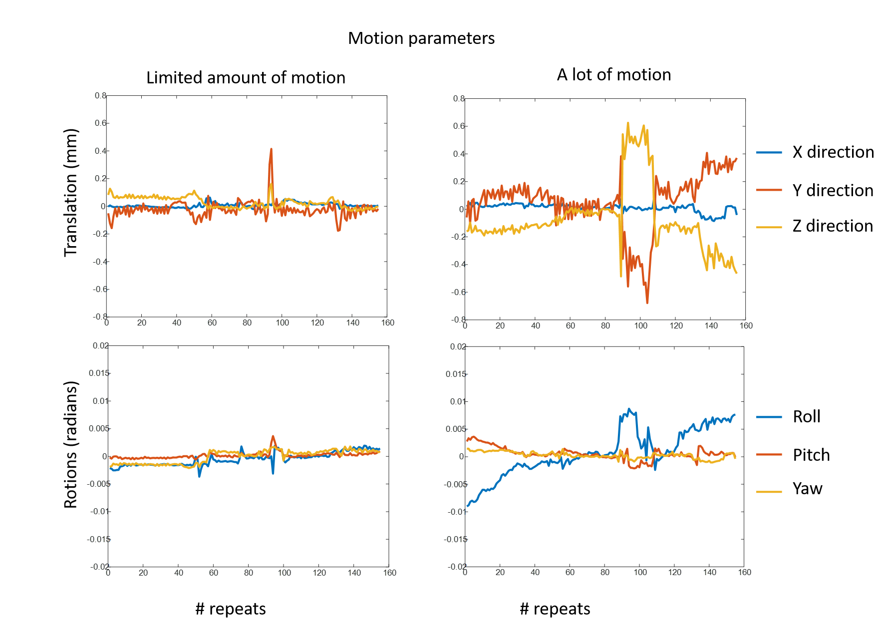
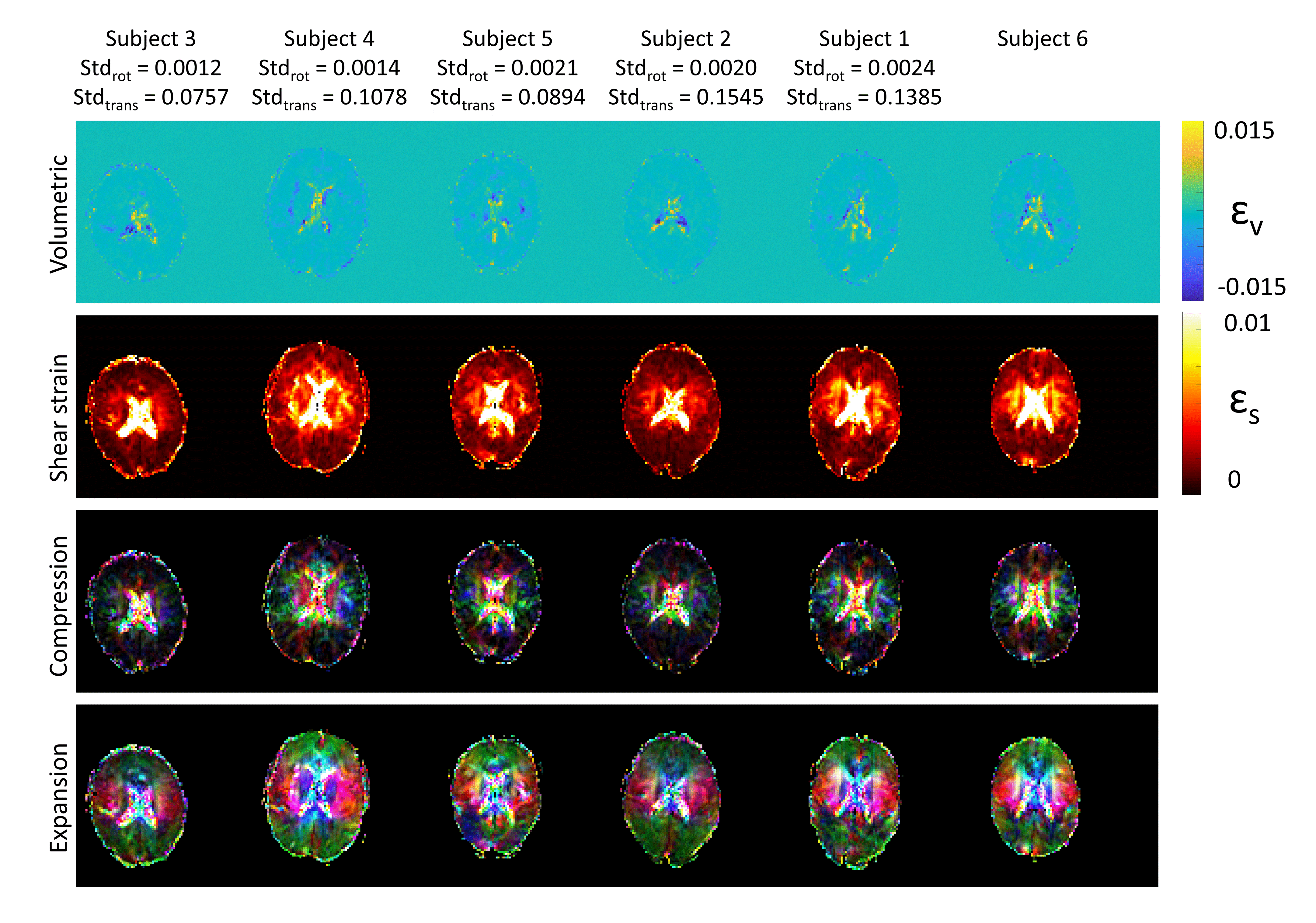
An example of movement during the MRI acquisition is shown in Figure 2. Figure 3 shows a representative example of the strain tensor maps for all participants at the moment of largest mean volumetric strain ordered based on the standard deviation of the motion parameters. Volumetric strain is mostly present at the periphery of the brain, shear strain is most pronounced in regions close to the ventricle. The compression map shows the funnel shaped pattern pointed towards the foramen magnum and the expansion map shows expansion in the opposite direction, reflecting the poison effect. Overall a similar pattern was shown for all participants, regardless the amount of motion. In Figure 4, these four maps are shown for the entire cardiac cycle to demonstrate the influence of the heartbeat on these four parameters in a representative participant.Discussion and Conclusion
By using a single-shot multi-slice DENSE sequence, brain motion maps were acquired from which strain maps could be derived on a voxel-wise level. Even though the pediatric participants moved during the MR acquisition, good quality strain maps were obtained with the expected patterns (as in adults)[9]. This demonstrates the robustness of this sequence against unwanted motion which will allow us to obtain information about the vasculature and the brain tissue’s mechanical properties after treatment of the posterior fossa tumor in this patient group.Acknowledgements
We would like to thank the participants and their parents for their participation to the SIMBA study. This research is funded by KiKa (#450) and NWO VICI: Seismology of the brain (#18674).References
1. Bell et al., A systematic review of factors related to children's quality of life and mental health after brain tumor. Psychooncology. 2018 2. Lassaletta et al., Functional and neuropsychological late outcomes in posterior fossa tumors in children. Childs Nerv Syst. 2015 3. Makale et al., Mechanisms of radiotherapy-associated cognitive disability in patients with brain tumours. Nat Rev Neurol. 2017 4. Yang et al., Aberrant Brain Activity at Early Delay Stage Post-radiotherapy as a Biomarker for Predicting Neurocognitive Dysfunction Late-Delayed in Patients With Nasopharyngeal Carcinoma. Front Neurol. 2019 5. Partanen et al., Early changes in white matter predict intellectual outcome in children treated for posterior fossa tumors. Neuroimage Clin. 2018 6. Adams et al. Quantifying cardiac-induced brain tissue expansion using DENSE, NMR in biomedicine, 2018, DOI: 10.1002/nbm.4050 7. Soellinger et al. 3D Cine displacement-encoded MRI of pulsatile brain motion, Magnetic Resonance in Medicine, 2009 8. Eichhorn et al. Characterization of children’s head motion for magnetic resonance imaging with and without general anesthesia, Frontiers in Radiology, 2021 9. Sloots et al. Strain Tensor Imaging: Cardiac-induced brain tissue deformation in humans quantified with high-field MRI, NeuroImage, 2021, https://doi.org/10.1016/j.neuroimage.2021.118078. 10. Jenkinson et al., Improved Optimisation for the Robust and Accurate Linear Registration and Motion Correction of Brain Images. NeuroImage, 2002Figures