2643
A deep-learning-based framework for spark artifacts detection and correction1Shanghai United Imaging Healthcare Co., Ltd., Shanghai, China
Synopsis
Keywords: Artifacts, Artifacts
Motivation: The occasional occurrence of spark artifacts in MRI could significantly hinder diagnosis. Previous whole K-space segmentation methods are unstable and require stringent intensity normalization.
Goal(s): In this study, we aim to improve the accuracy and stability of spark identification, especially for sparks with low intensities and near the K-space center.
Approach: We propose a two-step deep learning-based framework consisting of spark patch classification and patch-level spark segmentation, which are further corrected by ESPIRiT.
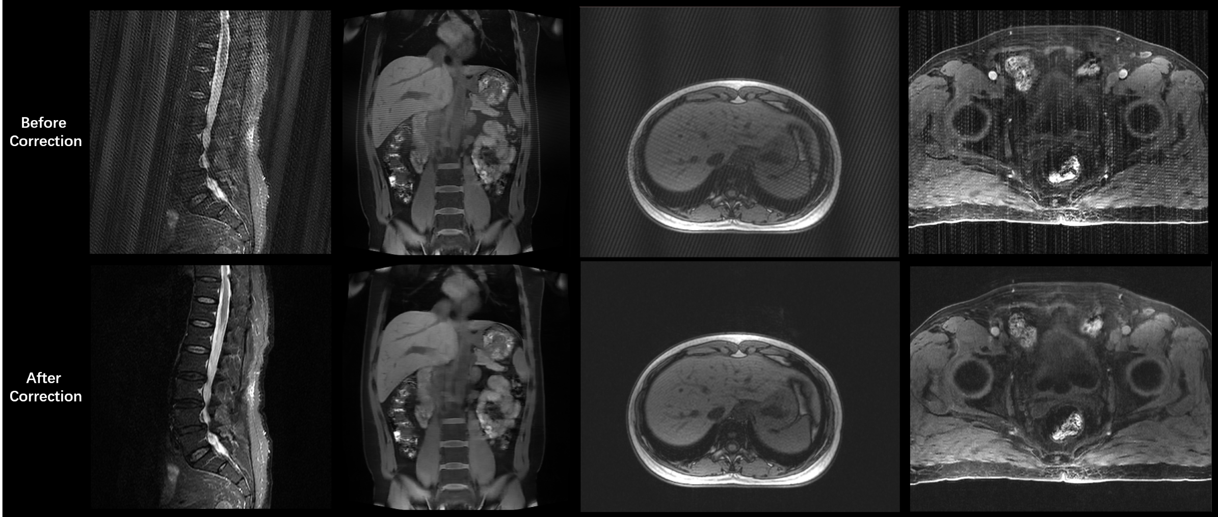
Results: The proposed methods are demonstrated to be effective and robust on various imaging protocols and body parts for different degrees of spark artifacts.
Impact: Incidental spark artifacts in MRI can significantly hinder diagnosis. We developed a deep-learning-based two-step framework for robust spark detection and correction, which has been validated to be effective on a variety of imaging protocols for different degrees of spark artifacts.
Introduction
The spark artifact (also known as spike artifact or herringbone artifact) is an MRI artifact related to one or a few aberrant data point(s) in K-space. The causes of spark artifacts include electromagnetic sparks by gradient coils or external electromagnetic interference. In image space, the regularly spaced stripes resemble the appearance of a fabric with a herringbone pattern. The artifact usually covers the entire image, thus significantly hindering the diagnosis. Previous studies have been proposed to correct spark artifacts for fMRI1, diffusion tensor imaging2, and TOF-MRA3. Methods including robust principal component analysis have been utilized for spark artifacts removal4. However, these conventional methods are usually hindered by their generalizability to various imaging protocols and degrees of spark. We propose a pipeline for universal spark detection and correction using a two-step deep-learning-based spark detection framework in the K-space. A classification network is trained to identify K-space patches with spark, and another segmentation network is trained to locate exact spark pixels. Then, the ESPIRiT method5 is utilized to fill in the spark pixels and the original under-sampled regions. Extensive experiments have demonstrated the accuracy and robustness of the proposed spark artifacts detection and correction framework.Methods
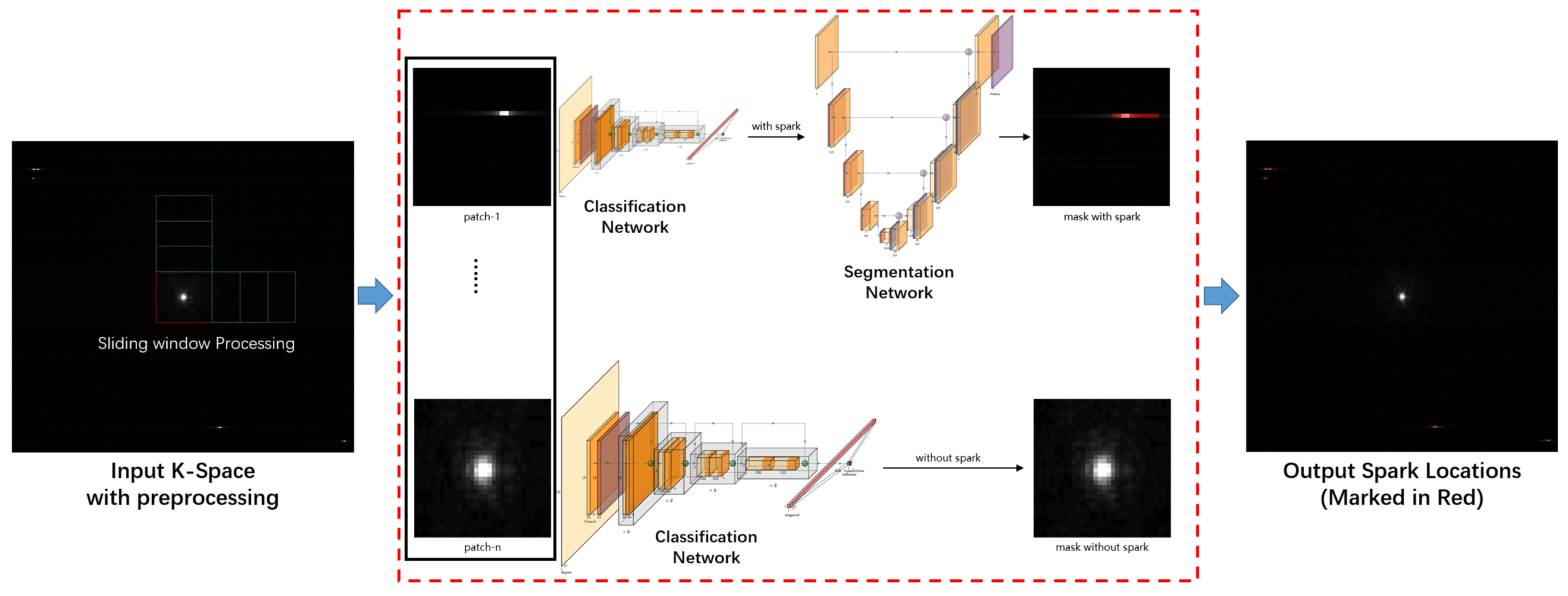
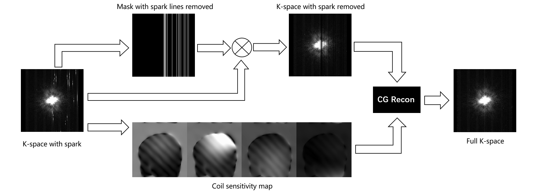
A deep learning-based two-step framework is proposed for spark detection (Figure 1). First, we apply a sliding window on the processed K-space and split the entire K-space into multiple patches. Inspired by a similar procedure in whole-slide imaging6, splitting the whole K-space into patches can significantly improve the accuracy of locating spark artifacts. A ResNet-based classification network is trained to predict whether each patch contains spark pixels7. Then, for those spark patches, a VB-net-based segmentation network is trained to predict accurate spark locations at the pixel-level8. The two-step predictions can be combined to locate spark pixels precisely in the original K-space. To mitigate the variations in K-space and spark intensities, we normalize the K-space using the intensities of the K-space center before spark detection. To correct the K-space with identified spark pixels, we multiply the raw K-space data with the post-processed spark mask, which sets the entire readout line with spark pixels as uncollected to improve the stability of spark correction. Then, we use the ESPIRiT method to correct the spark artifacts (Figure 2). The coil sensitivity maps are computed based on the reference line in the original K-space. The K-space data with spark removed and the estimated coil sensitivity maps are then input into the CG iterator. The ESPIRiT reconstruction also fills the un-acquired K-space data if acceleration acquisition is enabled, resulting in a fully sampled K-space.Results
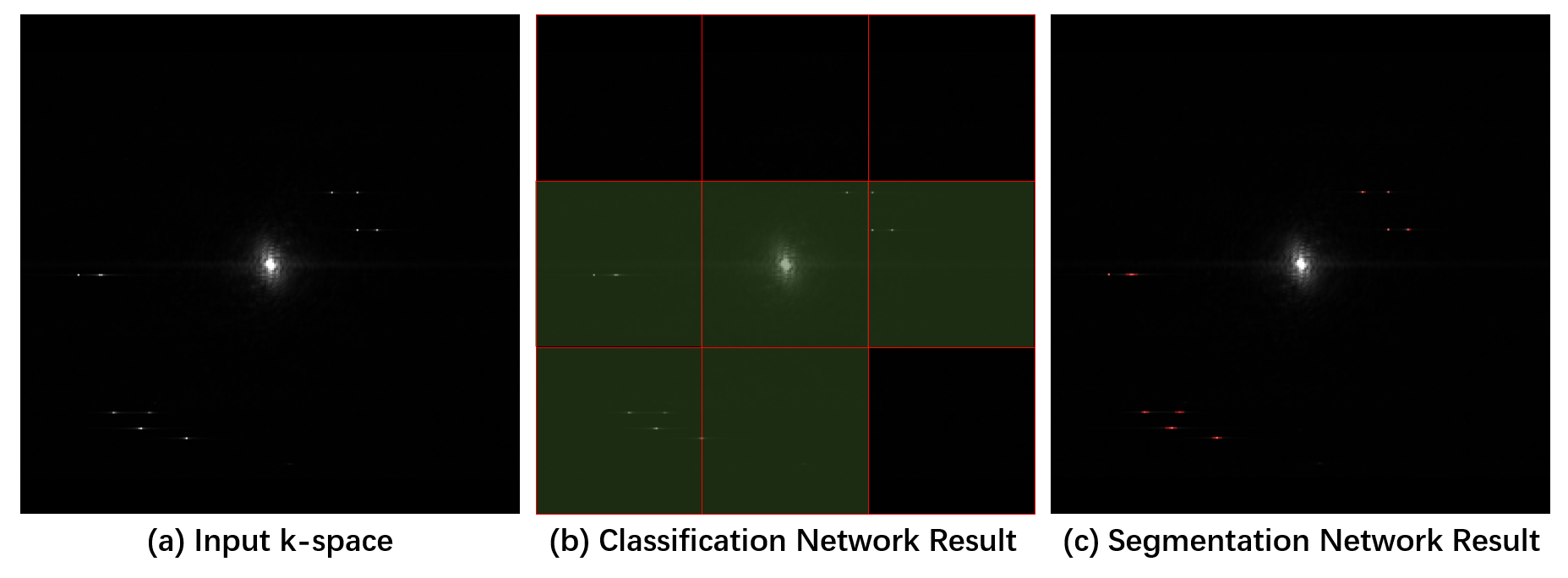
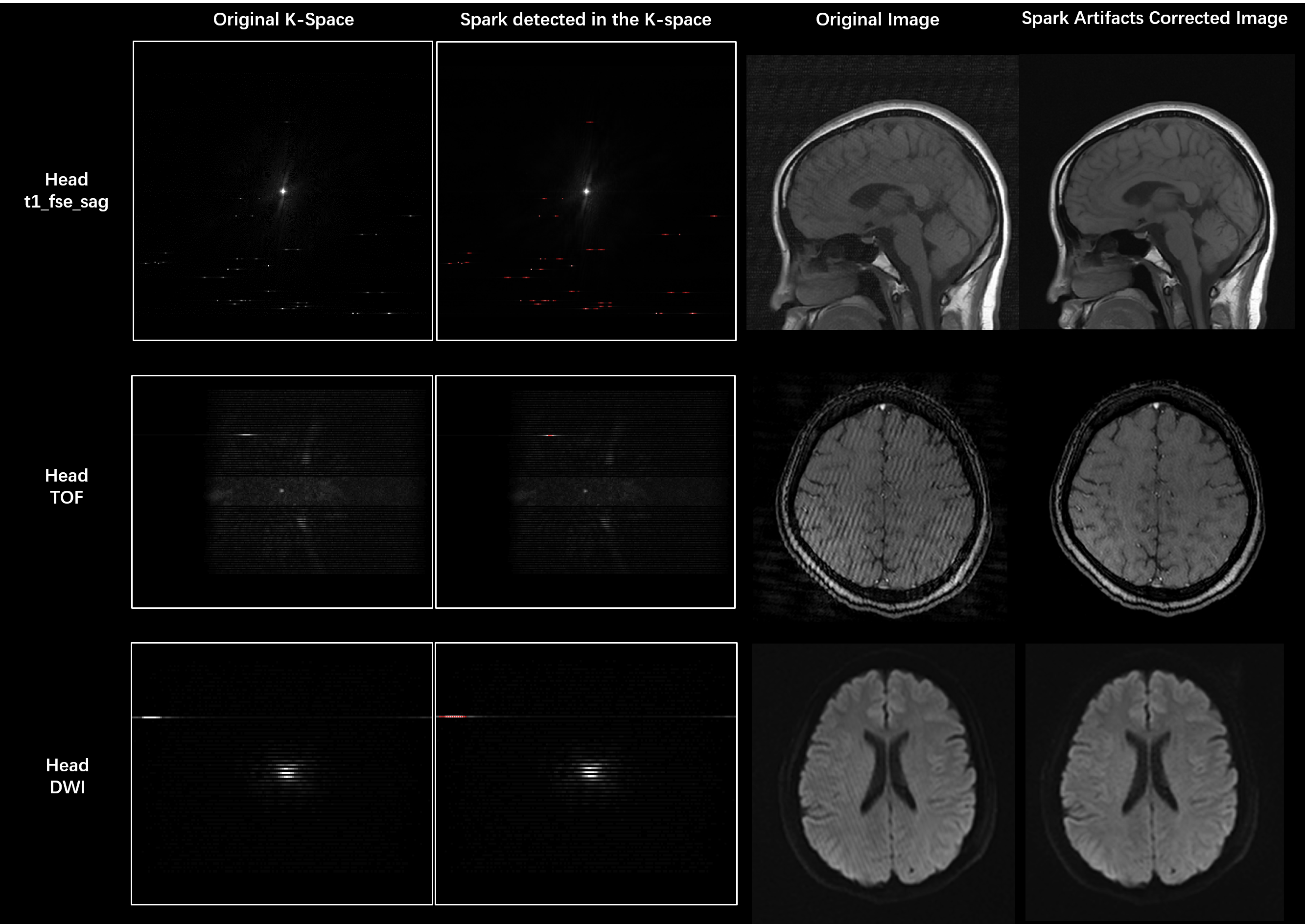
We collected 300 spark-free data from 10 healthy volunteers on 3T scanners (uMR 790/870/880, United Imaging Healthcare, China). 12,000 spark data with different degrees and patterns were simulated from the collected spark-free data as the training data. The classification network was trained with binary cross-entropy loss for 200 epochs, and the segmentation network was trained with Dice and binary cross-entropy loss for 200 epochs. The proposed methods were tested on retrospectively collected real spark data. The two-step spark patch identification and spark pixel location results are presented in Figure 3. With the spark pixels identified and accurately located in the K-space, the ESPIRiT method can fill the spark pixels and realize spark artifact correction (Figure 4). The proposed method has been validated to be effective on various imaging protocols and body parts for different degrees of spark artifacts (Figure 5).Conclusions
The proposed deep learning-based two-step framework can achieve accurate and robust detection of spark artifacts in the K-space, which can be further corrected by ESPIRiT to restore MRI images with diagnostic qualities.Acknowledgements
No acknowledgement found.References
1. Zhang X, Van De Moortele PF, Pfeuffer J, Hu X. Elimination of K-space spikes in fMRI data. Magnetic Resonance Imaging. 2001;19(7):1037-1041. doi:10.1016/S0730-725X(01)00428-3
2. Chavez S, Storey P, Graham S j. Robust correction of spike noise: Application to diffusion tensor imaging. Magnetic Resonance in Medicine. 2009;62(2):510-519. doi:10.1002/mrm.22019
3. Li N, Zhou S, Zhao G, Zhang Z, Xie Y, Liang X. Iterative stripe artifact correction framework for TOF-MRA. Computers in Biology and Medicine. 2021;134:104456. doi:10.1016/j.compbiomed.2021.104456
4. Campbell-Washburn AE, Atkinson D, Nagy Z, et al. Using the robust principal component analysis algorithm to remove RF spike artifacts from MR images. Magnetic Resonance in Medicine. 2016;75(6):2517-2525. doi:10.1002/mrm.25851
5. Uecker M, Lai P, Murphy MJ, et al. ESPIRiT—an eigenvalue approach to autocalibrating parallel MRI: Where SENSE meets GRAPPA. Magnetic Resonance in Medicine. 2014;71(3):990-1001. doi:10.1002/mrm.24751
6. Hou L, Samaras D, Kurc TM, Gao Y, Davis JE, Saltz JH. Patch-Based Convolutional Neural Network for Whole Slide Tissue Image Classification. In CVPR 2016:2424-2433.
7. He K, Zhang X, Ren S, Sun J. Deep Residual Learning for Image Recognition. December 2015. doi:10.48550/arXiv.1512.03385
8. Han M, Yao G, Zhang W, et al. Segmentation of CT Thoracic Organs by Multi-resolution VB-nets. In ISBI 2019.
Figures