2641
Reducing pulsation artifacts in 3D time-of-flight angiography at 7T using locally-scrambled ordering of the acquisition1Department of Brain Sciences, Weizmann Institute of Science, Rehovot, Israel, 2Life Sciences Core Facilities, Weizmann Institute of Science, Rehovot, Israel, 3Sackler Faculty of Medicine, Tel-Aviv University, Tel-Aviv, Israel, 4Department of Radiology, Tel Aviv Sourasky Medical Center, Tel-Aviv, Israel
Synopsis
Keywords: Artifacts, Velocity & Flow, acquisition order; angiography
Motivation: Non-contrast-enhanced time-of-flight (TOF) at 7T greatly improves the delineation of small vessels but is impaired by pulsation artifacts.
Goal(s): Our goal was to reduce pulsation artifacts in 3D TOF to better delineate vessels.
Approach: We recently developed “local-scrambling” which generates semi-random acquisition ordering to reduce artifacts from semi-periodic local signal fluctuations. This local-scrambling was tested in 3D TOF to reduce pulsatile blood flow artifacts in human scanning at 7T.
Results: Artifacts from pulsatile blood flow were significantly reduced using the new local-scrambling (of the 2D phase encodes), both for line-by-line and center-out acquisitions. The method can be of special interest for high-resolution angiography.
Impact: Increased resolution of non-contrast-enhanced time-of-flight (TOF) can provide more accurate vessels delineation. A new local-scrambling acquisition scheme can significantly improve 3D angiography by reducing interference noise and pulsation artifacts without requiring any changes to the reconstruction scheme.
Introduction
Non-contrast-enhanced time-of-flight (TOF) has proven especially beneficial at 7T due to the longer T1 relaxation at higher fields which provides better vessels contrast1,2. Moreover, increased SNR at 7T can be harnessed to increase the achievable resolution. However, one of the challenges still existing is artifacts from pulsatile blood flow, especially near major blood vessels3 — usually appearing as repeated dark and white stripes. Flow compensation approaches — based on gradient moment nulling techniques — were developed over the years3,4 but require additional time within the sequence. In a recent study5, we developed a new approach, “local-scrambling”, that generates a semi-randomized ordering with a controllable cutoff frequency above which the artifacts are drastically reduced. With this approach, the artifacts resulting from local fluctuations due to cardiac pulsation are significantly reduced. The benefit of this approach was demonstrated in T2* weighted 3D GRE5. In this study, we explore the ability of this local-scrambling approach to reduce the pulsatile blood flow artifacts in a 3D TOF acquisition at 7T. Since it was shown that center-out ordering can reduce motion related artifacts3, we examined our method in human scanning for both center-out and line-by-line ordering.Methods
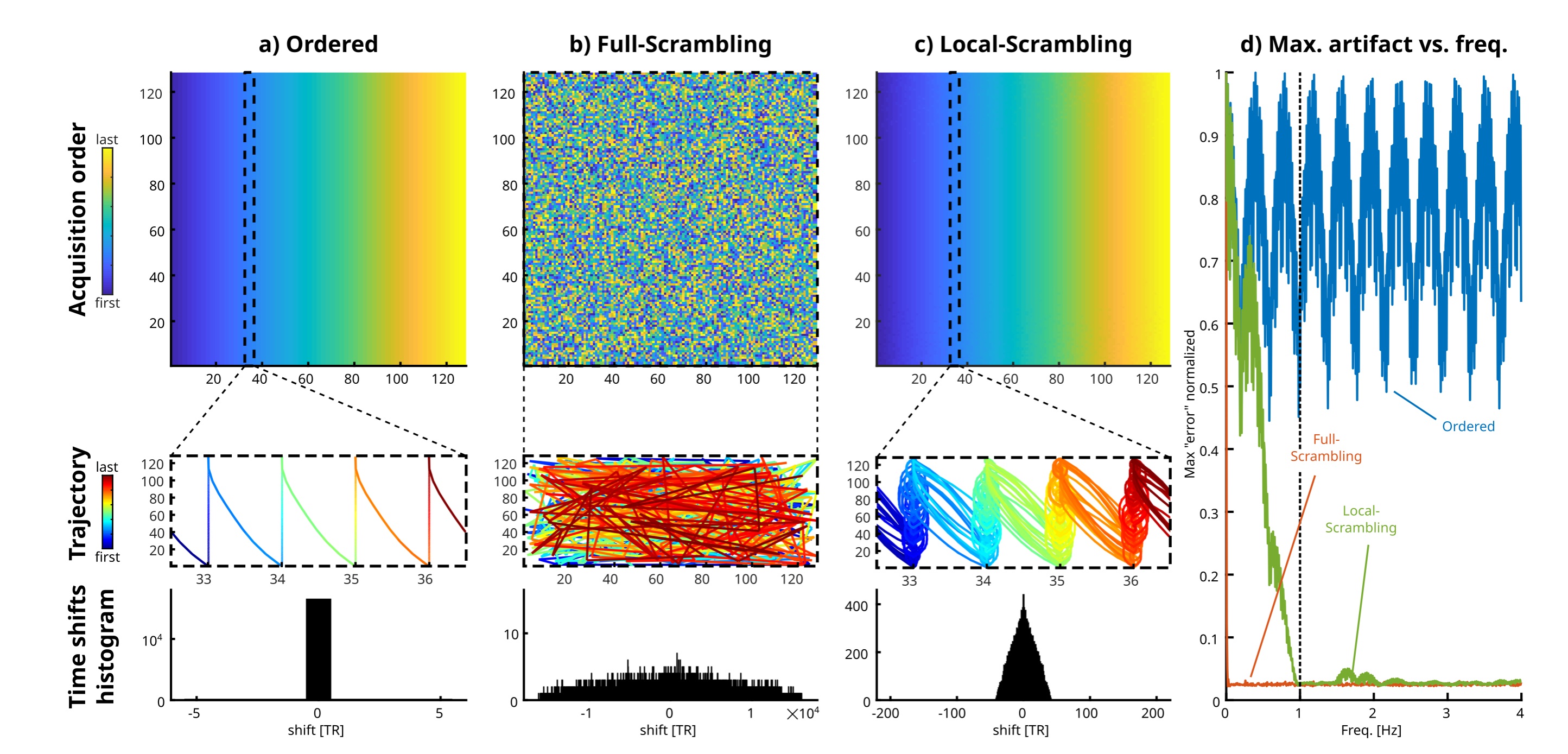
Local-scrambling ordering approach. Pulsatile blood flow is essentially a periodic physiological processes. Its periodicity can be broken by scrambling the chronological order in which k-space is acquired. However, complete random scrambling is prone to two major drawbacks - it may produce extra noise due to eddy currents in case of large changing jumps in k-space and it can result in noticeable noise due to global changes, e.g., slow subject movement during the scan. By performing local scrambling of the k-space sampling – i.e., switching the order of each planned sample with a randomly different one, but one that is not too far away in time – the above drawbacks are mitigated. This approach allows to define a cutoff frequency above which the scrambling is effective5. Fig. 1 illustrates the approach for a 2D case (128x128 phase encoded, TR=20 ms, and cutoff frequency of 1 Hz for illustrative purposes). The maximum simulated artifact per oscillation frequency is shown in Fig. 1d, demonstrating artifact suppression as function of frequency.Examined Ordering schemes. In this study, we scanned with Cartesian sampling, comparing line-by-line and center-out ordering schemes. Each scheme was scanned with and without local-scrambling of the order.
Simulations. To gain insight, simulations were performed with a point-distribution whose phase modulated with a frequency f. This was considered as a pure artifact source. The k-space signal of this artifact source was analytically “sampled” on a 2D Cartesian k-space grid for the orderings above and then reconstructed. This was repeated for different modulation frequencies.
Volunteer scanning. 3D TOF brain images of 2 healthy volunteers were acquired with the Nova 1Tx/32Rx head coil on a 7T Terra scanner (Siemens) with the following parameters: FOV 220x176x33 mm3, acquired matrix size 672x540x56 (kx×ky×kz) ,2 slabs, resolution 0.32x0.32x0.32 mm3, bandwidth per pixel 120 Hz, TR/TE 23/5.61ms, flip angle 20°, acceleration x3 using GRAPPA parallel acquisition (36 central lines fully sampled), phase encoding R-L direction. The actual number of acquired phase-encoding lines was 51x169 (kz×ky). Scan duration was 6:43 min.
Results
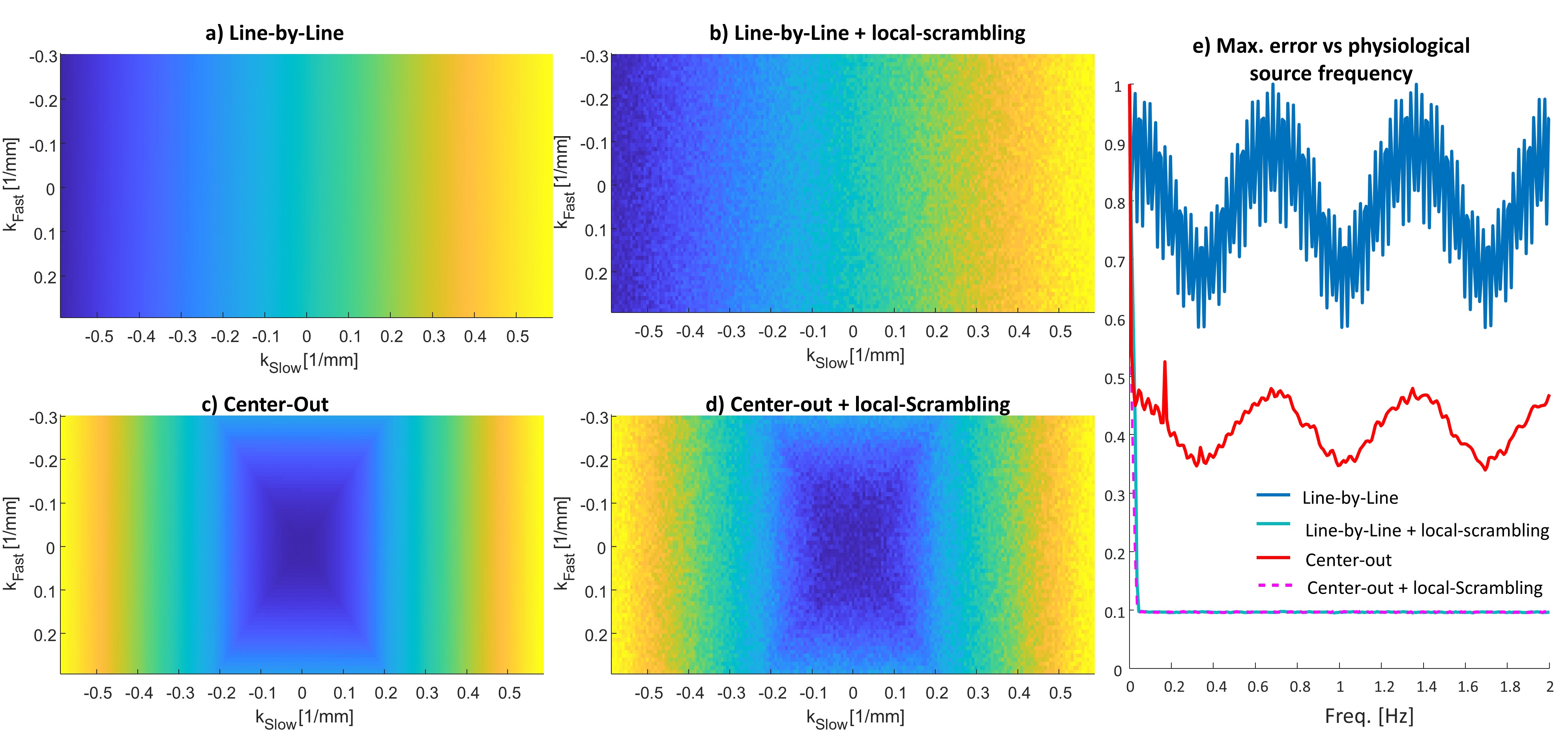
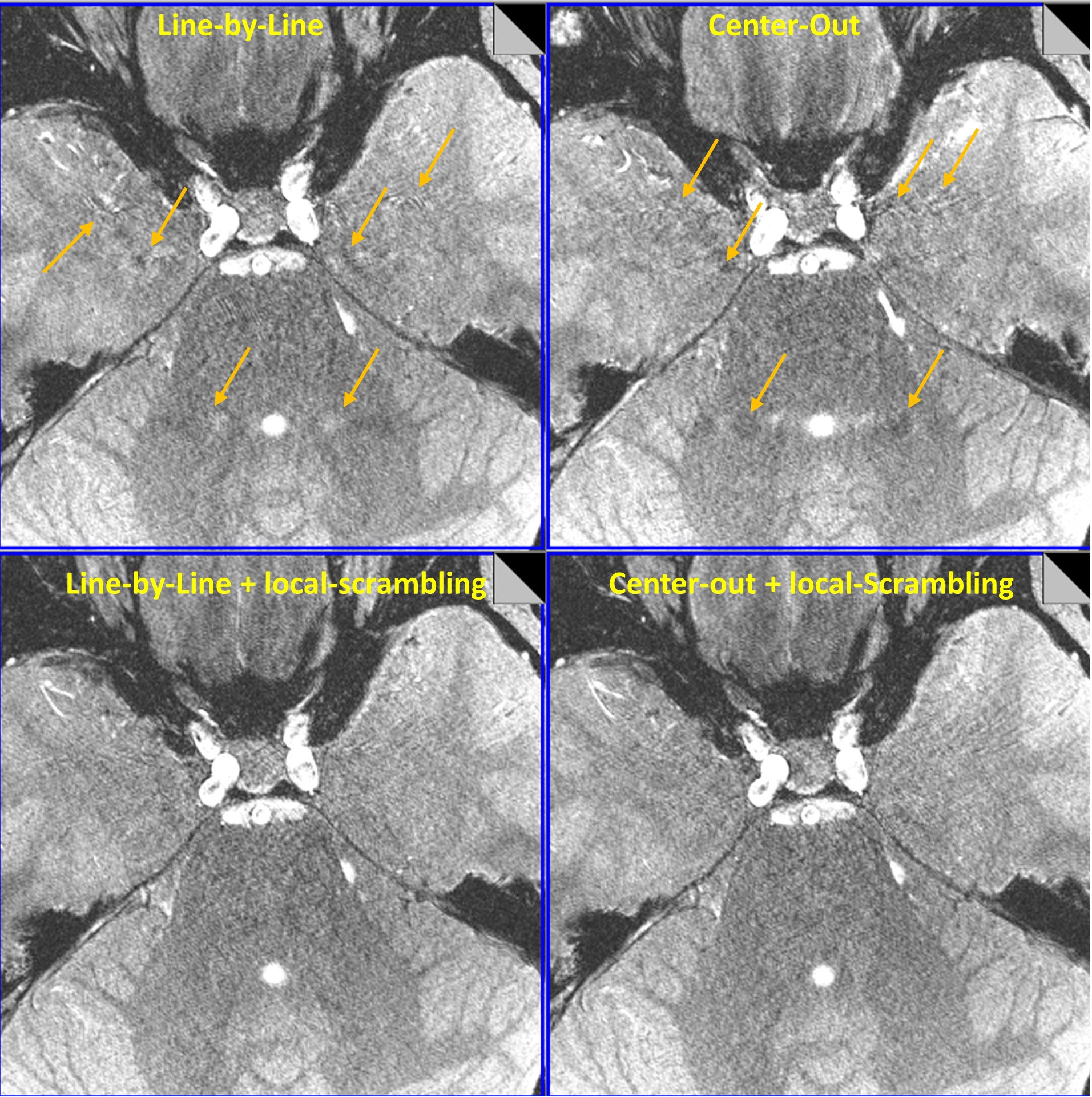
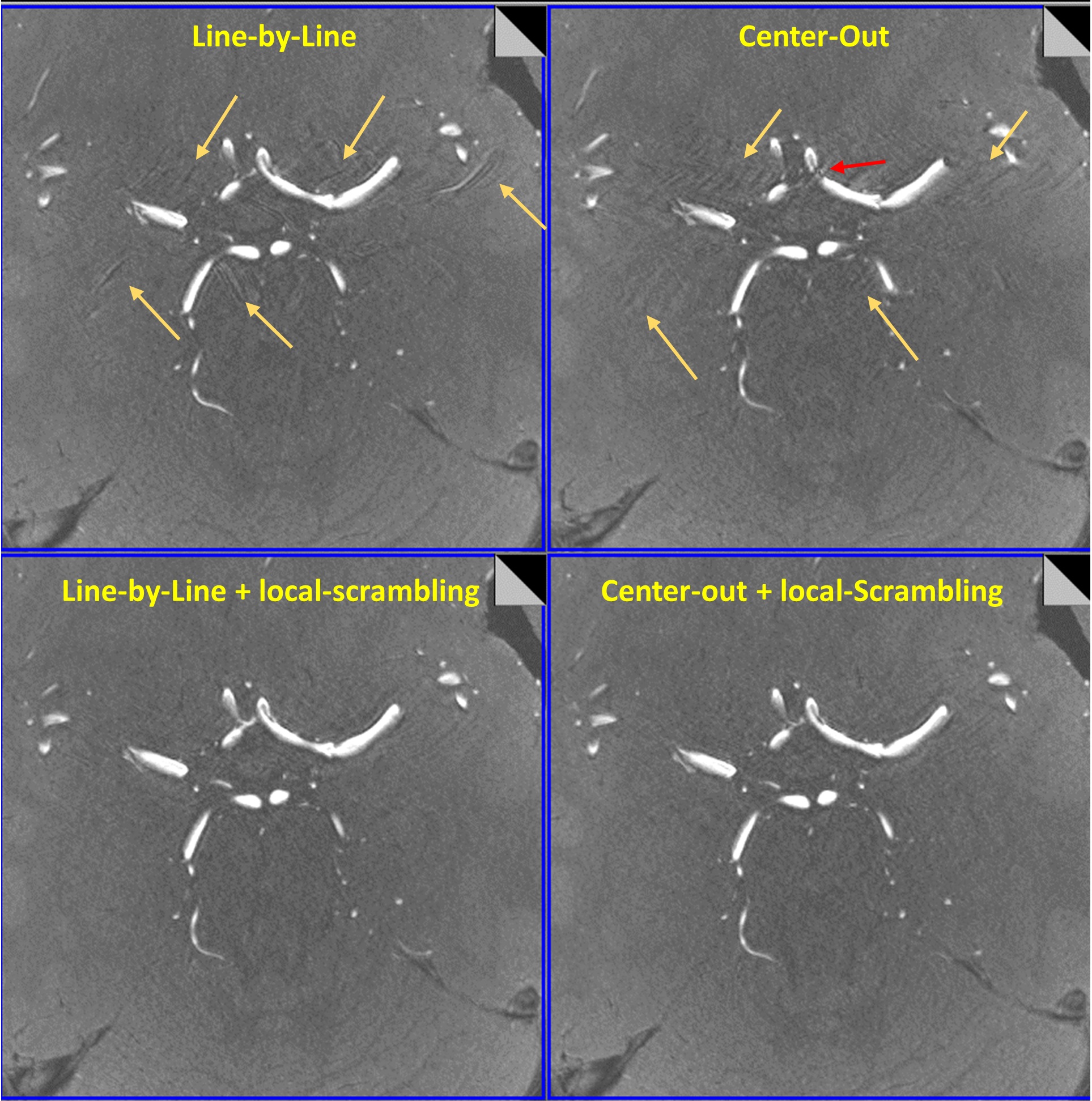
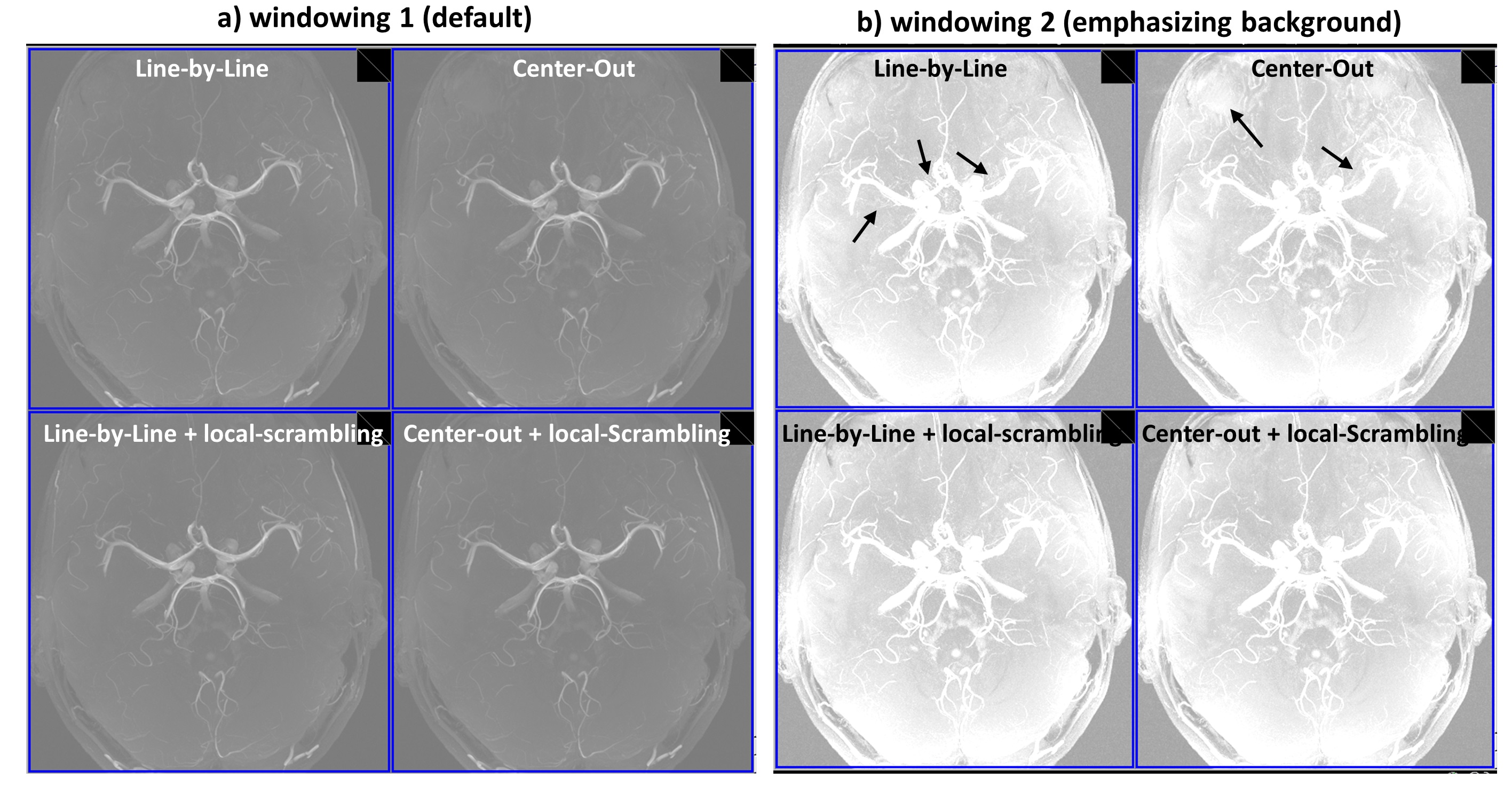
Fig. 2 shows the simulated artifact intensity for the four examined order schemes. As was reported in Ref. 3, the center-out ordering is expected to reduce the artifact intensity, but a reduction factor of only ~x2-3 was observed. When local-scrambling was applied to either Line-by-Line or Center-Out ordering, a ~x10 times reduction in the simulated artifact intensity was achieved, for the examined parameters. Figures 3,4 show two representative slices using the four acquisition schemes. The Line-by-Line and the center-out ordered schemes both resulted in artifacts, however, the artifacts spatial distribution is different. In both cases, the artifacts were drastically reduced when local-scrambling was applied. Fig. 5 shows Maximal Intensity Projection (MIP) images for each case. These also show reduced level of interference noise with the local-scrambling scheme, which can help in delineating the vessels.Conclusions
In this study, a significant reduction of artifacts due to pulsatile blood flow was achieved in TOF using a new semi-randomized local-scrambling ordering of the acquired 2D phase encodes. The local-scrambling improves the quality of the images for both line-by-line and center-out ordering. This approach can easily be implemented in the scanner without any changes to the reconstruction scheme. The method was found to be compatible with the state-of-the-art acceleration techniques - parallel acquisition and Compressed Sensing5. The method can be of special interest for high-resolution angiography and for a more accurate pathology demarcation.Acknowledgements
We are grateful to E. Tegareh and N. Oshri - for assistance in the human imaging scans. This research was generously supported by Joyce Eisenberg Keefer and Mel Keefer Career Development Chair for New Scientists, Sir Charles Clore Research Prize and Sara Z. de Usansky and Hinda Machesz Zalc Scheib. E. Furman-Haran holds the Calin and Elaine Rovinescu Research Fellow Chair for Brain Research at the Weizmann Institute of Science.References
[1] Schmitter, S., Bock, M., Johst, S., Auerbach, E. J., Uğurbil, K., & Van de Moortele, P. F. (2012). Contrast enhancement in TOF cerebral angiography at 7 T using saturation and MT pulses under SAR constraints: impact of VERSE and sparse pulses. Magnetic resonance in medicine, 68(1), 188-197.
[2] Saïb, G., Gras, V., Mauconduit, F., Boulant, N., Vignaud, A., Brugieres, P., ... & Amadon, A. (2019). Time-of-flight angiography at 7T using TONE double spokes with parallel transmission. Magnetic Resonance Imaging, 61, 104-115.
[3] Parker, D. L., Goodrich, K. C., Roberts, J. A., Chapman, B. E., Jeong, E. K., Kim, S. E., ... & Katzman, G. L. (2003). The need for phase‐encoding flow compensation in high‐resolution intracranial magnetic resonance angiography. Journal of Magnetic Resonance Imaging: An Official Journal of the International Society for Magnetic Resonance in Medicine, 18(1), 121-127.
[4] Nishimura, D. G., Jackson, J. I., & Pauly, J. M. (1991). On the nature and reduction of the displacement artifact in flow images. Magnetic resonance in medicine, 22(2), 481-492.
[5] Seginer, A., & Schmidt, R. (2023). Messing up to clean up: Semi‐randomized frequency selective space‐filling curves to suppress physiological signal fluctuations in MRI. Magnetic Resonance in Medicine.
Figures