2639
Dynamic distortion correction of ME-EPI using learning-based single-scan multi-echo blip up-down acquisition (ME-BUDA)1Department of Electronic Science, Xiamen University, Xiamen, China, 2Department of Magnetic Resonance Imaging, The First Affiliated Hospital of Zhengzhou University, Zhengzhou University, Zhengzhou, China, 3Clinical & Technical Solutions, Philips Healthcare, Shenzhen, China
Synopsis
Keywords: Artifacts, Artifacts, Distortion correction, ME-EPI
Motivation: Multi-echo (ME) fMRI approaches based on ME-EPI acquisition achieve higher BOLD sensitivity and reproducibility than traditional EPI. However, ME-EPI suffers from severe image distortion.
Goal(s): To develop a technique to correct ME-EPI distortion artifacts in single scan.
Approach: We developed a deep learning-based technique to correct ME-EPI distortion artifacts using single-scan multi-echo blip up-down acquisition (ME-BUDA). The proposed method was suitable for both spin- and gradient-echo-based EPI and was validated in simulation, human brain and additional dynamic imaging experiments.
Results: For simulation experiment, the PSNR and SSIM were 33.31 and 0.98, respectively. For in vivo dynamic imaging, the temporal SNR increased by 75%.
Impact: The ME-BUDA method can reliably correct the geometric distortion of dynamic ME-EPI images without additional information, ensuring the distortion-free, real-time, and high-quality ME-fMRI analysis of important function regions.
Introduction
In recent years, multi-echo functional MRI (ME-fMRI) relying on ME-EPI sequence has been used as a novel technique to improve the fidelity and interpretability of fMRI1. However, in order to correct the image distortion of ME-EPI, the current strategies still follow the technique of single-echo EPI. Those methods are based on blip up/down double scans2 or additional distortion-free scans3,4, and usually rely on time-consuming postprocessing, making them hard to use for real-time ME-fMRI analysis. In this work, blip up/down acquisition scheme was directly used in the continuous EPI trains to achieve single-scan distortion correction for ME-EPI without additional information. Synthetic data-driven deep learning was used to reconstruct distortion-free images.Methods
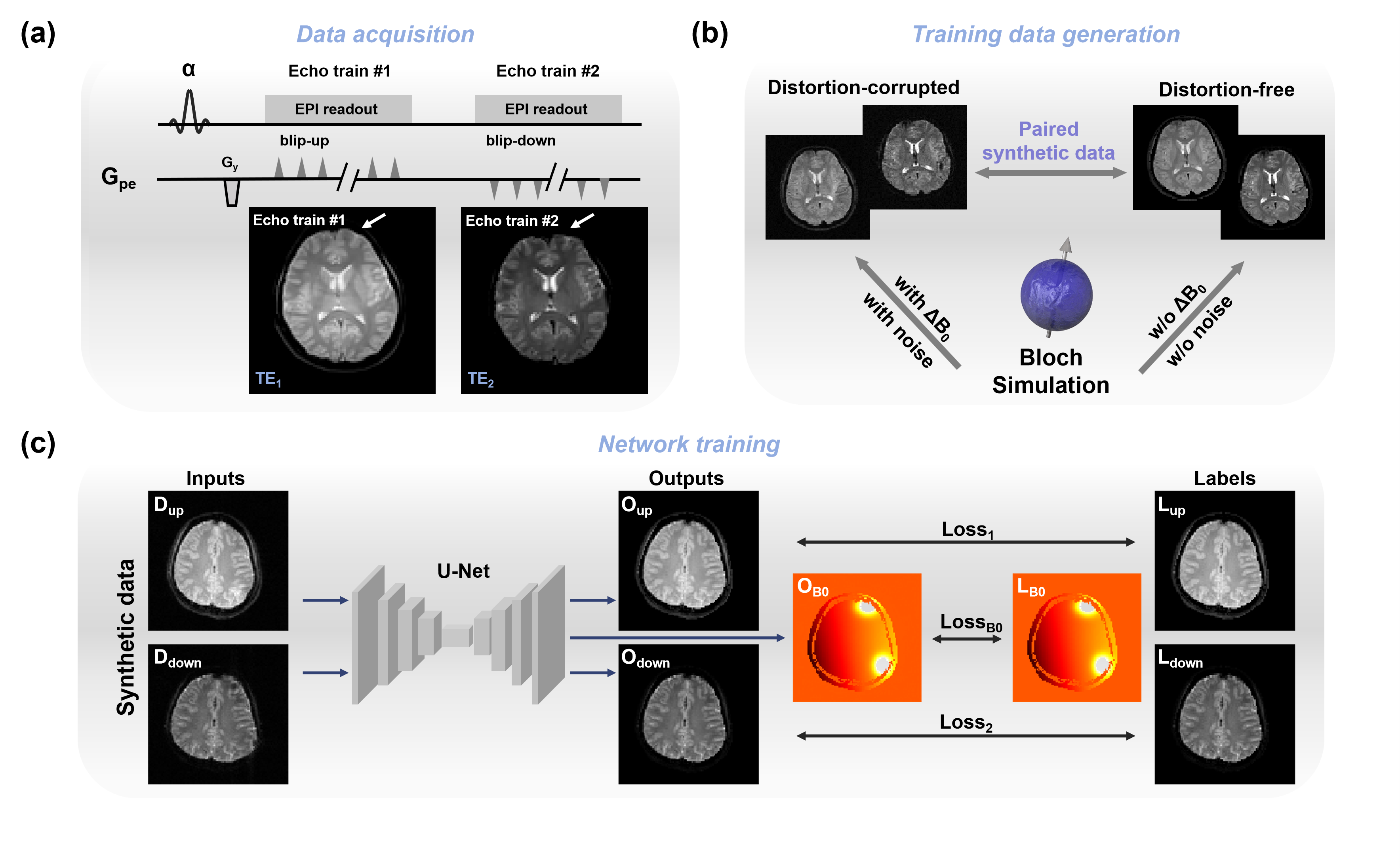
Framework: The overall framework of the ME-BUDA method is shown in Figure 1. Figure 1(a) illustrates the pulse sequence of GRE-EPI with ME-BUDA. Figure 1(b) displays the generation of paired training samples5,6. Non-ideal imaging condition (e.g., B0-inhomogeneity and noise) was considered in the Bloch simulation to obtain distortion-corrupted data as network inputs. In contrast, the distortion-free data did not contain noise to enhance the denoising capability of the network. Subsequently, a U-Net was trained using synthetic data and constrained by the ΔB0 field map used as a part of the loss function. Figure 1(c) shows the details of the network training procedures. The loss function:$$ Loss = \gamma\mid L_{up}-O_{up}\mid + \theta\mid L_{down}-O_{down}\mid +\lambda\mid L_{B0}-O_{B0} \mid $$ in which, Lup and Ldown are the synthetic distortion-free data of two echo trains, and Oup, Odown, and OB0 are the output of U-Net. LB0 is the ΔB0 map of distortion-corrupted data.
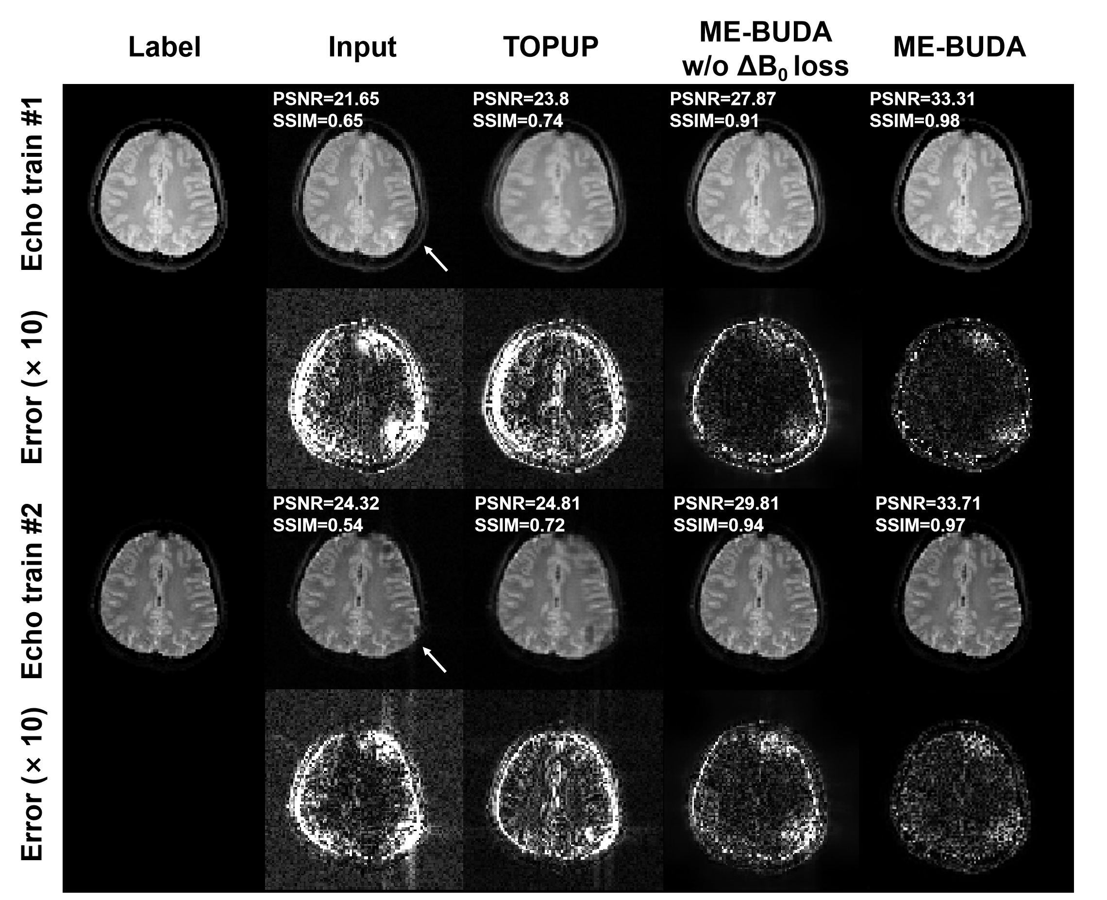
Simulation experiments: To assess the performance of the proposed method, a simulation experiment was performed. The double-scan-based TOPUP method2 and the proposed method with/without ΔB0 map constrained were compared. The peak signal-to-noise ratio (PSNR) and structural similarity index measure (SSIM) of the results were calculated.
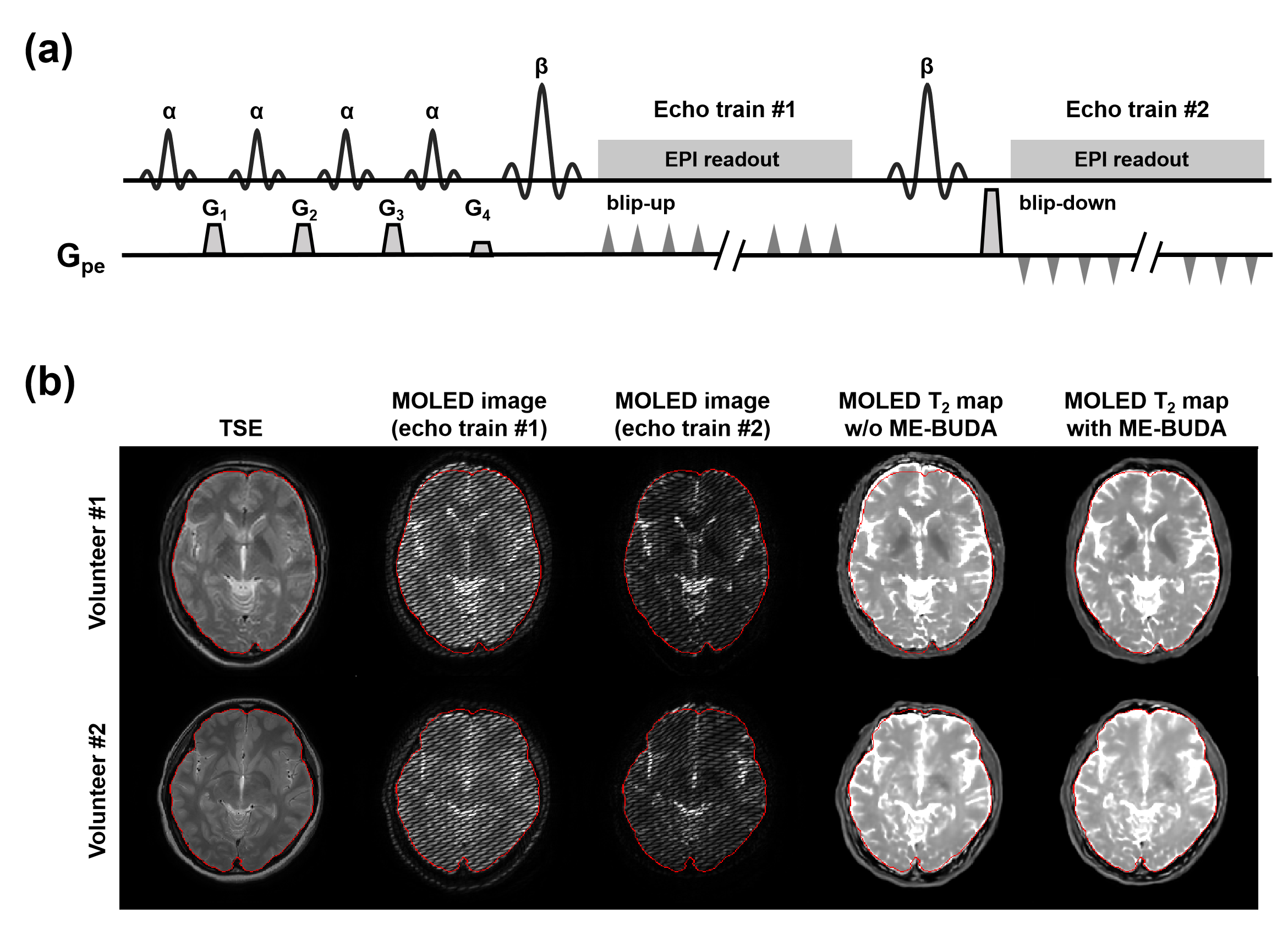
In vivo experiments: In vivo data were acquired from healthy volunteers on a whole-body MRI system at 3T with a 32-channel head coil (Ingenia-CX, Philips Healthcare). Experiment #1 was designed to evaluate the distortion correction capability of the ME-BUDA method in GRE-EPI. Participants were instructed to do four scans: distortion-free GRE, GRE-EPI without ME-BUDA, GRE-EPI with ME-BUDA and dynamic GRE-EPI imaging with ME-BUDA. The acquisition parameters were as follows: FOV = 22 × 22 cm2, voxel size = 2.3 × 2.3 × 3 mm2, GRAPPA factor = 2, TEs = 13.5/39.1 ms, TR = 6,000 ms, and slice number = 21. The parameters used in dynamic acquisition were the same as mentioned above, expect TEs = 10.2/29.3 ms, voxel size = 2.75 × 2.75 mm2, TR = 2,000 ms, scan number = 80. Experiment #2 was designed to assess the capability of the ME-BUDA method in multiple overlapping-echo detachment (MOLED) images7,8 based on spin-echo EPI to achieve rapid distortion-free T2 mapping. Participants were instructed to do three scans: distortion-free TSE, MOLED without ME-BUDA, MOLED with ME-BUDA. Figure 4(a) shows the pulse sequence diagram of MOLED.
Results
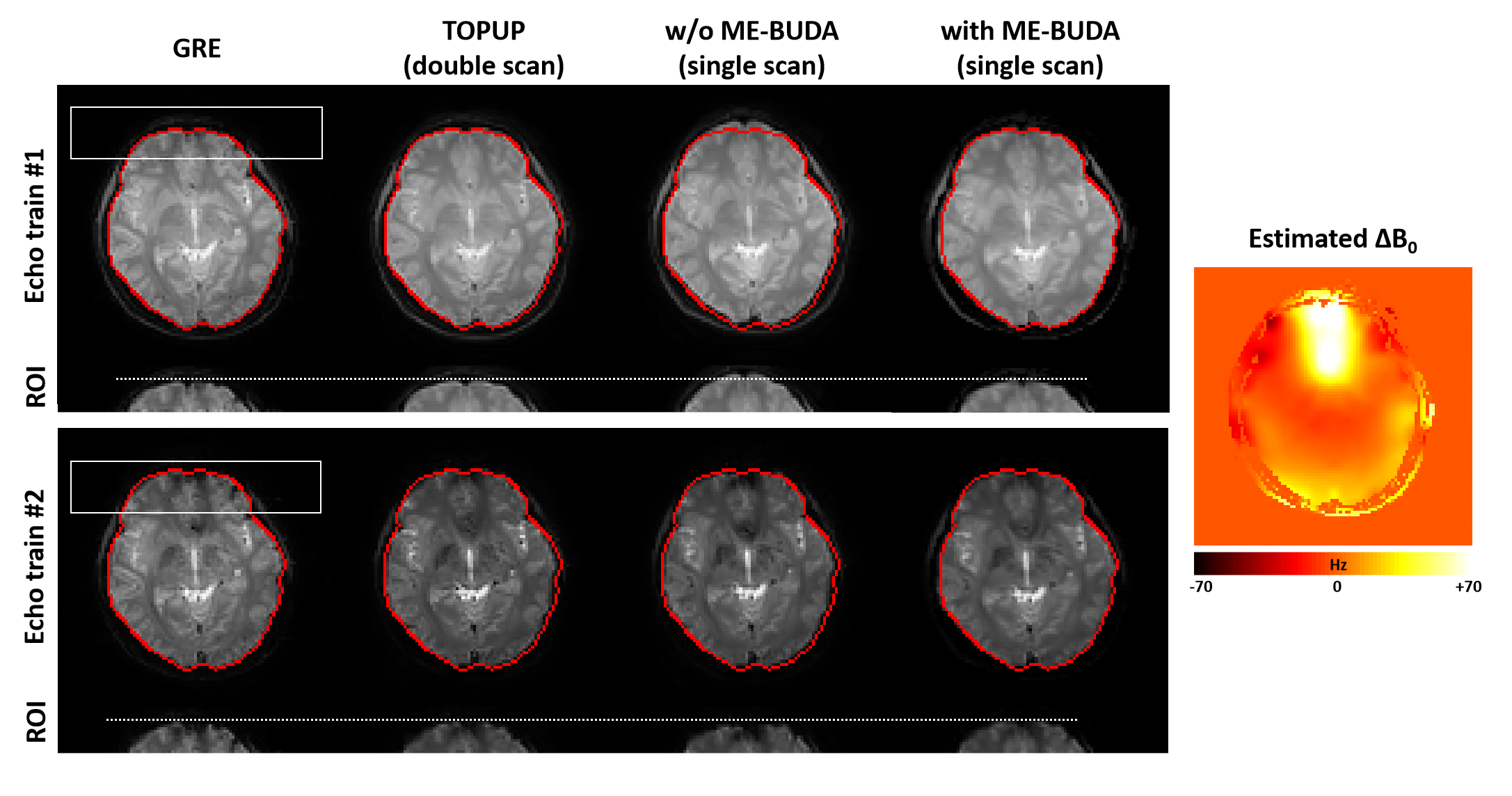
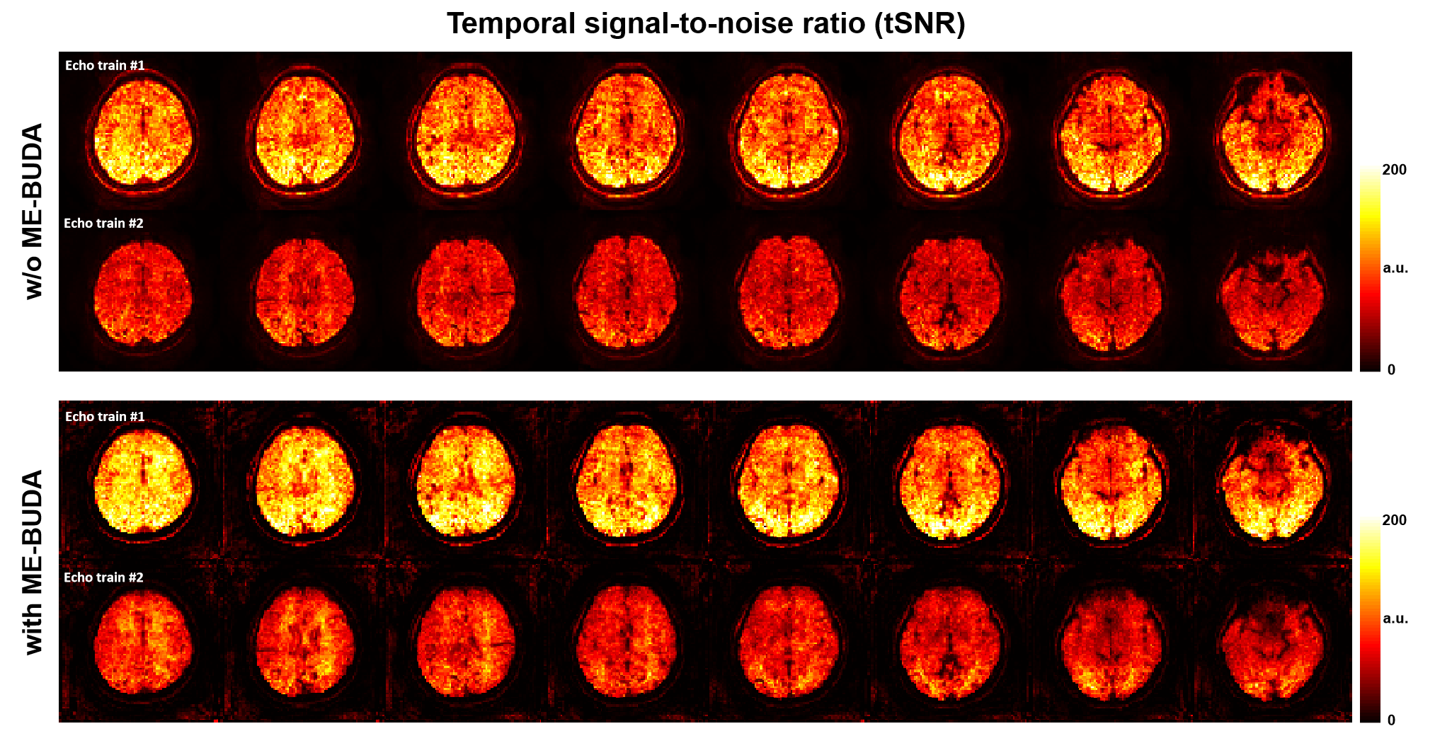
Figure 2 shows the correction results of the simulation experiment and the corresponding quantitative comparison. The PSNR/SSIM for the proposed method was 33.31/0.98 and 33.71/0.97 for echo train #1 and echo train #2, which was higher than 23.80/0.74 and 24.81/0.72 for TOPUP, 27.87/0.91 and 29.81/0.94 for the proposed method without ΔB0 map constrained. The correction results of in vivo experiment #1 are presented in Figure 3. The brain boundaries of ME-BUDA results are consistent with the distortion-free GRE and TOPUP results. Notably, the TOPUP method needs double scans in acquisition and is time-consuming in postprocessing. Figure 4 showcases the temporal signal-to-noise ratio (tSNR) maps calculated from dynamic imaging in experiment #1. For both echo trains, the tSNR of ME-BUDA results is much higher than the GRE-EPI without ME-BUDA in all regions. This result demonstrates the repeatability of the distortion correction capability of the proposed method and also shows its denoising capability. Figure 4(b) displays the correction results of the experiment #2. The MOLED T2 maps of two volunteers, each with a different phase-encoding direction, show the perfect correction of the proposed method compared to the TSE images.Discussion and conclusion
Experimental results reveal that ME-BUDA method, in combination with deep learning reconstruction, has the ability to correct geometric distortion without additional scans. It shows excellent performance in the correction of geometric distortion in ME-EPI, whether it is gradient-echo or spin-echo EPI. Furthermore, the higher tSNR observed in GRE-EPI with ME-BUDA shows enhanced stability compared with the GRE-EPI without ME-BUDA. These results highlight the potential of ME-BUDA to provide distortion-free and high-quality ME-EPI images for ME-fMRI.Acknowledgements
This work was supported in part by the National Natural Science Foundation of China under grant numbers 82071913 and 22161142024.References
1. Kundu P, Voon V, Balchandani P, Lombardo MV, Poser BA, Bandettini PA. Multi-echo fMRI: a review of applications in fMRI denoising and analysis of BOLD signals. Neuroimage. 2017;154:59-80.
2. Andersson J, Skare S, Ashburner J. How to correct susceptibility distortions in spin-echo echo-planar images: application to diffusion tensor imaging. Neuroimage. 2003;20:870-888.
3. Matakos A, Balter J, Cao Y. Estimation of geometrically undistorted B0 inhomogeneity maps. Phys Med Biol. 2014;59(17):4945-4959.
4. Hu ZX, Wang YS, Zhang Z, et al. Distortion correction of single-shot EPI enabled by deep-learning. Neuroimage. 2020;221:117170.
5. Yang QQ, Wang Z, Guo KY, Cai CB, Qu XB. Physics-driven synthetic data learning for biomedical magnetic resonance: The imaging physics-based data synthesis paradigm for artificial intelligence. IEEE Signal Process Mag. 2023;40(2):129-140.
6. Yang QQ, Lin YH, Wang JC, et al. Model-based synthetic data-driven learning (MOST-DL): Application in single-shot T2 mapping with severe head motion using overlapping-echo acquisition. IEEE Trans Med Imaging. 2022;41:3167-3181.
7. Cai CB, Wang C, Zeng YQ, et al. Single-shot T2 mapping using overlapping-echo detachment planar imaging and a deep convolutional neural network. Magn Reson Med. 2018;80:2202-2214.
8. Zhang J, Wu J, Chen SJ, et al. Robust single-shot T2 mapping via multiple overlapping-echo acquisition and deep neural network. IEEE Trans Med Imaging. 2019;38:1801-1811.
Figures