2638
Feasibility Assessment of Measuring ADC of Meningeal Lymphatic Vessels using High-Resolution MUSE DWI and PROPELLER DWI1Ph.D. program in Electrical and Communication Engineering, Feng Chia University, Taichung, Taiwan, 2Department of Medical Imaging, Yuanlin Christian Hospital, Changhua, Taiwan, 3Department of Medical Imaging, Changhua Christian Hospital, Changhua, Taiwan, 4Department of Medical Imaging, Chung Shan Medical University Hospital, Taichung, Taiwan, 5Department of Biomedical Imaging and Radiological Science, China Medical University, Taichung, Taiwan, 6Department of Biomedical Engineering, The Chinese University of Hong Kong, Hong Kong, Hong Kong, 7Department of Medical Imaging, China Medical University Hsinchu Hospital, Hsinchu, Taiwan, 8Department of Radiology, China Medical University Hospital, Taichung, Taiwan, 9Department of Automatic Control Engineering, Feng Chia University, Taichung, Taiwan
Synopsis
Keywords: Vessels, Diffusion/other diffusion imaging techniques, MUSE, PROPELLER, Meningeal lymphatics vessels
Motivation: Is it possible to water diffusion be measured in meningeal lymphatic vessels (MLVs)?
Goal(s): Diffusion-weighted MRI is employed to assess the circulatory condition of MLVs, eliminating the need for MRI with contrast agent administration, which typically requires monitoring for over 4 hours.
Approach: Water diffusion measurements in MLVs were conducted using MUSE DWI and PROPELLER DWI, which offer low image distortion and high resolution.
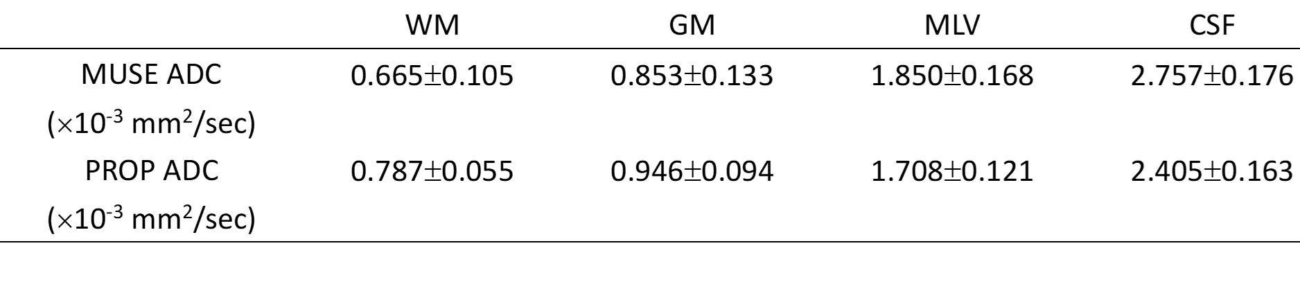
Results: Our results indicate that MLVs were clearly visible on high spatial resolution DWI and ADC maps, and the ADC values of MLVs are higher than those of GM and WM but lower than CSF.
Impact: Since evaluating the circulatory condition of meningeal lymphatic vessels takes over 4 hours by contrast agent injection, our study investigates the feasibility of assessing the ADC of meningeal lymphatic vessels using high-resolution MUSE DWI and PROPELLER DWI.
Introduction
The lymphatic system is a circulation that contributes to facilitating the clearance of excess fluid and macromolecules from the interstitium. It accomplishes this by passing through lymph nodes to remove bacteria, abnormal cells, and other matter [1]. Recently, the brain glymphatic system (GS) [2] and the meningeal lymphatic vessels (MLVs) [3] have been discovered. The brain GS collects cerebrospinal fluid (CSF) from the subarachnoid space and brain interstitial fluid (ISF) through aquaporin-4 (AQP4) water channels. The meningeal lymphatic vessels (MLVs) situated in the dorsal and basal regions serve as downstream channels, responsible for draining ISF, macromolecules, and immune cells out of the cranial cavity. Additionally, they play a crucial role in regulating immune responses in the brain. Recent research has shown that ISF, CSF, MLVs, and the brain GS are important factors impacting brain homeostasis [4].Using 3D T2-Fluid Attenuated Inversion Recovery (FLAIR) magnetic resonance imaging, dural lymphatic structures were demonstrated along the dural venous sinuses in the dorsal regions and along the cranial nerves in the ventral regions of the human brain [5]. The function of MLVs and the flux rate of cerebrospinal fluid to the parasagittal dura have been evaluated using the propagation of tracer with multi-phase T1WI after the injection of a contrast agent [6]. However, the observation period in multi-phase T1WIs exceeds 4 hours due to the extremely slow fluid drain in the MLVs. The function of MLVs can potentially be evaluated using echo-planar diffusion-weighted imaging (EP-DWI) since the slow lymphatic drainage is sensitive to water diffusion. Unfortunately, due to the characteristics of low resolution and image distortion, EP-DWI cannot be effectively applied to the MLVs. However, 2D spatially-selective RF and Multiplexed Sensitivity Encoding (MUSE) technique [7] and PROPELLER [8] can provide diffusion weighted image (DWI) with higher spatial resolution and slight image distortion. In this study, MUSE DWI and PROPELLER DWI with high resolution were used to investigate water diffusion in the MLVs.
Materials and Methods
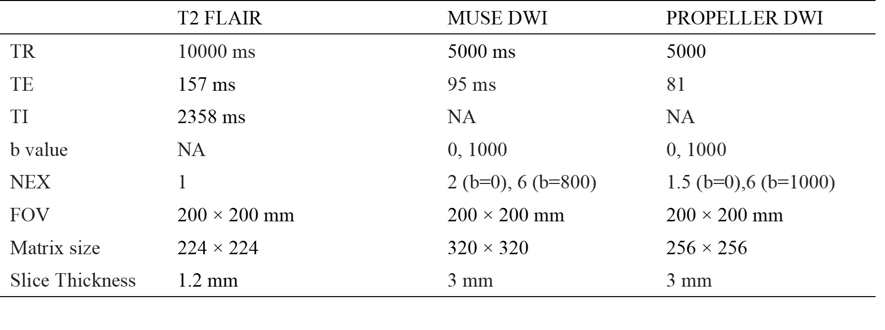
Volunteers: This study conveniently recruited 3 male volunteers aged 23 to 28 years old, who did not exhibit any symptoms related to brain and cerebral circulation. MRI scans: MR studies were conducted using 3.0T scanners (SIGNA Architect, GE Healthcare) with head and neck coils. Table 1 presents the protocols for T2-FLAIR, MUSE DWI, and PROPELLER DWI sequences. Data processing: ADC maps were generated by pixel-by-pixel computation from DWI images using the formula SI=SIb0 × e-bD. ADC values were measured within regions of interest (ROI) encompassing the MLVs, gray matter, white matter, and CSF. Mean values and standard deviations were calculated for the analysis. The signal-to-noise ratio (SNR) was determined by dividing the signal mean of the brain parenchyma by the standard deviation of the air area.Results
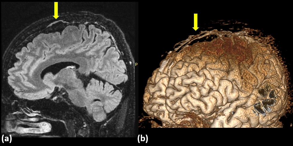
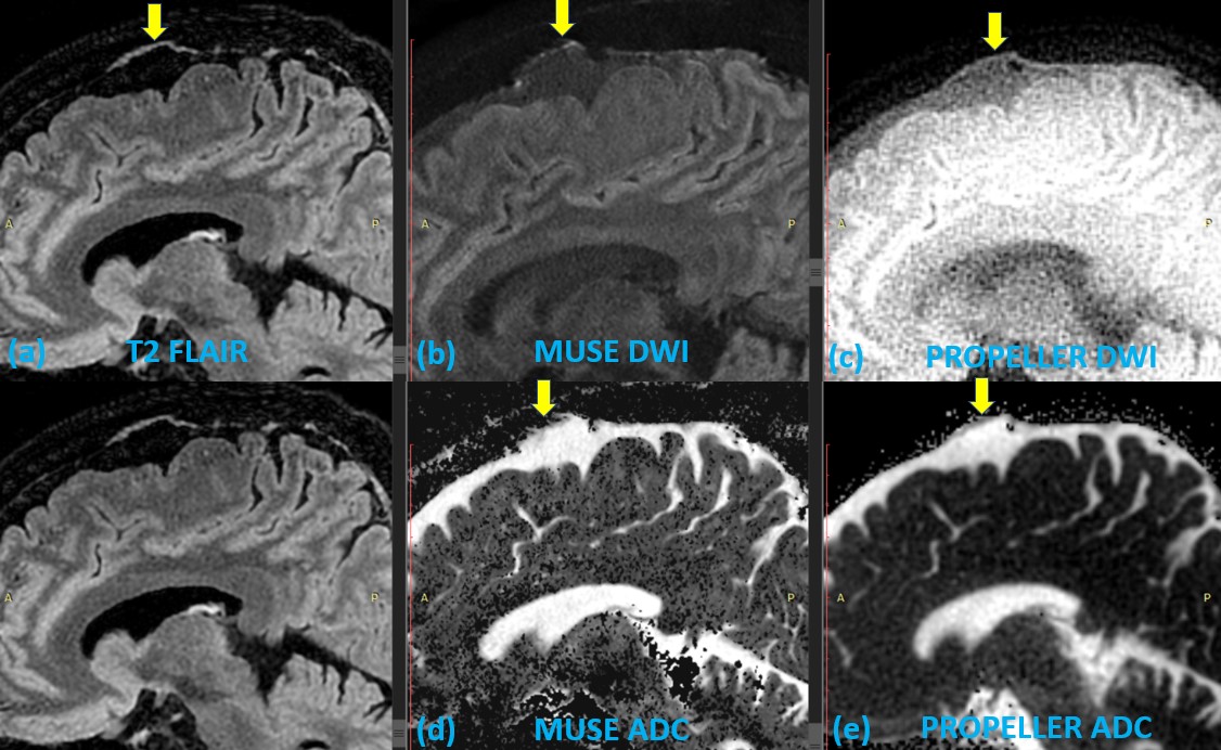
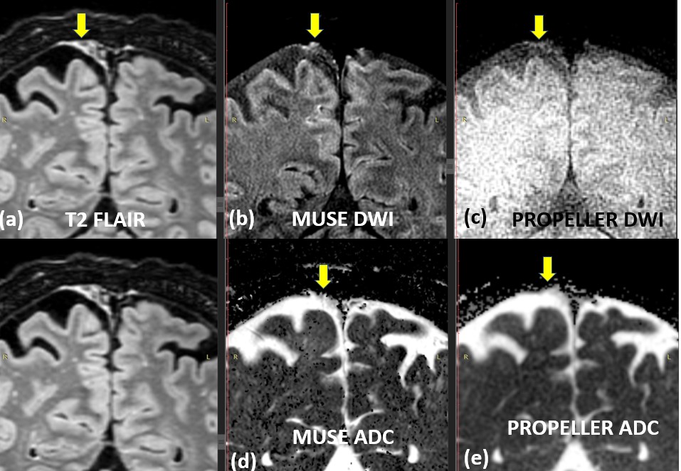
The high-resolution T2 FLAIR image and 3D reconstruction were shown in Figure 1. Figure 2 displayed sagittal view images, including T2 FLAIR, MUSE DWI, PROPELLER DWI, MUSE ADC map, and PROPELLER ADC map. Figure 3 displayed the coronal view images. Table 2 listed the DWI SNR for MUSE and PROPELLER, while Table 3 showed ADC values (mean ± SD) of WM, GM, MLV, and CSF.Discussion
Because the thickness of the MLV is thin (0.5~1.5 mm), a high-resolution MRI is necessary for observing MLVs. In this study, DWI with a voxel size of 0.78×0.78×3 mm was used to evaluate water diffusion in MLVs. Our results showed that MLVs were clearly visible on high spatial resolution DWI and ADC map, corresponding to the T2 FLAIR image, in both MUSE and PROPELLER imaging. The study demonstrated the feasibility of assessing ADC measurement in MLVs using MUSE DWI and PROPELLER DWI, which have high spatial resolution and minimal image distortion. The ADC values of MLVs are intermediate between those of brain parenchyma (GM and WM) and CSF, as diffusion is restricted in bound waters such as solids tissue or macromolecules. The CSF-ISF-CSF washout, originating from the perivenous space, eventually drains into the MLVs with thin film structure [9]. Consequently, the concentration of macromolecules in MLVs is higher than that in CSF, due to macromolecular accumulation and the narrow space within MLVs. In conclusion, the high resolution of MUSE DWI and PROPELLER DWI allows for the measurement of diffusion in MLVs, and the ADC values of MLVs are higher than those of GM and WM but lower than CSF.Acknowledgements
Supported by the Taiwan Ministry of Science and Technology under grants 110-2221-E-035-016 and Taiwan National Science and Technology Council under grants 111-2314-B-035 -001 -MY3.References
- Secker GA, Harvey NL. VEGFR signaling during lymphatic vascular development: From progenitor cells to functional vessels. Dev Dyn. 2015 Mar;244(3):323-31. doi: 10.1002/dvdy.24227. Epub 2014 Dec 4. PMID: 25399804.
- Iliff JJ, Wang M, Liao Y, Plogg BA, Peng W, Gundersen GA, et al. A paravascular pathway facilitates csf flow through the brain parenchyma and the clearance of interstitial solutes, including amyloid beta. Sci Transl Med. 2012;4:147ra111
- Louveau A, Smirnov I, Keyes TJ, Eccles JD, Rouhani SJ, Peske JD, Derecki NC, Castle D, Mandell JW, Lee KS, Harris TH, Kipnis J. Structural and functional features of central nervous system lymphatic vessels. Nature. 2015 Jul 16;523(7560):337-41. doi: 10.1038/nature14432. Epub 2015 Jun 1. Erratum in: Nature. 2016 May 12;533(7602):278. PMID: 26030524; PMCID: PMC4506234.
- Louveau, A., Smirnov, I., Keyes, T. et al. Structural and functional features of central nervous system lymphatic vessels. Nature 523, 337–341 (2015). https://doi.org/10.1038/nature14432.
- Albayram, M.S., Smith, G., Tufan, F. et al. Non-invasive MR imaging of human brain lymphatic networks with connections to cervical lymph nodes. Nat Commun 13, 203 (2022). https://doi.org/10.1038/s41467-021-27887-0
- Ringstad, G., Eide, P.K. Cerebrospinal fluid tracer efflux to parasagittal dura in humans. Nat Commun 11, 354 (2020). https://doi.org/10.1038/s41467-019-14195-x
- Chen NK, Guidon A, Chang HC, Song AW. A robust multi-shot scan strategy for high-resolution diffusion weighted MRI enabled by multiplexed sensitivity-encoding (MUSE). Neuroimage. 2013 May 15;72:41-7. doi: 10.1016/j.neuroimage.2013.01.038. Epub 2013 Jan 28. PMID: 23370063; PMCID: PMC3602151.
- Pipe JG, Farthing VG, Forbes KP. Multishot diffusion-weighted FSE using PROPELLER MRI. Magn Reson Med. 2002 Jan;47(1):42-52. doi: 10.1002/mrm.10014. Erratum in: Magn Reson Med 2002 Mar;47(3):621. PMID: 11754441.
- Yankova G, Bogomyakova O, Tulupov A. The glymphatic system and meningeal lymphatics of the brain: new understanding of brain clearance. Rev Neurosci. 2021 Feb 23;32(7):693-705. doi: 10.1515/revneuro-2020-0106. PMID: 33618444.
Figures