2637
Evaluation of the glymphatic system in patients with bipolar disorder using the DTI-ALPS method1Department of Radiology, Juntendo University Graduate School of Medicine, Tokyo, Japan, 2Department of Innovative Biomedical Visualization, Nagoya University Graduate School of Medicine, Aichi, Japan, 3Department of Psychiatry and Behavioral Science, Juntendo University Graduate School of Medicine, Tokyo, Japan
Synopsis
Keywords: Neurofluids, Psychiatric Disorders, Glymphatic system
Motivation: Our previous study revealed abnormalities in aquaporin 4, which works at the cerebrospinal–interstitial fluid exchange in the postmortem brains of patients with bipolar disorder (BP). However, glymphatic system (GS) alterations in BP are still unclear.
Goal(s): This study aims to assess GS function in BP patients.
Approach: We used diffusion tensor image analysis along the perivascular space (DTI–ALPS) method in BP subjects and healthy controls (HCs).
Results: The ALPS index of BP subjects was significantly lower than that of HCs. We also found associations between the ALPS index and BP risk. Thus, per our findings, BP patients could have GS dysfunction.
Impact: This study suggests the potential for GS dysfunction in BP patients. Our findings could support the fact that BP patients are more likely to experience dementia since they have an Alzheimer’s disease-like pathology that causes amyloid β accumulation.
INTRODUCTION
Bipolar disorder (BP) is a condition characterized by alternating mental states that are polar opposites of each other. These include "manic" states marked by significantly elevated moods and "depressive" states characterized by decreased motivation and sadness1. It often occurs at a young age2 and persists for long periods. It has been reported that BP patients are more likely to develop dementia and speculated that the background is the Alzheimer’s disease-like pathology of amyloid β (Aβ) accumulation in BP3-5. Besides, Iwamoto et al. revealed abnormalities in aquaporin 4, which works at the cerebrospinal–interstitial fluid junction in the postmortem brains of BP patients, through gene expression analyses of postmortem brains 6,7. However, the GS alteration in BP is still not clear. Then, the diffusion tensor image analysis along the perivascular space (DTI–ALPS) method is suggested to evaluate the interstitial fluid dynamics7,8. This study aims to assess GS function using the ALPS index in BP patients.METHODS
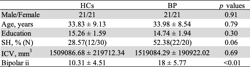
Subjects and MRI data acquisitionWe used an open database known as the UCLA Consortium for Neuropsychiatric Phenomics LA5c Study9. In this study, the data of 42 BP patients and 42 age-and-sex-matched healthy controls (HCs) were used. Fig.1 shows the study participants’ demographic characteristics. We also used the data of the Scale for Traits that Increase the Risk for Bipolar II Disorder (https://nda.nih.gov/data_structure.html?short_name=stirbd01) , which measures mood liability, energy activity, daydreaming, and social anxiety in all subjects. A higher score on this test means a higher risk of BP II. DWI data were collected from all participants who underwent using a 3T MRI scanner (Siemens, TrioTim) with the following parameters: TR/TE = 7000/92.6ms, b value = 0, 1000s/mm2, 65 directions with acquisition matrix = 96 x 96, voxel size=2 × 2 × 2 mm³, and total scan time = 10 minutes.
DWI preprocessing
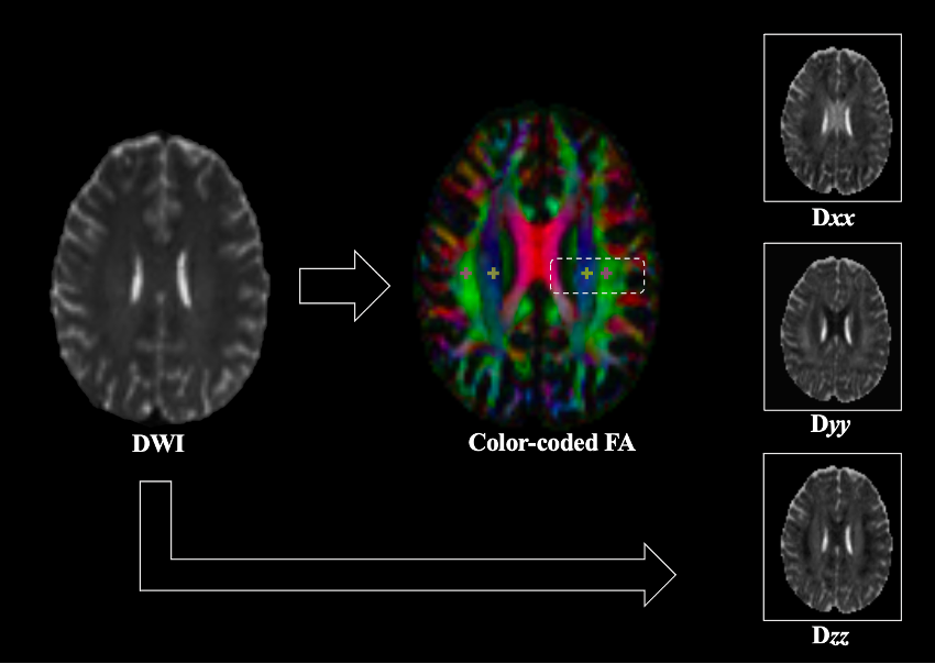
DWI data were processed using the MRtrix3.010 as follows: denoise11, remove Gibbs artifact12, eddy-current distortions, bias-field using the ANTs option. Diffusivity maps of each subject were obtained in the direction of the x-axis (right–left; Dxx), y-axis (anterior–posterior; Dyy), and z-axis (inferior–superior; Dzz) using the FMRIB Software Library (version 6.0.)13.
ALPS index calculation
We placed 5-mm cross-shaped region of interests (ROIs) in the projection and the association areas at the level of the lateral ventricle bodies using the color fractional anisotropy (FA) map of each subject (Fig.2). Then, the ALPS index was calculated as a ratio of the mean x-axis diffusivity in the projection area (Dxxproj) and x-axis diffusivity in the association area (Dxxassoc) to the mean of the y-axis (Dyyproj) and the z-axis diffusivity in the association area (Dzzaccoc) as follows7. ALPS index = (Dxxproj + Dxxassoc) / (Dyyproj + Dzzassoc). Then, we calculated the mean ALPS index of the left and right hemispheres.
Statistical analysis
Statistical analyses were conducted using SPSS 29.0 (IBM Corporation). The mean ALPS indexes in the HCs and BP groups were compared using the generalized linear model and age, sex, smoking history, and intracranial volume as independent variables. Then, partial correlation analyses between the ALPS index and the Scale for Traits that Increase the Risk for Bipolar II Disorder (https://nda.nih.gov/data_structure.html?short_name=stirbd01) were evaluated, adjusting for age, sex, smoking history, and intracranial volume in all participants. A p-value of <0.05 was considered statistically significant.
RESULTS
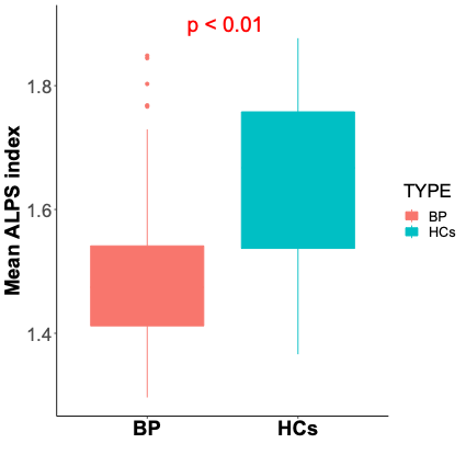
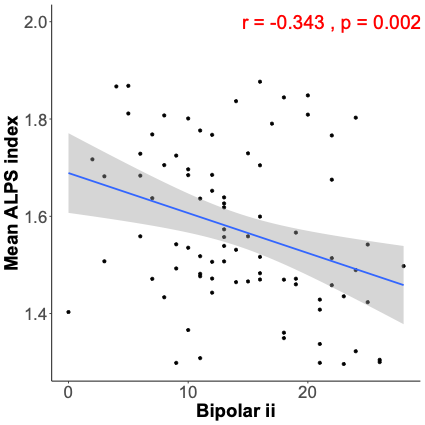
The mean ALPS index in the BP group was significantly lower than that in the HCs (p < 0.001, Fig.3). Partial correlation coefficients were observed for the significant associations between the mean ALPS index and the Scale for Traits that Increase the Risk of Bipolar II Disorder (r = −0.343, p = 0.002, Fig.4).DISCUSSION
The findings of this study demonstrate that BP subjects have a significantly lower ALPS index than HCs. Additionally, this study showed associations between the mean ALPS index and the risk of BP. These results suggest the potential for the dysfunction of interstitial fluid dynamics in BP. Previous studies have shown higher ratios of CSF isoforms Aβ42/38 and Aβ42/40 in patients with BP compared to HCs4 and lower plasma-Aβ42 and higher Aβ40/42 ratios in patients with BP compared to HCs, which are similar to findings of pathophysiology on Alzheimer’s disease5. Our findings may support these previous studies showing that BP has the background of Aβ accumulation due to GS impairment.Acknowledgements
This work was supported by JSPS KAKENHI Grant Number 20K16737, the Japan Agency for Medical Research and Development (AMED) under Grant Number JP21wm0425006, and Grants-in-Aid for Transformative Research Areas ― Platforms for Advanced Technologies and Research Resources “Advanced Bioimaging Support.”References
- Rajkowska G, Halaris A, Selemon LD. Reductions in neuronal and glial density characterize the dorsolateral prefrontal cortex in bipolar disorder. Biol Psychiatry. 2001;49(9):741-752.
- Vieta E, Salagre E, Grande I, et al. Early intervention in bipolar disorder. Am J Psychiatry. 2018;175(5):411-426.
- Knorr U, Simonsen AH, Jensen CS, et al. Alzheimer’s disease related biomarkers in bipolar disorder - A longitudinal one-year case-control study. J Affect Disord. 2022;297:623-633.
- Jakobsson J, Zetterberg H, Blennow K, et al. Altered concentrations of amyloid precursor protein metabolites in the cerebrospinal fluid of patients with bipolar disorder. Neuropsychopharmacol. 2013;38(4):664-672.
- Piccinni A, Veltri A, Vizzaccaro C, et al. Plasma amyloid-β levels in drug-resistant bipolar depressed patients receiving electroconvulsive therapy. Neuropsychobiology. 2013;67(4):185-191.
- Iwamoto K, Kakiuchi C, Bundo M, et al. Molecular characterization of bipolar disorder by comparing gene expression profiles of postmortem brains of major mental disorders. Mol Psychiatry. 2004;9(4):406-416.
- Taoka T, Masutani Y, Kawai H, et al. Evaluation of glymphatic system activity with the diffusion MR technique: Diffusion tensor image analysis along the perivascular space (DTI-ALPS) in Alzheimer's disease cases. Jpn J Radiol. 2017;35(4):172-178.
- Kamagata K, Andica C, Takabayashi K, et al. Association of MRI indices of glymphatic system with amyloid deposition and cognition in mild cognitive impairment and Alzheimer disease. Neurology. 2022;99(24):e2648-e2660.
- Poldrack RA, Congdon E, Triplett W, et al. A phenome-wide examination of neural and cognitive function. Sci Data. 2016;3:160110.
- Tournier JD, Smith R, Raffelt D, et al. MRtrix3: A fast, flexible and open software framework for medical image processing and visualisation. NeuroImage. 2019;202:116137.
- Veraart J, Novikov DS, Christiaens D, et al. Denoising of diffusion MRI using random matrix theory. NeuroImage. 2016;142:394-406.
- Kellner E, Dhital B, Kiselev VG, et al. Gibbs-ringing artifact removal based on local subvoxel-shifts. Magn Reson Med. 2016;76(5):1574-1581.
- Jenkinson M, Beckmann CF, Behrens TE, et al. FSL. NeuroImage. 2012;62(2):782-790.
Figures