2636
Validation of diffusivity analysis along the perivascular space (ALPS) index as a biomarker for vascular cognitive impairment and dementia1Laboratory of FMRI Technology (LOFT), Mark & Mary Stevens Neuroimaging and Informatics Institute, University of Southern California, Los Angeles, CA, United States, 2Radiology and Biomedical Imaging, University of California, San Francisco, San Francisco, CA, United States, 3Neurosurgery, Stanford University, Stanford, CA, United States, 4Department of Neurology, University of California, Davis, Davis, CA, United States, 5The Mind Research Network, Albuquerque, NM, United States, 6Neurology, Keck School of Medicine, University of Southern California, Los Angeles, CA, United States, 7Radiology, Johns Hopkins University School of Medicine, Baltimore, MD, United States, 8Biomedical Engineering, Illinois Institute of Technology, Chicago, IL, United States, 9Diagnostic Radiology and Nuclear Medicine, Rush University, Chicago, IL, United States, 10University of California, Davis, Davis, CA, United States, 11Neuroscience, University of Kentucky, Lexington, KY, United States, 12Population Health Sciences and Glenn Biggs Institute for Neurodegenerative Diseases, University of Texas Health Science Center at San Antonio, San Antonio, TX, United States, 13Neuroimage Analytics Laboratory and Glenn Biggs Institute Neuroimaging Core, Glenn Biggs Institute for Neurodegenerative Diseases, University of Texas Health Science Center at San Antonio, San Antonio, TX, United States, 14Neurology, University of California, San Francisco, San Francisco, CA, United States, 15Neurology, Massachusetts General Hospital, Boston, MA, United States, 16Radiology, Harvard Medical School, Boston, MA, United States, 17Athinoula A. Martinos Center for Biomedical Imaging, Massachusetts General Hospital, Charlestown, MA, United States, 18Computer Science and AI Lab, Massachusetts Institute of Technology, Cambridge, MA, United States
Synopsis
Keywords: Neurofluids, Diffusion Tensor Imaging, glymphatic system, cerebral small vessel disease (cSVD), vascular cognitive impairment and dementia (VCID))
Motivation: To test the validity of the ALPS index as a biomarker for VCID
Goal(s): To test our hypothesis that ALPS index is an independent biomarker for the cognitive decline in cSVD.
Approach: Participants from MarkVCID consortium underwent baseline and follow-up MRI examinations and clinical evaluations of cognitive function
Results: We found the baseline ALPS index was correlated with the existing biomarkers of cSVD and VCID, and was independently associated with the baseline cognitive performance
Impact: Our study provides the clinical validation of the ALPS index as a sensitive and independent biomarker for the cognitive function of cSVD related VCID.
Abstract
Introduction: The diffusion tensor analysis along the perivascular space (DTI-ALPS) method was recently proposed to non-invasively evaluate the glymphatic clearance function1. We recently demonstrated that the index of diffusivity along the perivascular space (ALPS index) has favorable cross-vendor reproductivity and test-retest repeatability using a cohort dataset in the MarkVCID consortium2. To further validate the ALPS index as a biomarker for vascular cognitive impairment and dementia (VCID), we investigate the correlation of the ALPS index with existing biomarkers of cerebral small vessel disease (cSVD), followed by the association of the ALPS index with cognitive performance in the MarkVCID cohort.Methods: The participants and MRI data used in this study were acquired as part of the MarkVCID consortium, which consisted of seven sites. A total of 578 participants (age: 72.5±7.2 years old, 232 Male) at risk of cSVD who received comprehensive clinical and cognitive evaluations and underwent baseline MRI examinations were included in this study. Follow-up MRI examinations and cognitive assessments were conducted on 273 participants. The Montreal Cognitive Assessment (MoCA) score, Principal Component Analysis derived General Cognitive Function (GCF_PCA) score3, and a composite score of Executive Function (EFC) based on Item Response Theory4 were used to evaluate the cognitive performance. Structural MRI and DTI data were acquired by using 3.0T MR scanners at each of the 7 sites. The acquisition protocol of DTI used a single-shell (b=1000 s/mm2), 40-direction diffusion sequence with a voxel size of 2.0x2.0x2.0 mm3 and six b=0 s/mm2. A separate scan using a reverse-polarity phase encoding gradient was acquired for correcting image distortions. The DTI data were processed by using an in-house automatic processing pipeline with FMRIB Software Library 6.0.62 (https://github.com/gbarisano/alps/). The mean free water (mFW) and peak width of skeletonized mean diffusivity (PSMD) were computed in the white matter (WM). The ALPS index was defined as the average of bilateral ALPS indices which were calculated by the ratio of mean of x-axis diffusivity in the ROIs of projection fibers (Dxxproj) and x-axis diffusivity in the ROIs of association fibers (Dxxassoc) to the mean of y-axis diffusivity in the ROIs of projection fibers (Dyyproj) and z-axis diffusivity in the ROIs of association fibers (Dzzassoc)1. A few ROIs that covered the enlarged lateral ventricle or thickened cortex were manually amended to improve the accuracy of ALPS index calculation. The WM hyperintensity volumes (WMHV) were calculated on FLAIR images and normalized by intracranial volume (ICV). Univariate correlation (Pearson or Spearman) was used to examine the associations between imaging markers (ALPS index, mFW, PSMD, and WMHV). Linear regression models were used to evaluate the associations of baseline ALPS index with baseline and longitudinal changes of cognitive outcomes, regressing out three types of covariates: 1) age, sex, and education (model 1), 2) added vascular risk factors (VRFs), including diabetes, hypertension and smoking (model 2), 3) further added mFW, PSMD and WMHV (model 3). SAS 9.4 software was used for all statistical analyses, and P<0.05 was regarded as statistical significance.
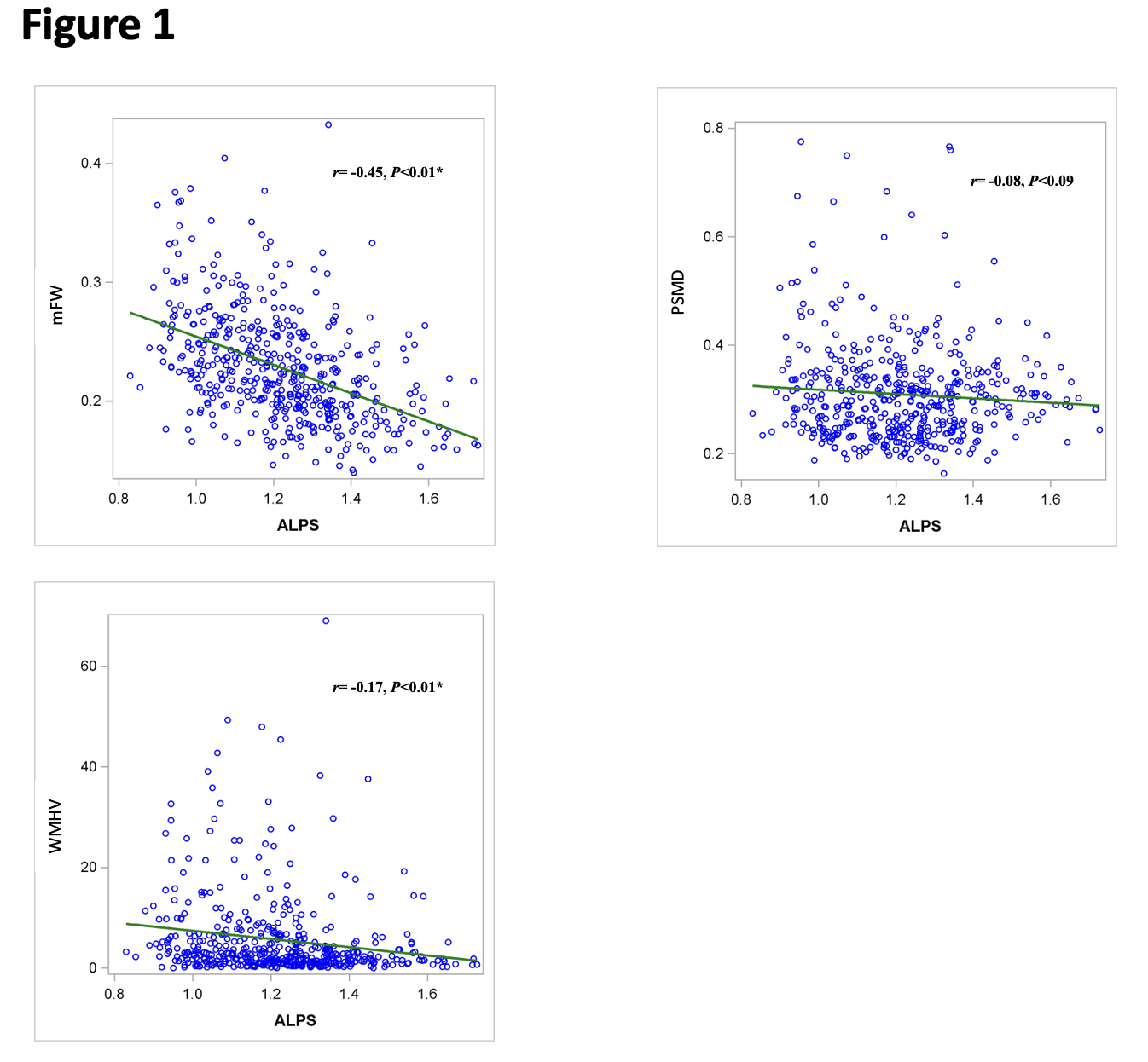
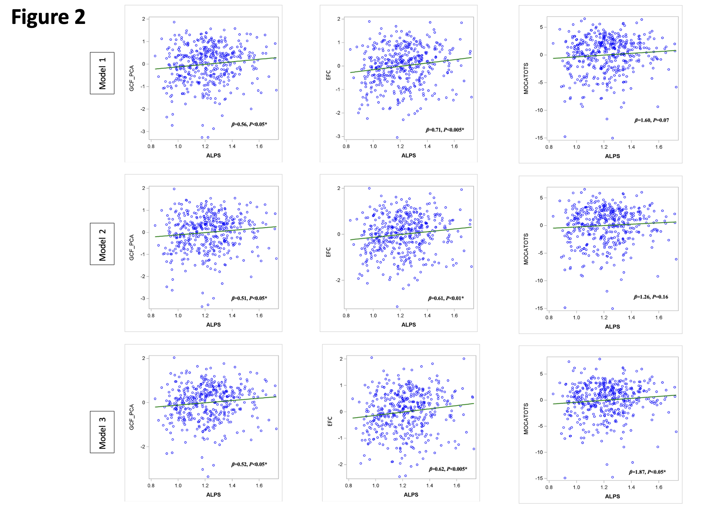
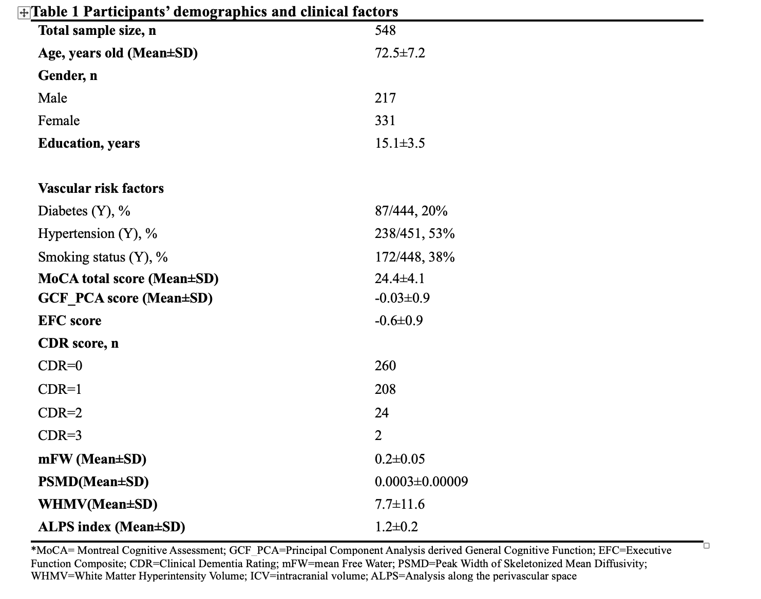
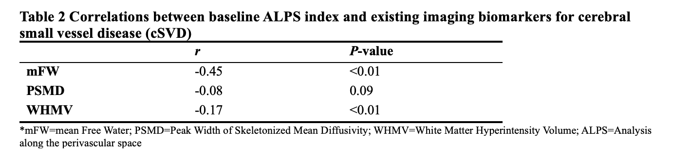
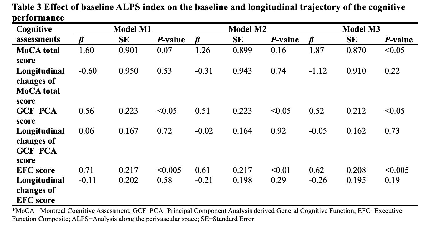
Results: Table 1 shows the demographic and clinical information of the participants. Table 2 and Figure 1 show that the baseline ALPS index was negatively correlated with mFW (r=-0.45, P<0.01), and WMHV (r=-0.17, P<0.01). In regression model 1 and model 2, the baseline ALPS index was significantly associated with the baseline GCF_PCA score (β =0.56, P<0.05 and β =0.51, P<0.05) and EFC score (β =0.71, P<0.005 and β =0.61, P<0.01), but not significantly associated with the baseline MoCA total score. In regression model 3, which controlled the effects of existing imaging biomarkers on cognitive performance, the associations between the baseline ALPS index and the baseline MoCA total score (β=1.87, P<0.05), GCF_PCA score (β =0.52, P<0.05) and EFC score (β =0.62, P<0.005) were significant (See Table 3 and Figure 2). However, we didn’t detect significant correlations between the baseline ALPS index and the longitudinal changes of MoCA total score, GCF_PCA score, or EFC score in all regression models.
Discussion: The present study provides the clinical validation of the ALPS index for cSVD-related VCID. The baseline ALPS index was significantly correlated with existing biomarkers of cSVD-related VCID, i.e. mFW, PSMD, and WMHV, which is consistent with previous studies5–8. Additionally, we detected the association between the baseline ALPS index and baseline cognitive performances after adjusting for the demographics, VRFs, and existing biomarkers of cSVD and VCID, suggesting that the ALPS index is an independent contributor to the cognitive decline in cSVD.
Conclusions: ALPS index appears to be a sensitive and independent biomarker for the cognitive function of cSVD related VCID.
Acknowledgements
The authors thank the participants in this study and the support of MarkVCID consortium. This study was supported by NIH grants UF1-NS100614, U24NS1 00591, UF1NS100599, UF1NS100588, UF1NS100608, UF1NS100605, UF1NS100606, UF1NS100598, P30AG 010129, P30AG072946, P30AG066530, UF1NS125513, P30AG066546.References
1. Taoka T, Masutani Y, Kawai H, et al. Evaluation of glymphatic system activity with the diffusion MR technique: diffusion tensor image analysis along the perivascular space (DTI-ALPS) in Alzheimer’s disease cases. Jpn J Radiol. 2017;35(4):172–178.
2. Liu X, Barisano G, Shao X, et al. Cross-Vendor Test-Retest Validation of Diffusion Tensor Image Analysis along the Perivascular Space (DTI-ALPS) for Evaluating Glymphatic System Function. Aging Dis. 2023;14.
3. Cowman M, Lonergan E, Burke T, et al. Evidence supporting the use of a brief cognitive assessment in routine clinical assessment for psychosis. Schizophrenia. 2022;8(1):113.
4. Staffaroni AM, Asken BM, Casaletto KB, et al. Development and validation of the Uniform Data Set (v3.0) executive function composite score (UDS3-EF). Alzheimers Dement. 2021;17(4):574-583.
5. Low A, Mak E, Stefaniak JD, et al. Peak Width of Skeletonized Mean Diffusivity as a Marker of Diffuse Cerebrovascular Damage. Front Neurosci.2020;14:238.
6. Maillard P, Hillmer LJ, Lu H, et al. (2022). MRI free water as a biomarker for cognitive performance: Validation in the MarkVCID consortium. Alzheimer Dement (Amst). 2022;14(1):e12362.
7. Ke Z, Mo Y, Li J, et al. Glymphatic Dysfunction Mediates the Influence of White Matter Hyperintensities on Episodic Memory in Cerebral Small Vessel Disease. Brain Sci. 2022;12:1611.
8. Kamagata K, Andica C, Takabayashi K, et al. (2022). Association of MRI Indices of Glymphatic System With Amyloid Deposition and Cognition in Mild Cognitive Impairment and Alzheimer Disease. Neurology.2022;99(24):e2648–e2660.
Figures