2634
Reduced Glymphatic System Activity Induces Elevated GABA Level in Hepatic Encephalopathy Patients: A Potential MRI-Based Mechanism1Guizhou Provincial People’s Hospital, Guiyang, China, 2GE HealthCare MR Research, Beijing, China
Synopsis
Keywords: Structural Connectivity, Diffusion/other diffusion imaging techniques, ALPS; GABA; hepatic encephalopathy
Motivation: Hepatic encephalopathy (HE) is a severe complication of decompensated cirrhosis. Understanding the pathogenesis is crucial for developing effective treatments and improving outcomes.
Goal(s): This study delved into the HE pathogenesis by employing diffusion tensor imaging analysis focused on the perivascular space (DTI-ALPS), alongside the quantification of GABA levels in the thalamus of patients with cirrhosis.
Approach: 48 patients with decompensated cirrhosis were enrolled, with all of them undergoing DTI and MRS assessments.
Results: We found that patients with HE exhibited impaired glymphatic system function and increased levels of the inhibitory neurotransmitter GABA in the thalamus, with a noteworthy correlation found between them.
Impact: The discovery of decreased glymphatic system activity and increased GABA levels may hold significant promise in understanding the potential mechanisms behind HE patients. These findings could contribute to the development of targeted therapies for HE and improve patient care and outcomes.
Introduction
Hepatic encephalopathy (HE) is a neuropsychiatric syndrome resulting from severe hepatic dysfunction or abnormal portosystemic shunts (PSSs), causing metabolic disorder1. Associated with poor outcomes, it represents a major complication of decompensated cirrhosis2. The underlying mechanisms of HE remain unclear although factors such as metabolic dysfunction, blood-brain barrier disruption, and increased inhibitory neurotransmitter secretion have been covered1.Recent research indicates the potential involvement of the glymphatic system and alterations in gamma-aminobutyric acid (GABA) levels in neuropsychiatric disorders3,4. However, these findings primarily originate from studies conducted on animal models and other metabolic disorders and are deficient in clinical investigations utilizing MRI in patients with cirrhotic HE. Our study employs diffusion tensor imaging (DTI) to analyze the perivascular space (ALPS index), reflecting the glymphatic system activity. We also use magnetic resonance spectroscopy (MRS) to measure thalamic GABA levels, offering insights into HE pathogenesis in cirrhotic patients.
Materials and Methods
PatientsThe study obtained approval from the Ethics Committee, and informed consent was acquired from all 48 decompensated cirrhosis patients. These patients were categorized into three groups: liver cirrhosis (LC, n=22), mild hepatic encephalopathy (MHE, n=19), and hepatic encephalopathy (HE, n=7).
MRI Acquisition
Participants underwent MRI using a 3.0T GE 750W Discovery scanner with a 48-channel head and neck coil. T1-weighted sagittal structural images were acquired using the 3D-EFGRE BRAVO sequence (1-mm isotropic resolution, FOV=25.6cm, Matrix=256×256, slice=192, flip angle=12°, TR=6.622 ms, TE=2.164 ms, Acceleration factor=2). GABA levels were assessed by MEGA-PRESS (TR/TE=1800/68 ms, TA=8 min24s, spectral width=5000 Hz, number of points=4096) from 20mm voxel thickness. DTI data was acquired with 64 diffusion directions EPI impulse sequence (TR/TE=9700/74.2 ms, Matrix=128×128, 3mm thickness, FOV=25.6cm, no gap, slice=47, b-value=1000, Acceleration=2). Data meeting quality criteria were selected for inclusion. Following the export of data, adjustments for spectral frequency and phase were performed. Voxel co-registration with the T1 structural phase was carried out using water and creatine (Cr) signals as references. GABA and glutamate/glutamine (GLX) concentrations were quantified in SI units normalized to water content (GABA and GLX/water) and represented as integer ratios to chromium (GABA/Cr and GLX/Cr). MRS data with line motion artifacts or insufficient water suppression were excluded.
Data Analysis
DTI was processed with FSL version 6.0 (www.fmrib.ox.ac.uk/fsl)5, and MEGA-PRESS data were analyzed using GANNET 3.1 (http://gabamrs.org)6. Continuous variables were expressed as mean ± SD or median (25th-75th percentile) and analyzed using Student's t-test or ANOVA. Categorical data, presented as numbers (percentages), were analyzed with the Chi-square test. Spearman correlation was adopted to assess relationships between variables, with significance at P<0.05.
Results
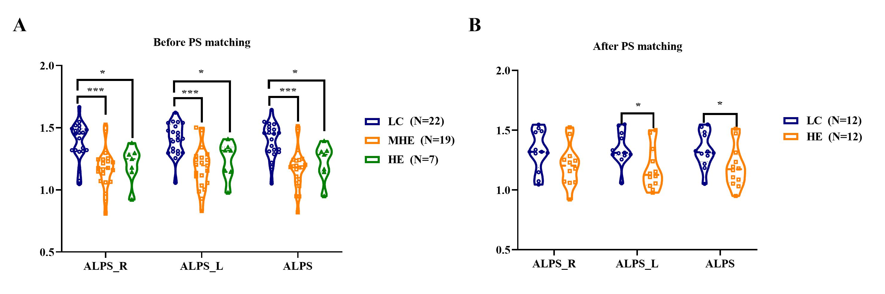
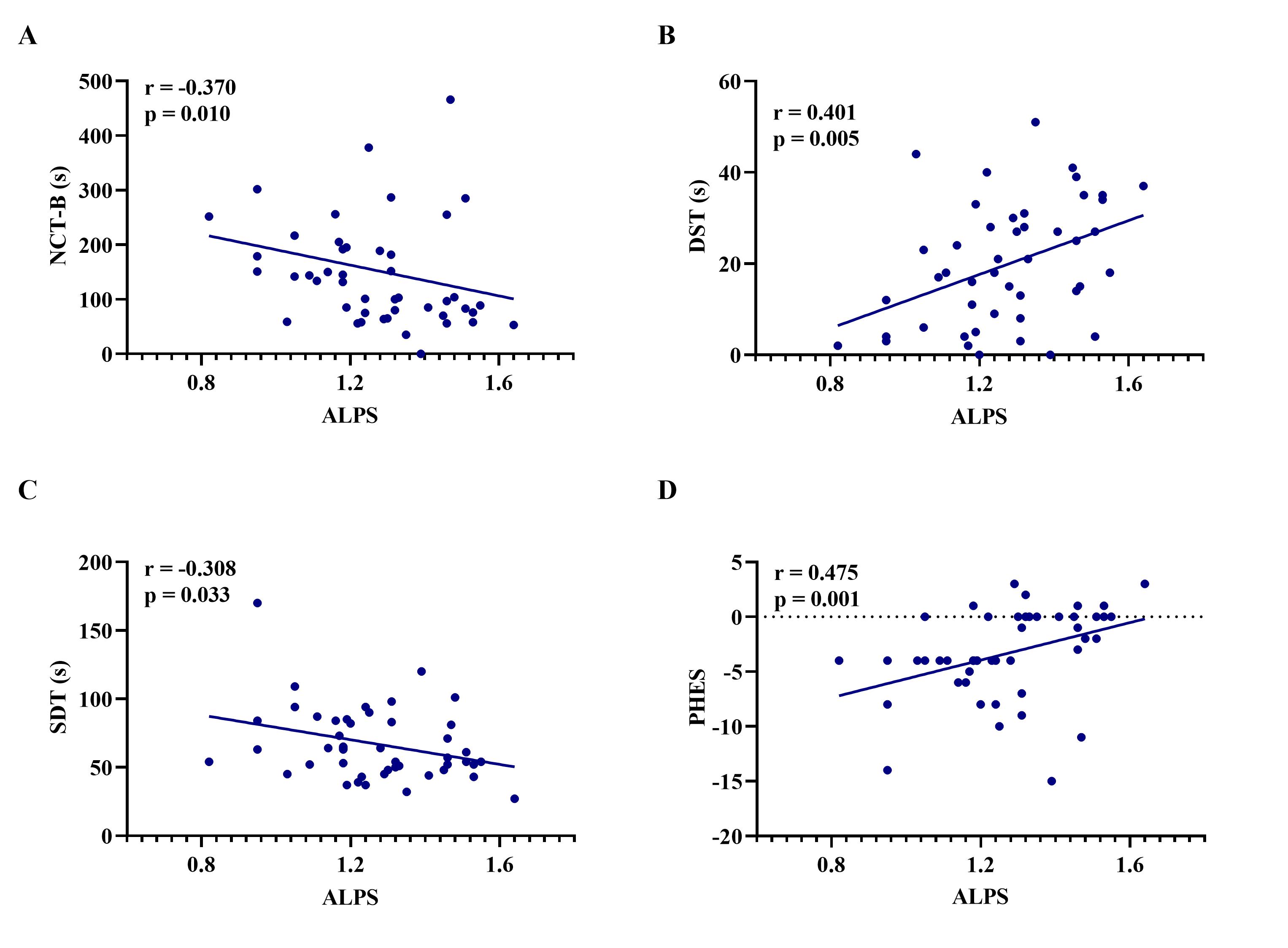
Among the three groups, there were no significant differences in sex, education years, and Child-Pugh scores. However, significant differences were found in age, PHES score, and animal naming test. Specifically, the HE group was older with lower PHES and animal naming test scores compared to the LC group.The MHE (1.21±0.23) and HE groups (1.20±0.26) showed significantly lower ALPS index levels than the LC Group (1.47±0.35) (P<0.05), even after conducting propensity score (PS) matching for sex, age, and education years (Figure 1). Meanwhile, the ALPS index was significantly correlated with psychometric test scores (NCT-B, DST, SDT, PHES, P<0.05) (Figure 2).
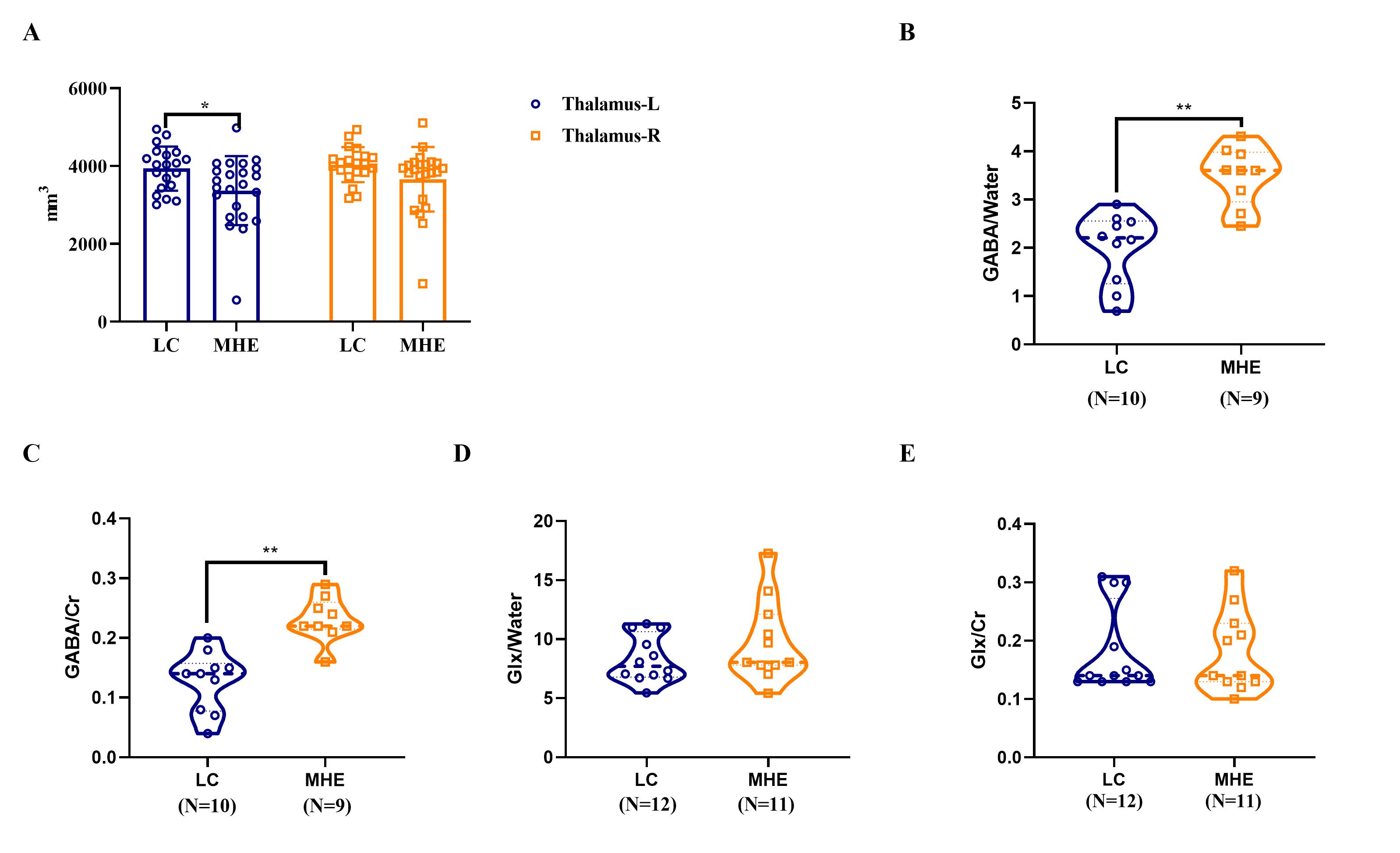
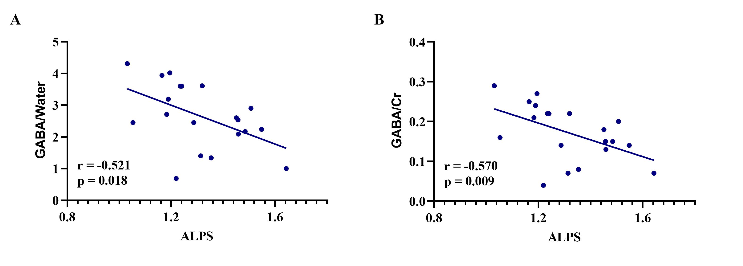
Furthermore, we conducted an analysis of the thalamic volume and the thalamic GABA levels. The results showed that the volume of the thalamus was decreased, along with elevated ratios of thalamus GABA/Water and GABA/Cr in the MHE group (P<0.05) (Figure 3). Finally, a strong negative correlation was observed between the ALPS index and GABA levels (Figure 4).
Discussion and Conclusion
Our study advances the understanding of HE in cirrhotic patients, revealing significant findings. Firstly, we identified glymphatic system dysfunction in cirrhotic HE patients, as reflected by the ALPS index, which showed a notable correlation with cognitive test scores. This suggests that impaired glymphatic system function likely contributes to HE development.Additionally, we observed elevated GABA/Water and GABA/Cr levels in the thalamus of cirrhotic HE patients. Notably, these GABA levels displayed a negative correlation with the ALPS index. These findings support the hypothesis that glymphatic system dysfunction in cirrhotic patients leads to the accumulation of neural waste, including GABA, in the brain. This, in turn, features prominently in the pathogenesis of HE 7.
Our study provides vital insights into the potential mechanisms underlying HE in cirrhotic patients. The recognition of glymphatic dysfunction, along with its correlation with cognitive test scores, implies that enhancing the glymphatic pathway for efficient brain waste clearance could bear important clinical implications for the management and treatment of HE3,4.
In conclusion, our research underscores the significance of glymphatic system dysfunction and altered GABA levels in cirrhotic HE pathogenesis. Further research is needed to explore the therapeutic potential of targeting the glymphatic system for HE management and to uncover the precise mechanisms involved in the accumulation of GABA and other neurotoxins in the brain.
Acknowledgements
We would like to acknowledge the Guizhou senior innovative talent project (Grant numbers [QKHPTRC-GCC [2022]041-1]).References
1. European Association for the Study of the Liver. Electronic address eee, European Association for the Study of the L. EASL Clinical Practice Guidelines on the management of hepatic encephalopathy. J Hepatol. 2022;77:807-824. doi: 10.1016/j.jhep.2022.06.001
2. Tapper EB, Parikh ND. Diagnosis and Management of Cirrhosis and Its Complications: A Review. JAMA. 2023;329:1589-1602. doi: 10.1001/jama.2023.5997
3. Sepehrinezhad A, Stolze Larsen F, Ashayeri Ahmadabad R, Shahbazi A, Sahab Negah S. The Glymphatic System May Play a Vital Role in the Pathogenesis of Hepatic Encephalopathy: A Narrative Review. Cells. 2023;12. doi: 10.3390/cells12070979
4. Backstrom T, Turkmen S, Das R, Doverskog M, Blackburn TP. The GABA system, a new target for medications against cognitive impairment-Associated with neuroactive steroids. J Intern Med. 2023;294:281-294. doi: 10.1111/joim.13705
5. Jenkinson M, Beckmann CF, Behrens TE, Woolrich MW, Smith SM. Fsl. Neuroimage. 2012;62:782-790. doi: 10.1016/j.neuroimage.2011.09.015
6. Edden RA, Puts NA, Harris AD, Barker PB, Evans CJ. Gannet: A batch-processing tool for the quantitative analysis of gamma-aminobutyric acid-edited MR spectroscopy spectra. J Magn Reson Imaging. 2014;40:1445-1452. doi: 10.1002/jmri.24478
7. Hadjihambi A, Harrison IF, Costas-Rodriguez M, Vanhaecke F, Arias N, Gallego-Duran R, Mastitskaya S, Hosford PS, Olde Damink SWM, Davies N, et al. Impaired brain glymphatic flow in experimental hepatic encephalopathy. J Hepatol. 2019;70:40-49. doi: 10.1016/j.jhep.2018.08.021
Figures