2633
Alteration in asymmetry of glymphatic system function in patients with Self-limited epilepsy with centrotemporal spikes1Department of Radiology, the first affiliated hospital of Xi'an Jiaotong University, Xi‘an, China
Synopsis
Keywords: Epilepsy, Diffusion/other diffusion imaging techniques
Motivation: Self-limited epilepsy with centrotemporal spikes (SLECTS) is the most prevalent type of idiopathic childhood epilepsy. However, the underlying pathophysiologic mechanisms of SLECTS is largely unknown.
Goal(s): Studies on the glymphatic system function in SLECTS patients may contribute to better understanding of the associated mechanisms.
Approach: Diffusion tensor imaging analysis along the perivascular space (DTI-ALPS) was used to evaluate the glymphatic system function.
Results: The SLECTS patients exhibited altered DTI-ALPS index. Altered DTI-ALPS index were more severe in ipsilateral than in the contralateral hemisphere. Moreover, RSLECTS (Right SLECTS ) and LSLECTS (Left SLECTS) patients exhibited different change patterns of the glymphatic system.
Impact: This study indicating that the glymphatic system may be a separate system in the left and right hemispheres. Patients with RSLECTS and LSLECTS exhibited different pattern of glymphatic system changes. Our research may contribute to elucidating the pathophysiologic of SLECTS.
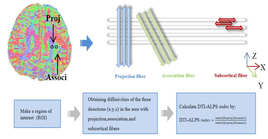
Methods:The local institutional review board approved this study and written informed consents were obtained from parents of the children. Subjects: Children diagnosed with SLECTS according diagnostic criteria defined by the International League Against Epilepsy[8].MRI protocols: All MR examinations were performed using a 3T scanner (Signa HDxt, GE Healthcare, Milwaukee, Wisconsin) with 8-channel head coil. The parameters of DTI sequence were as follows: TR/TE=11000ms/69.5ms, b values=0, 1000, thickness=2.5 mm, acquisition matrix =128×128, field of view=240×240mm2. Data analysis: The DTI-ALPS index were calculated for the right (right ALPS index) and left (left ALPS index) hemispheres respectively(Figure 1). An asymmetry index (AI) was calculated by AI=(Left-Right)/[(Left+Right)×0.5] to represent the asymmetric pattern. Meanwhile, correlation of DTI-ALPS index in both hemispheres with age, seizure duration and epileptic frequency were respectively explored via pearson’s coefficient(r).
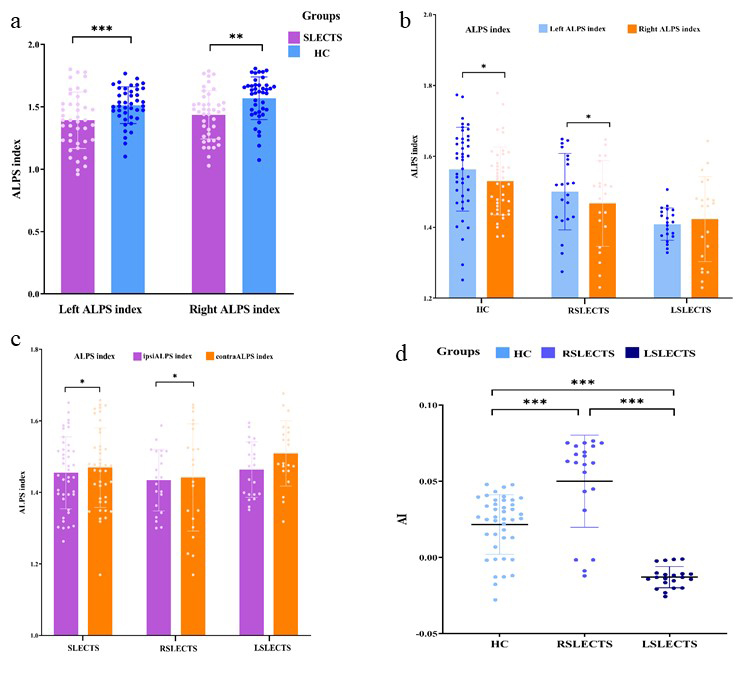
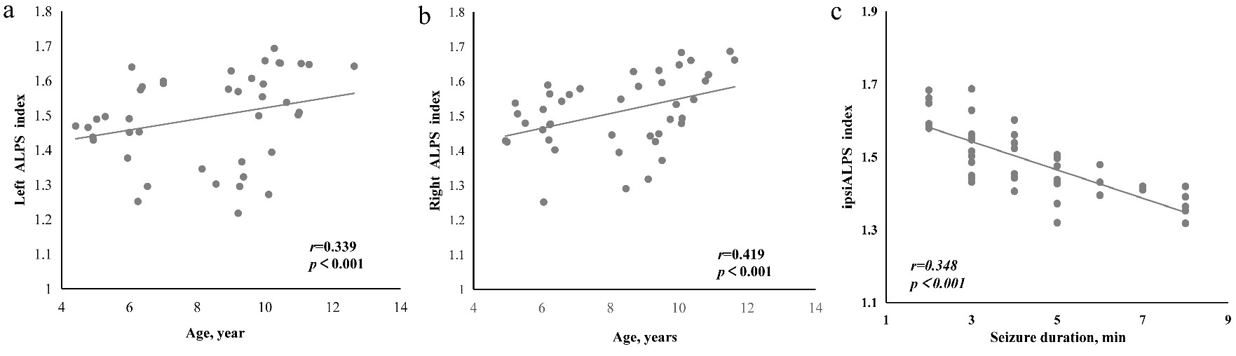
Results:A total of 42 SLECTS and 42 health controls (HC) were enrolled and the demographic information is shown in Table 1. All participants are right-handed. Leftward asymmetry in DTI-ALPS was found in HC and RSLECTS patients. Both left DTI-ALPS index and right DTI-ALPS index of SLECTS patients were significantly decreased. Compared to contralateral DTI-ALPS index, the ipsilateral DTI-ALPS index was significantly decreased in SLECTS and RSLECT patients (Figure 2). The DTI-ALPS index positively correlated with age in HC. Meanwhile, DTI-ALPS index gradually decreased with seizure duration (Figure 3).
Discussion:In this study, SLECTS patients exhibited altered DTI-ALPS index, which could be triggered by glymphatic system dysfunction[9]. Altered DTI-ALPS index were more severe in ipsilateral than in the contralateral hemisphere. With prolonged seizures duration and increased seizures frequency, more metabolic products would be released, resulting in more severe blood brain barrier dysfunction leading to the dysfunction of glymphatic system[10]. Sustained hypertension after onset of seizure would weaken arterial pulsation, thus affecting the glymphatic system function by altering neurovascular coupling also an important reason[11]. In childhood, the blood brain barrier is underdeveloped, and as they grow, the function of the blood–brain barrier develops, so DTI-ALPS index positively correlated with age[12].
Conclusion:The SLECTS patients exhibited altered DTI-ALPS index, which could be triggered by glymphatic system function. Altered DTI-ALPS index were more severe in ipsilateral than in the contralateral hemisphere. Moreover, RSLECTS and LSLECTS patients exhibited different change patterns of the glymphatic system.
Acknowledgements
This study was supported by National Natural Science Foundation of China(82272618, 81971581) ,Natural Science Basic Research Plan of Shannxi(2022JQ-811) ,the Institutional Foundation of the First Affiliated Hospital of Xi'an Jiaotong University(2021ZYTS-04). Please address correspondence to Jian Yang, e-mail: yj1118@mail.xjtu.edu.cn and Xianjun Li, e-mail: xianj.li@mail.xjtu.edu.cn.References
1.Lohela TJ, Lilius TO, Nedergaard M. The glymphatic system: implications for drugs for central nervous system diseases. Nature reviews Drug discovery 2022, 21(10): 763-779.
2.Lee HJ, Lee DA, Shin KJ, Park KM. Glymphatic system dysfunction in patients with juvenile myoclonic epilepsy. Journal of neurology 2022, 269(4): 2133-2139.
3.Lee DA, Lee J, Park KM. Glymphatic system impairment in patients with status epilepticus. Neuroradiology 2022, 64(12): 2335-2342.
4.Kim J, Lee DA, Lee HJ, Park KM. Glymphatic system dysfunction in patients with occipital lobe epilepsy. Journal of neuroimaging : official journal of the American Society of Neuroimaging 2023, 33(3): 455-461.
5.Panayiotopoulos CP, Michael M, Sanders S, Valeta T, Koutroumanidis M. Benign childhood focal epilepsies: assessment of established and newly recognized syndromes. Brain : a journal of neurology 2008, 131(Pt 9): 2264-2286.
6.Taoka T, Masutani Y, Kawai H, Nakane T, Matsuoka K, Yasuno F, et al. Evaluation of glymphatic system activity with the diffusion MR technique: diffusion tensor image analysis along the perivascular space (DTI-ALPS) in Alzheimer's disease cases. Japanese journal of radiology 2017, 35(4): 172-178.
7.Feldman RE, Rutland JW, Fields MC, Marcuse LV, Pawha PS, Delman BN, et al. Quantification of perivascular spaces at 7T: A potential MRI biomarker for epilepsy. Seizure 2018, 54: 11-18.
8.Riney K, Bogacz A, Somerville E, Hirsch E, Nabbout R, Scheffer IE, et al. International League Against Epilepsy classification and definition of epilepsy syndromes with onset at a variable age: position statement by the ILAE Task Force on Nosology and Definitions. Epilepsia 2022, 63(6): 1443-1474.
9.Rabinovitch A, Aviram I, Biton Y, Braunstein D. Explaining recent postictal epilepsy EEG results by the G-lymphatic clearance hypothesis. Medical hypotheses 2020, 137: 109600.
10.Hanael E, Veksler R, Friedman A, Bar-Klein G, Senatorov VV, Jr., Kaufer D, et al. Blood-brain barrier dysfunction in canine epileptic seizures detected by dynamic contrast-enhanced magnetic resonance imaging. Epilepsia 2019, 60(5): 1005-1016.
11.Liu C, Habib T, Salimeen M, Pradhan A, Singh M, Wang M, et al. Quantification of visible Virchow-Robin spaces for detecting the functional status of the glymphatic system in children with newly diagnosed idiopathic generalized epilepsy. Seizure 2020, 78: 12-17.
12.Saunders NR, Liddelow SA, Dziegielewska KM. Barrier mechanisms in the developing brain. Frontiers in pharmacology 2012, 3: 46.
Figures