2630
Diffusion tensor imaging analysis along the perivascular space revealed the dysfunction of glymphatic system in narcolepsy type1 patients1Department of Radiology, The Second Clinical Medical College of Nanchang University, Nanchang,China, China, 2MR Research, GE Healthcare, Beijing, China, Beijing, China, China
Synopsis
Keywords: Other Neurodegeneration, Diffusion Tensor Imaging, sleep, narcolepsy, glymphatic system
Motivation: The glymphatic system plays an important role in sleep. However, it’s unknown if the glymphatic system was impaired in narcolepsy type1 (NT1) patients.
Goal(s): Investigate if the glymphatic system was impaired and the correlation between dysfunction of glymphatic system and clinical symptom.
Approach: 31 NT1 and 23 healthy controls (HC) were enrolled. DTI-ALPS was used to assess the function of glymphatic system and compared between NT1 and HC. The correlation between DTI-ALPS index and clinical symptom was analyzed.
Results: The DTI-ALPS index was significantly lower in NT1 than in HC and was significantly correlated with the percentage of N3 stage sleep in NT1.
Impact: We firstly reported the dysfunction of glymphatic system in narcolepsy type1 patients and its correlation with clinical symptom. Our study suggested the glymphatic system should be taken into consideration for the treatment of narcolepsy type1 patients.
Introduction
Narcolepsy is a sleep-wake disorder characterized by excessive daytime sleepiness (EDS) and abnormal rapid eye movement (REM) sleep manifestations[1] . The prevalence of narcolepsy is approximately 0.025% to 0.05%[2]. Narcolepsy is divided into type 1 (NT1) and type 2 (NT2) according to the presence of cataplexy and the level of HCRT-1 in the cerebrospinal fluid (CSF). NT1 has reduced or lower CSF HCRT-1 levels that may lead to various functional impairments including motor, emotional, cognitive, metabolic, autonomic [3]. The impairment of glymphatic system function is closely associated with the cognitive function, executive function and memory of patients[4]. However, it’s unknown if the dysfunction of glymphatic system was presented and correlated with clinical symptom in NT1 patients.Taoka et al., [5] proposed the use of diffusion tensor imaging (DTI) analysis along the perivascular space (DTI-ALPS) to invasively evaluate the function of glymphatic system without the injection of contrast agency. DTI-ALPS has been used for study the alteration of glymphatic system in Parkinson’s disease with REM as well as other diseases including Alzheimer's disease, multiple sclerosis, and epilepsy [5-9]. With the promising results, this study aimed to evaluate the glymphatic system function in patients with NT1 using DTI-ALPS and compared to healthy controls. We also analyzed the correlations between DTI-ALPS index and clinical characteristics of patients with NT1.
Methods and Materials
Participant31 NT1 patients and 23 sex- and age-matched healthy volunteers were enrolled. The clinical symptom of NT1 patients was assessed using overnight polysomnography and multiple sleep latency test.
MRI acquisition
DTI was performed at a 3T MRI scanner (SIGNA Architect; GE Healthcare, CA, USA) with 19-channel phased-array head coil. The imaging parameters were TR/TE: 8441/95.5ms, FOV: 240mm×240mm, matrix size: 112×112, slice thickness: 3.0mm, number of slices: 34, b value: 0 and 1000 s/mm2, number of excitations: 1, number of diffusion gradient directions: 64.
DTI-ALPS processing
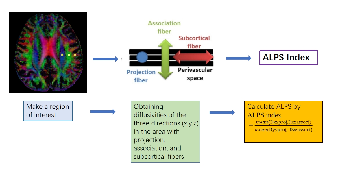
As shown in Figure 1, the color map obtained from DTI was used for the delineation of region of interest (ROI). The diffusivity parameters of projection and association fibers was extracted from DTI and used for calculating ALPS index with the following formula[5]:ALPS index = mean (Dxproj, Dxassoc) / mean (Dyproj, Dzassoc).Dxxproj: diffusivity along the x-axis in the projection fiber; Dxxassoci: diffusivity along the x-axis in the association fiber; Dyyproj: diffusivity along the y-axis in the projection fiber; Dzzassoci: diffusivity along the z-axis in the association fiber.
Statistical analysis
Mann-Whitney test was used to compare DTI-ALPS index between NT1 patients and healthy controls. Spearman rank correlation was used to analyze the relationship between DTI-ALPS index and clinical symptom. P<0.05 was considered significant.
Result
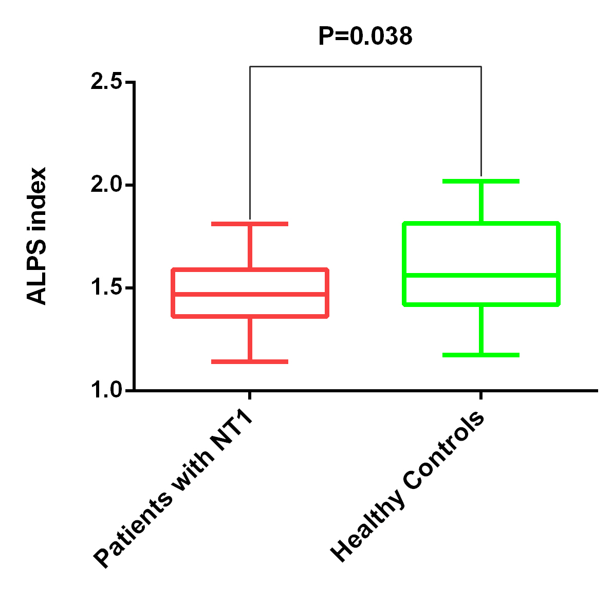
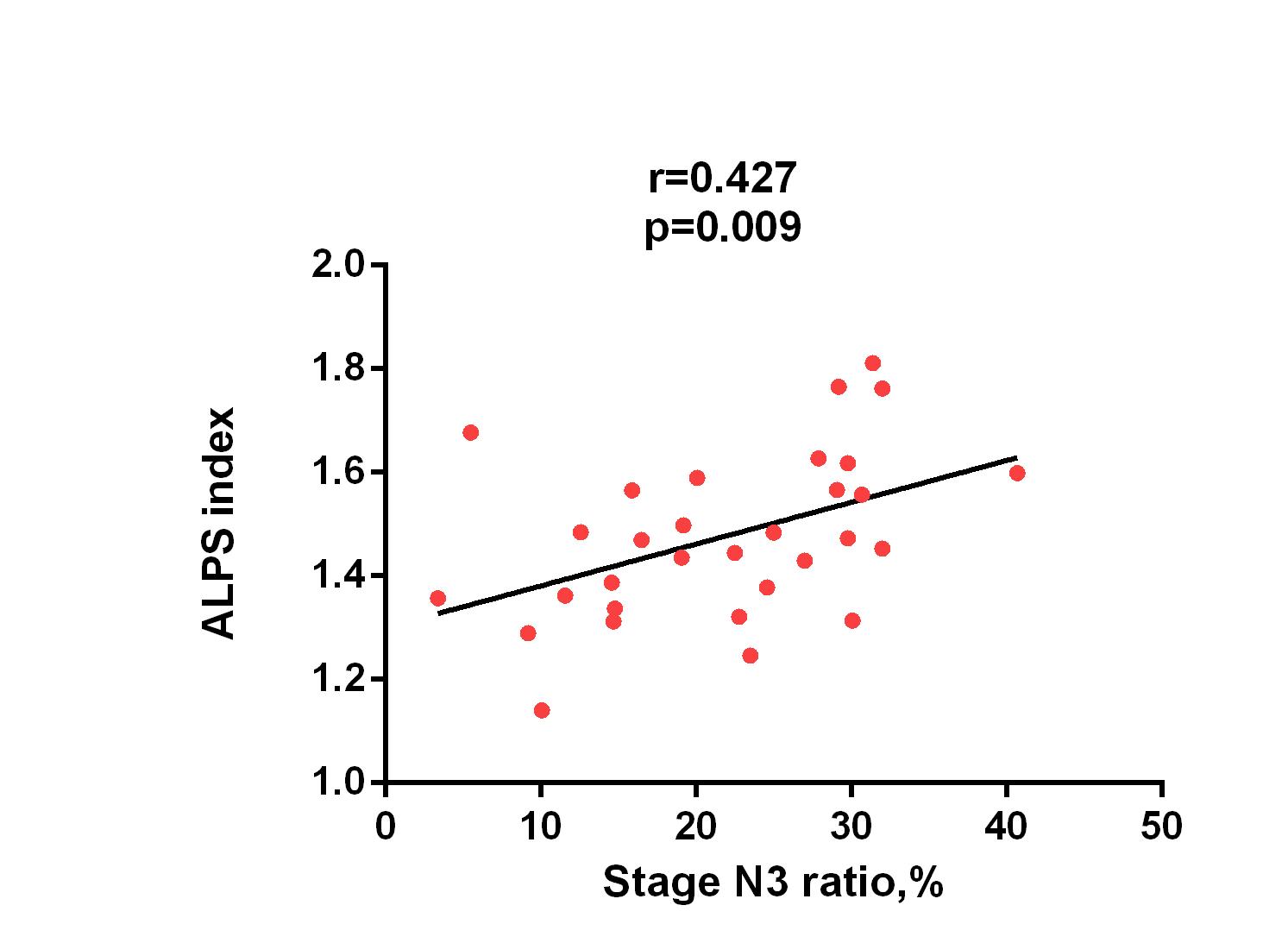
As shown in Figure 2, the DTI-ALPS index was significantly lower in NT1 patients than in healthy controls (1.479±0.156 vs 1.603±0.242; P=0.038). In NT1 patients, the DTI-ALPS index was significantly correlated with the percentage of N3 stage sleep (Figure 3; r=0.459, P=0.009). No significantly correlation between DTI-ALPS index and other clinical symptom (e.g., WASO, course and ESS score) was shown in NT1 patients. We observed the diffusivity along the z-axis in projection fibers was significantly faster in NT1 patients than in healthy controls (10.20±0.83 × 10⁻³ mm2/s vs 1.07±0.62 × 10⁻³ mm2/s; P=0.013). However, this parameter was not significantly correlated with clinical symptom in NT1 patients.Discussion and Conclusion
This study investigated the alterations of glymphatic system in NT1 patients using DTI-ALPS and its correlation with clinical symptom. Previous studies had reported the dysfunction of glymphatic system would result with low DTI-ALPS [9]. Our results showed NT1 patients had significantly lower DTI-ALPS index than healthy controls indicating the impairment of glymphatic system. The DTI-ALPS index was significantly correlated with the ratio of N3 stage sleep which reflected the dysfunction of glymphatic system may be one reason of abnormal sleep in NT1 patients. In conclusion, DTI-ALPS revealed the glymphatic system was impaired and the dysfunction of glymphatic system may be one of the causes of abnormal sleep in NT1 patients.Acknowledgements
No acknowledgement found.References
[1] DAUVILLIERS Y, ARNULF I, MIGNOT E. Narcolepsy with cataplexy [J]. Lancet, 2007, 369(9560): 499-511.
[2] LONGSTRETH WT JR K T, TON TG H A, VAN BELLE G. Longstreth WT Jr, Koepsell TD, Ton TG, Hendrickson AF, van Belle G. The epidemiology of narcolepsy. [J]. Sleep, 2007.
[3] BASSETTI C A-O, ADAMANTIDIS A, BURDAKOV D A-O, et al. Narcolepsy - clinical spectrum, aetiopathophysiology, diagnosis and treatment [J]. Nature Reviews Neurology, 2019, 15(9)(519-539). [4] THIERNO M BAH D A S, ASEEL H IBRAHIM , JUSTIN S CETAS , NABIL J ALKAYED Fluid dynamics in aging-related dementias [J]. Neurobiology of Disease, 2023, 177(2).
[5] TAOKA T, MASUTANI Y, KAWAI H, et al. Evaluation of glymphatic system activity with the diffusion MR technique: diffusion tensor image analysis along the perivascular space (DTI-ALPS) in Alzheimer's disease cases [J]. Jpn J Radiol, 2017, 35(4): 172-8.
[6] SI X, GUO T, WANG Z, et al. Neuroimaging evidence of glymphatic system dysfunction in possible REM sleep behavior disorder and Parkinson's disease [J]. NPJ Parkinsons Dis, 2022, 8(1): 54.
[7] KIM S T, KIM S E, LEE D A, et al. Anti-seizure medication response and the glymphatic system in patients with focal epilepsy [J]. Eur J Neurol, 2023.
[8] BAE Y J, KIM J M, CHOI B S, et al. Altered Brain Glymphatic Flow at Diffusion-Tensor MRI in Rapid Eye Movement Sleep Behavior Disorder [J]. Radiology, 2023, 307(5): e221848.
Figures