2629
Association of Cognitive Reserve and Glymphatic Function in Mild Cognitive Impairment Patients Using DTI-ALPS1Second Clinical School, Lanzhou University, Lanzhou, China, 2Department of Magnetic Resonance, Lanzhou University Second Hospital, Lanzhou, China, 3Philips Healthcare, Xi'an, China, 4Philips Healthcare, Shanghai, China, 5Gansu Province Clinical Research Center for Functional and Molecular Imaging, Lanzhou University Second Hospital, Lanzhou, China
Synopsis
Keywords: Dementia, Diffusion Tensor Imaging
Motivation: Cognitive reserve (CR) could moderate the cognitive decline. But the association between the glymphatic function and CR in mild cognitive impairment (MCI) remains unclear.
Goal(s): To evaluate the association between CR and glymphatic function.
Approach: By using diffusion tensor imaging along the perivascular space (DTI-ALPS) to acquire an index (ALPS-index) in MCI and normal controls (NCs). The education level was used as a representation of CR. We used mediator analysis to explore the association among ALPS-index, CR and cognitive function.
Results: MCI exhibited decreased glymphatic activity than NCs and CR mediates the link between the glymphatic system and cognition.
Impact: MCI patients exhibited decreased glymphatic activity compared to NCs. Our study confirms the protective effect of CR in MCI patients, and this protective effect can against the cognitive impairment by improving CR.
Introduction
The glymphatic system is a highly organized fluid-clearance pathway involving the movement of cerebrospinal fluid (CSF) alongside the perivascular space (1). It has been evaluated by a non-invasive diffusion tensor image-based method to measure diffusivity along the perivascular space (ALPS) (2). While the ALPS-index measures diffusivity in the direction of the perivascular space in the periventricular white matter, it has been proposed to be an indirect indicator of the state of glymphatic function (3). As the ALPS-index has also been positively correlated with Mini-Mental State Examination (MMSE) score, the integrity of glymphatic function has been linked to the prediction of cognition in AD and mild cognitive impairment (MCI) patients (4). Several studies have shown that MCI patients with higher levels of cognitive reserve (CR) are significantly more likely to reverse to normal cognition than to progress to dementia (5) and high CR is a positive role in alleviating cognitive decline (6). Neuroimaging studies have also confirmed that CR is associated with increased connectivity in cognitive control networks (7) and CR could modulate the cortical architecture, white matter macromolecular volume and cerebral blood flow to be protective against the cognitive impairment effect in MCI patients (8-10). But the association between the glymphatic function and CR in mild cognitive impairment (MCI) patients remain unclear. This study aimed to evaluate glymphatic function in MCI patients by using diffusion tensor imaging (DTI) along the perivascular space (DTI-ALPS) method and to explore the association between CR and glymphatic function.Methods
In this study, 54 MCI patients and 31 normal controls (NCs) were enrolled from Lanzhou university second hospital to evaluate the bilateral diffusivity along the perivascular spaces as well as projection fibers and association fibers separately, and to acquire an index for diffusivity along the perivascular space (ALPS-index). DTI scans were obtained using a 3.0T scanner (Ingenia CX, Philips Healthcare, the Netherlands) with 32-channel head coil. The parameters of the DTI sequence were as follows: TR= 5000 ms, TE= 102 ms, spatial resolution = 2x2x2 mm3, b-value = 1000s/mm2 along 120 gradient directions. The year of education was used as a representation of CR and cognitive function were measured with the mini mental state examinations (MMSE) and the montreal cognitive assessment (MoCA). The ALPS-index was calculated using an in-house software which developed by Philips. Then, the ALPS-index and CR were compared between groups. Correlation analysis among the ALPS-index, cognitive function and CR were conducted. We used mediator analysis to explore the ALPS-index as predictor, CR as mediators, and cognitive test scores as outcomes.Results
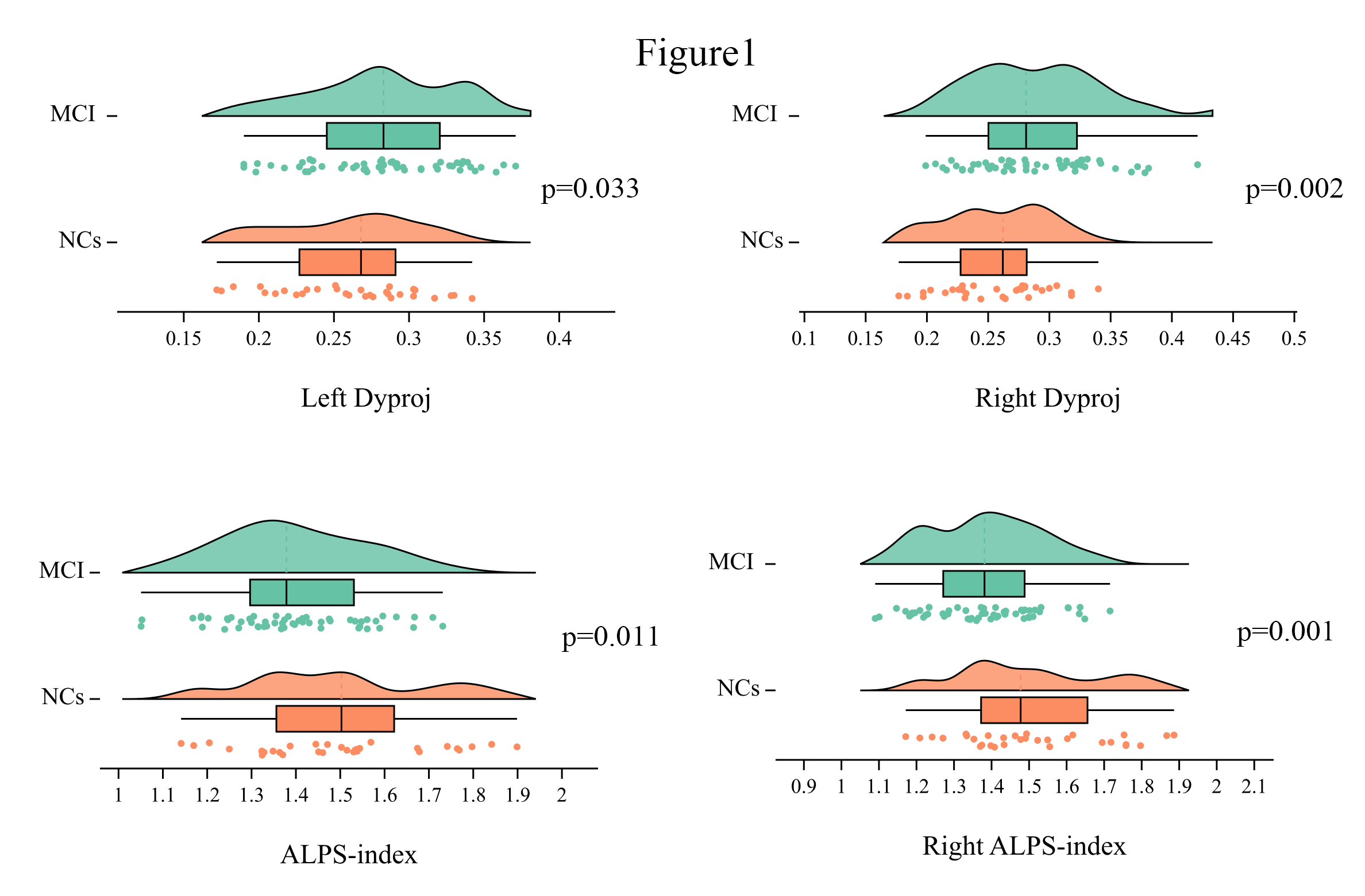
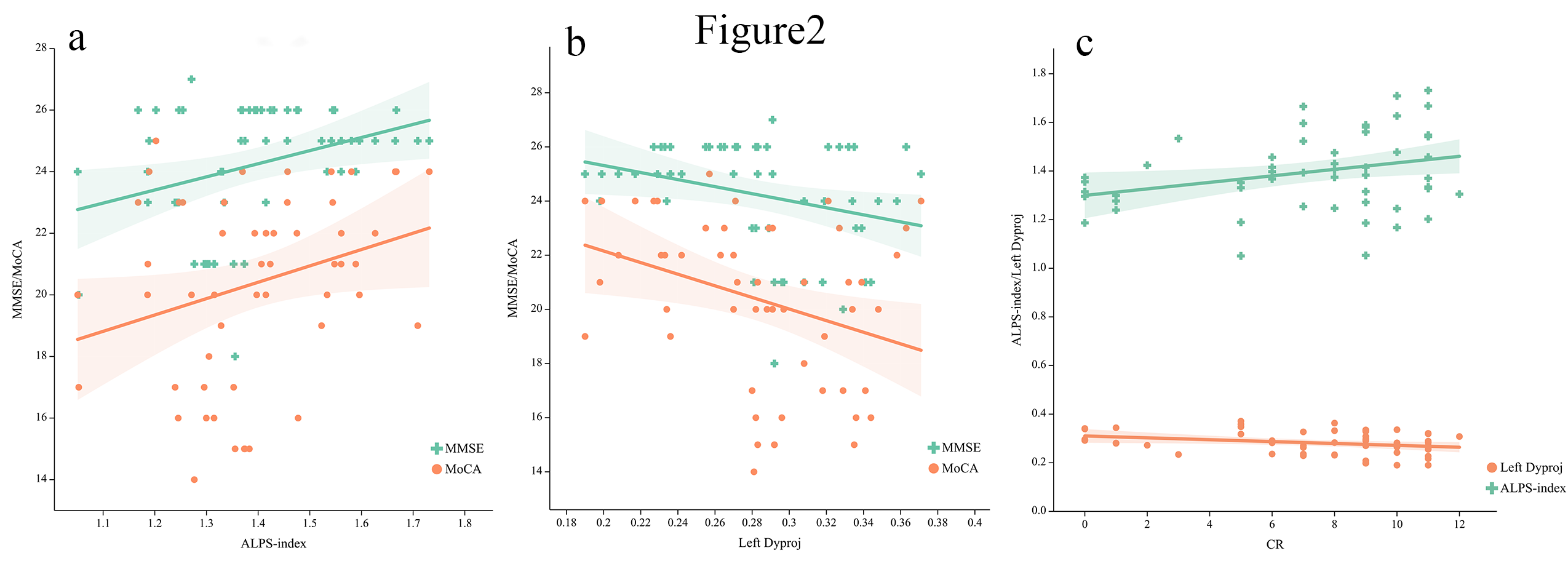
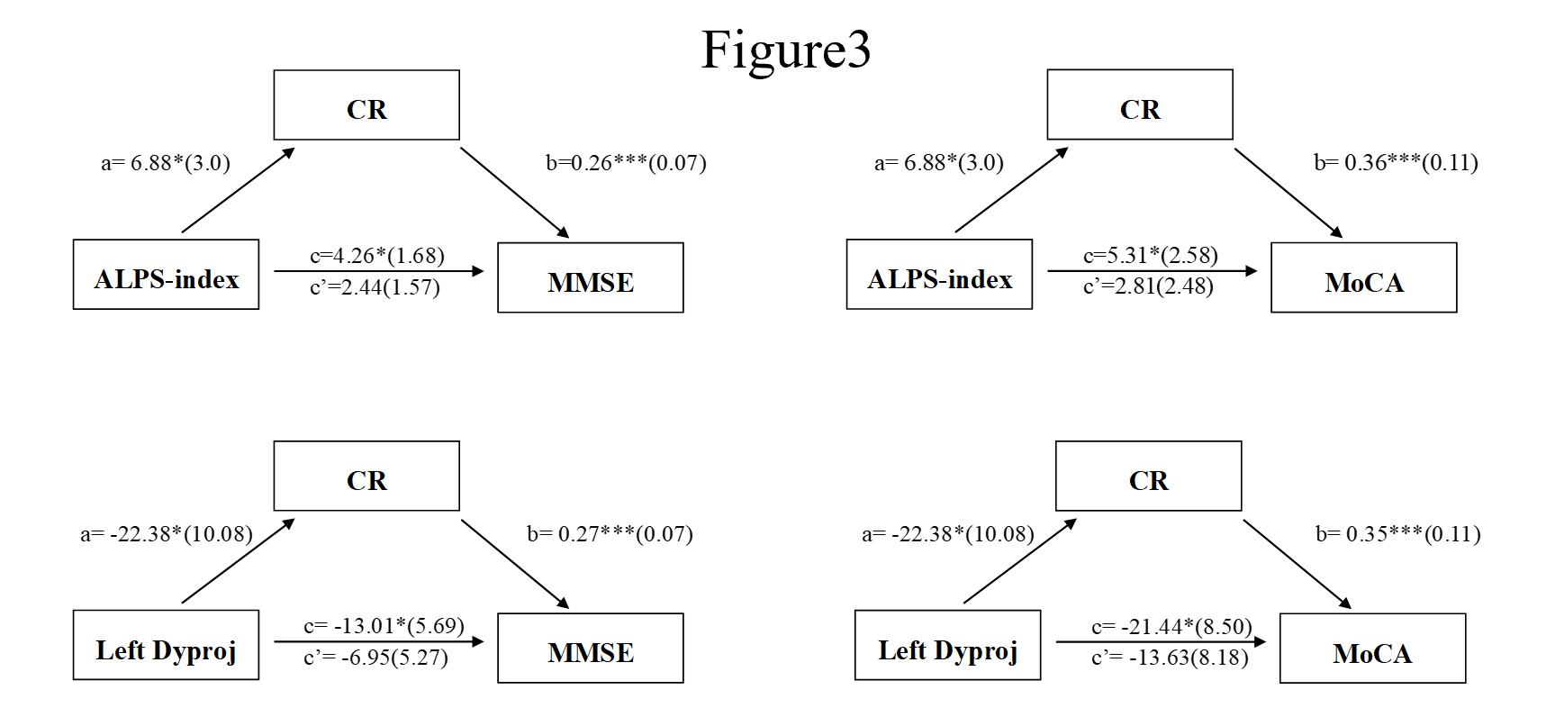
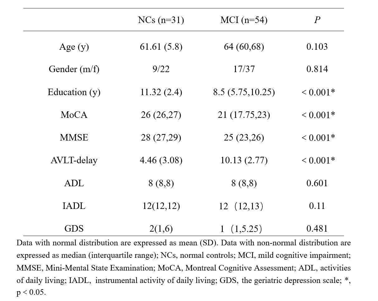
Table 1 showed the demographic characteristics of the enrolled cohort. As shown in figure 1, MCI group had significantly lower right ALPS-index and whole brain ALPS-index, but higher bilateral diffusivity along the y-axis in projection fiber area (Dyproj) than NCs group. The left Dyproj was negatively correlated with MoCA, MMSE and CR. The whole brain ALPS-index were positively with MoCA, MMSE and CR (figure 2). Mediation analysis suggested that CR significantly mediated the main effect of the relationship between ALPS-index and cognitive function (figure 3)Discussion
Our study found that the ALPS-index of the NCs was significantly higher than that of the MCI group, and was positively correlated with cognitive function scores. The result suggests that the glymphatic clearance of amyloid is related to cognitive function in MCI patients. Secondly, we found that MCI patients with higher CR were able to tolerate more decreased glymphatic activity, confirming the protective effect of CR on cognitive function. What’s more, CR has also been shown to modulate glymphatic activity to be protective against the cognitive impairment in MCI patients.Conclusion
MCI patients exhibited decreased glymphatic activity than NCs. CR mediates the link between the glymphatic system and cognition.Acknowledgements
No acknowledgement found.References
1. Nedergaard M, Goldman SA. Glymphatic Failure as a Final Common Pathway to Dementia. Science (New York, NY) (2020) 370(6512):50-6. Epub 2020/10/03. doi: 10.1126/science.abb8739.
2. Taoka T, Masutani Y, Kawai H, Nakane T, Matsuoka K, Yasuno F, et al. Evaluation of Glymphatic System Activity with the Diffusion Mr Technique: Diffusion Tensor Image Analysis Along the Perivascular Space (Dti-Alps) in Alzheimer's Disease Cases. Japanese journal of radiology (2017) 35(4):172-8. doi: 10.1007/s11604-017-0617-z.
3. Carlstrom LP, Eltanahy A, Perry A, Rabinstein AA, Elder BD, Morris JM, et al. A Clinical Primer for the Glymphatic System. Brain (2022) 145(3):843-57. Epub 2021/12/11. doi: 10.1093/brain/awab428.
4. Kamagata K, Andica C, Takabayashi K, Saito Y, Taoka T, Nozaki H, et al. Association of Mri Indices of Glymphatic System with Amyloid Deposition and Cognition in Mild Cognitive Impairment and Alzheimer Disease. Neurology (2022) 99(24):e2648-60. Epub 2022/09/20. doi: 10.1212/wnl.0000000000201300.
5. Iraniparast M, Shi Y, Wu Y, Zeng L, Maxwell CJ, Kryscio RJ, et al. Cognitive Reserve and Mild Cognitive Impairment. Neurology (2022) 98(11):e1114-e23. doi: 10.1212/wnl.0000000000200051.
6. Corbo I, Marselli G, Di Ciero V, Casagrande M. The Protective Role of Cognitive Reserve in Mild Cognitive Impairment: A Systematic Review. Journal of clinical medicine (2023) 12(5). doi: 10.3390/jcm12051759.
7. Franzmeier N, Caballero MÁA, Taylor ANW, Simon-Vermot L, Buerger K, Ertl-Wagner B, et al. Resting-State Global Functional Connectivity as a Biomarker of Cognitive Reserve in Mild Cognitive Impairment. Brain Imaging and Behavior (2017) 11(2):368-82. doi: 10.1007/s11682-016-9599-1.
8. Serra L, Giancaterino G, Giulietti G, Petrosini L, Di Domenico C, Marra C, et al. Cognitive Reserve Modulates Brain Structure and Cortical Architecture in the Alzheimer's Disease. Journal of Alzheimer's disease : JAD (2022) 89(3):811-24. doi: 10.3233/jad-220377.
9. Fingerhut H, Gozdas E, Hosseini S. Quantitative Mri Evidence for Cognitive Reserve in Healthy Elders and Prodromal Alzheimer's Disease. Journal of Alzheimer's disease : JAD (2022) 89(3):849-63. doi: 10.3233/jad-220197.
10. Brenner E, Thomas K, Weigand A, Edwards L, Edmonds E, Bondi M, et al. Cognitive Reserve Moderates the Association between Cerebral Blood Flow and Language Performance in Older Adults with Mild Cognitive Impairment. Neurobiology of aging (2023) 125:83-9. doi: 10.1016/j.neurobiolaging.2023.01.012.
Figures