2628
Investigating the characteristics of the region-of-interest for calculating the diffusion tensor imaging along perivascular spaces index1Department of Neuroradiology, University Medical Center Schleswig-Holstein (UKSH), Luebeck, Germany
Synopsis
Keywords: Neurofluids, Neurofluids, glymphatics, DTI-ALPS
Motivation: Several parameters affect diffusion tensor imaging along the perivascular spaces (DTI-ALPS) index calculation. However, the impact of ROI size, shape, and relative orientation to the lateral ventricle remains unclear, potentially hindering cross-study comparability.
Goal(s): To investigate the influence of ROI characteristics in estimating the DTI-ALPS index.
Approach: Differences in DTI-ALPS index and correlations among 8 ROI types (size, shape, and orientation) were investigated.
Results: DTI-ALPS index calculation depends on ROI size, shape, and orientation. Single-voxel and parallel rectangular ROIs exhibit strong agreement; parallel-parallel and orthogonal-orthogonal ROIs show excellent correlation. However, larger, rectangular ROIs with orthogonal placement result in significant differences and poor correlation.
Impact: Our analysis shows that ROI size, shape, and relative orientation to the lateral ventricle impact DTI-ALPS index calculation. Therefore, in addition to imaging parameters, ROI characteristics also need to be considered for comparison among studies.
Introduction
Diffusion tensor imaging along perivascular spaces (DTI-ALPS) aims to evaluate glymphatic system function noninvasively1. Recently, studies have observed alterations in glymphatic function in several illnesses, such as Alzheimer’s1–3 and Parkinson’s disease4–6. However, analysis of the DTI-ALPS index has shown inconsistencies that can limit comparability with other studies. Therefore, effort is being invested to improve reproducibility and to harmonize calculation of the DTI-ALPS7,8. Here, we investigate how the size, shape, and relative orientation (to the lateral ventricle) of the region-of-interest (ROI) impact DTI-ALPS index calculation.Methods
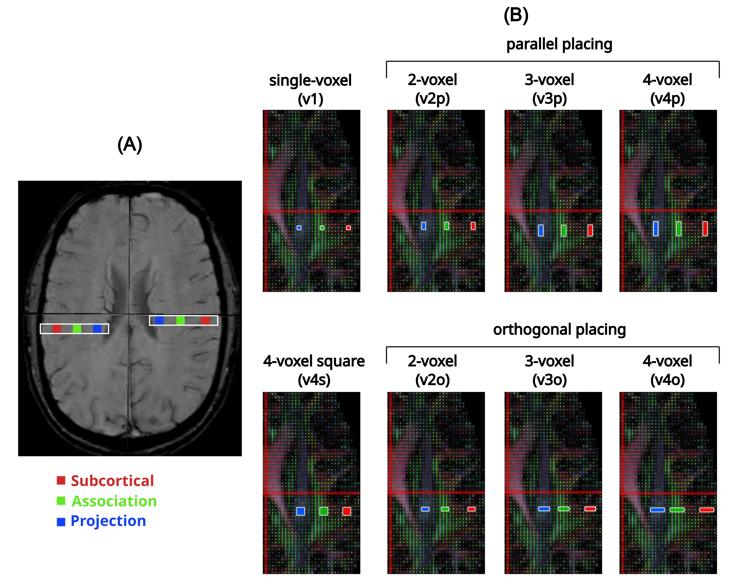
Eight volunteers (5 female, 3 male; aged 22.9±0.78 years) provided informed consent and were examined by DTI using a 3T MRI scanner (MAGNETOM Vida, Siemens Healthineers, Germany). The imaging parameters were: TE/TR=92/4400ms, FOV=200x200mm2, voxel size=2x2x2mm3, b-values= 0 and 1000s/mm2, and 64 diffusion directions. Simultaneous multi-slice imaging with a total acceleration factor of 4 was used. The total duration of the DTI scan was 5:30 min. Eight ROIs were placed next to the lateral ventricle in the projection, association, and subcortical areas in a single slice according to a color-coded fractional anisotropy map by referring to susceptibility-weighted images, using DSI-Studio. ROIs consisted of a single voxel (v1), 2, 3, and 4 voxels (with volumes of 8, 16, 24, and 32 mm3, respectively) and were placed parallel (v2p, v3p, v4p) and orthogonal (v2o, v3o, v4o) to the lateral ventricle, and as a 4-voxel ROI in a square configuration (v4s), as shown in Fig. 1. From the tensor matrix, x-, y-, and z-axis diffusivities were obtained, and the DTI-ALPS index was calculated as mean(DxProj,DxAssoc)/mean(DyProj,DzAssoc)1. Measurement differences and agreement were investigated using t-test and intraclass correlation coefficient (ICC). As there was no significant difference in the DTI-ALPS between the left and right hemispheres, we considered two values per participant for the analysis.Results
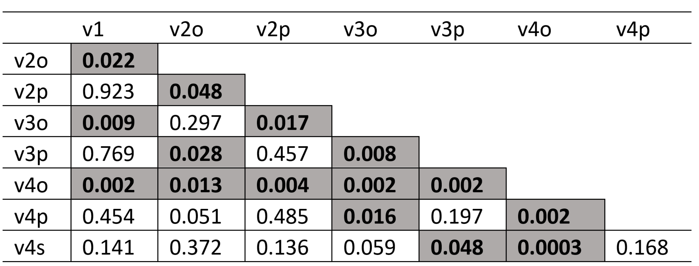
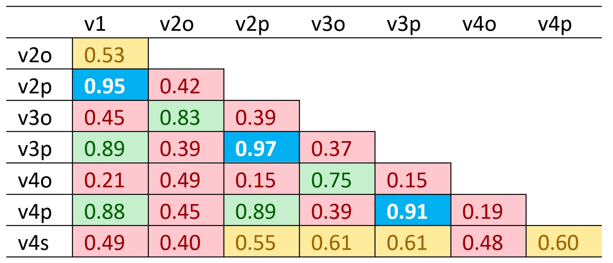
Significant differences were found between parallel and orthogonal ROI placements (p<0.05). The single-voxel ROI was not significantly different from the parallel and square ROIs (see Table 1). In Table 2, the ICC showed that reliability of the single-voxel measurement was poor compared to the v4s and orthogonal placement (ICC<0.5). However, it correlated well with parallel voxel positioning (0.75<ICC<0.9). Reliability is generally poor when the ROIs are placed orthogonal to the lateral ventricle (red in Table 2). Moderate (0.5<ICC<0.75) to excellent (ICC>0.9) reliability was shown when voxels were positioned parallel to the lateral ventricle.Discussion
Comparing the DTI-ALPS indexes from different imaging sequences does not make sense without harmonization7,8. The same would apply when different ROIs and/or software are used for the analysis. Calculation of the DTI-ALPS index has been explored using various ROI sizes and shapes, including spherical, square or rectangular, and single-voxel. A previous study reported moderate to good agreement in test-retest using spherical, cubic, and square ROIs7. However, correlations and differences among different ROI configurations have not been studied yet. Here, we observed significant differences between the DTI-ALPS indexes when the ROIs were placed parallel or orthogonal to the lateral ventricle. Those differences were reduced, in some cases, with increasing ROI volume. Additionally, we chose to include single-voxel ROIs in our analysis because they are most commonly used in publications employing DSI-Studio9,10. These small ROIs are often preferred for their precision in selecting specific areas. However, single-voxel ROIs might not be representative of the diffusion ALPS. Here, ROIs of 2 or more voxels appeared to be more reliable than a single voxel for analysis. Single-voxel DTI-ALPS indexes are significantly different from the ROIs perpendicular to the lateral ventricle, with good ICC for parallel ROIs, but not for square ones. However, we cannot extrapolate these results to spherical ROIs. Spherical ROIs were not included in this analysis because it was challenging to apply spherical ROIs with consistent diameters using DSI-Studio. Additionally, spherical ROIs include more than 1 slice, with partial-volume effects that have not been considered yet.Conclusion
There is no general agreement on the optimal ROI shape, size, and orientation for calculating the DTI-ALPS index. When comparing DTI-ALPS indexes from different imaging sequences (TE, number of diffusion directions), imaging plane or using various ROIs, it is important to consider factors such as ROI shape, size, and relative orientation to the lateral ventricle, too. These factors can significantly impact the DTI-ALPS index. Therefore, the ROI characteristics should be mentioned in publications and be considered when interpreting the results.Acknowledgements
The project was supported by the University of Luebeck in the form of a doctoral scholarship for J.C. RudolfReferences
(1) Taoka, T.; Masutani, Y.; Kawai, H.; Nakane, T.; Matsuoka, K.; Yasuno, F.; Kishimoto, T.; Naganawa, S. Evaluation of Glymphatic System Activity with the Diffusion MR Technique: Diffusion Tensor Image Analysis along the Perivascular Space (DTI-ALPS) in Alzheimer’s Disease Cases. Jpn. J. Radiol. 2017, 35 (4), 172–178. https://doi.org/10.1007/s11604-017-0617-z.
(2) Chang, H.-I.; Huang, C.-W.; Hsu, S.-W.; Huang, S.-H.; Lin, K.-J.; Ho, T.-Y.; Ma, M.-C.; Hsiao, W.-C.; Chang, C.-C. Gray Matter Reserve Determines Glymphatic System Function in Young-Onset Alzheimer’s Disease: Evidenced by DTI-ALPS and Compared with Age-Matched Controls. Psychiatry Clin. Neurosci. 2023. https://doi.org/10.1111/pcn.13557.
(3) Ota, M.; Sato, N.; Nakaya, M.; Shigemoto, Y.; Kimura, Y.; Chiba, E.; Yokoi, Y.; Tsukamoto, T.; Matsuda, H. Relationships Between the Deposition of Amyloid-β and Tau Protein and Glymphatic System Activity in Alzheimer’s Disease: Diffusion Tensor Image Study. J. Alzheimers Dis. JAD 2022, 90 (1), 295–303. https://doi.org/10.3233/JAD-220534.
(4) McKnight, C. D.; Trujillo, P.; Lopez, A. M.; Petersen, K.; Considine, C.; Lin, Y.-C.; Yan, Y.; Kang, H.; Donahue, M. J.; Claassen, D. O. Diffusion along Perivascular Spaces Reveals Evidence Supportive of Glymphatic Function Impairment in Parkinson Disease. Parkinsonism Relat. Disord. 2021, 89, 98–104. https://doi.org/10.1016/j.parkreldis.2021.06.004.
(5) Cai, X.; Chen, Z.; He, C.; Zhang, P.; Nie, K.; Qiu, Y.; Wang, L.; Wang, L.; Jing, P.; Zhang, Y. Diffusion along Perivascular Spaces Provides Evidence Interlinking Compromised Glymphatic Function with Aging in Parkinson’s Disease. CNS Neurosci. Ther. 2022, cns.13984. https://doi.org/10.1111/cns.13984.
(6) Chen, H.-L.; Chen, P.-C.; Lu, C.-H.; Tsai, N.-W.; Yu, C.-C.; Chou, K.-H.; Lai, Y.-R.; Taoka, T.; Lin, W.-C. Associations among Cognitive Functions, Plasma DNA, and Diffusion Tensor Image along the Perivascular Space (DTI-ALPS) in Patients with Parkinson’s Disease. Oxid. Med. Cell. Longev. 2021, 2021, 4034509. https://doi.org/10.1155/2021/4034509.
(7) Taoka, T.; Ito, R.; Nakamichi, R.; Kamagata, K.; Sakai, M.; Kawai, H.; Nakane, T.; Abe, T.; Ichikawa, K.; Kikuta, J.; Aoki, S.; Naganawa, S. Reproducibility of Diffusion Tensor Image Analysis along the Perivascular Space (DTI-ALPS) for Evaluating Interstitial Fluid Diffusivity and Glymphatic Function: CHanges in Alps Index on Multiple conditiON acquIsition eXperiment (CHAMONIX) Study. Jpn. J. Radiol. 2022, 40 (2), 147–158. https://doi.org/10.1007/s11604-021-01187-5.
(8) Saito, Y.; Kamagata, K.; Andica, C.; Taoka, T.; Tuerxun, R.; Uchida, W.; Takabayashi, K.; Owaki, M.; Yoshida, S.; Yamazaki, K.; Naganawa, S.; Aoki, S. Multisite Harmonization of Diffusion Tensor Image Analysis along the Perivascular Space Using the COMBined Association Test. Jpn. J. Radiol. 2023. https://doi.org/10.1007/s11604-023-01432-z.
(9) Lee, D. A.; Park, B. S.; Park, S.; Lee, Y. J.; Ko, J.; Park, K. M. Glymphatic System Function in Patients with Transient Global Amnesia. J. Integr. Neurosci. 2022, 21 (4), 117. https://doi.org/10.31083/j.jin2104117.
(10) Lee, H.-J.; Lee, D. A.; Shin, K. J.; Park, K. M. Glymphatic System Dysfunction in Patients with Juvenile Myoclonic Epilepsy. J. Neurol. 2022, 269 (4), 2133–2139. https://doi.org/10.1007/s00415-021-10799-w.
Figures