2627
Glymphatic dysfunction in non-dialysis, hemodialysis and peritoneal dialysis patients with ESRD is associated with cognitive decline1Department of Radiology, Beijing Friendship Hospital, Capital Medical University, Beijing, China, 2MR Research, GE Healthcare, Beijing, China, Beijing, China
Synopsis
Keywords: White Matter, Diffusion Tensor Imaging
Motivation: The glymphatic function has not yet been explored in non-dialysis,HD and PD patients with ESRD.
Goal(s): Our goal was to explore the pattern of glymphatic function in ESRD patients and its relationship with cognitive decline.
Approach: Diffusion tensor imaging along the perivascular space (DTI-ALPS) index methods was used to investigating brain glymphatic dysfunction in ESRD patients with non-dialysis, HD and PD patients using. The relationship between Montreal Cognitive Assessment (MoCA) and changes in DTI-ALPS index was measured.
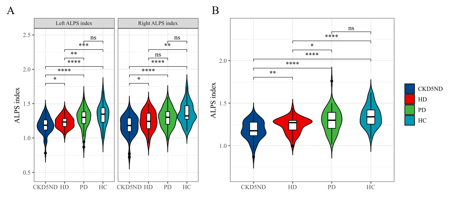
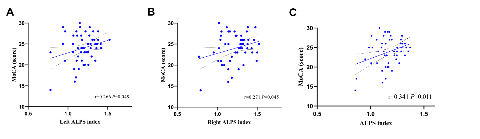
Results: Compared with HD and PD, ESRD patients with non-dialysis showed lower DTI-ALPS index.Changed DTI-ALPS index associated with cognitive decline in ESRD patients with non-dialysis.
Impact: Our findings reveal a greater degree of aberrant glymphatic functionality in ESRD patients with non-dialysis than HD and PD, which may offer new insights to the effectiveness of dialysis treatment.
Introduction and Purpose
End-stage renal disease (ESRD), defined as the presence of impaired renal function (estimated glomerular filtration rate(eGFR) <60 mL/min/1.73m2 ) or proteinuria (urine albumin-to-creatinine ratio < 30 mg/g), has become a globally recognized public health problem1. ESRD patients experience cognitive decline early in life, which is particularly severe in patients with ESRD stage 5. Since the vascular and neurodegenerative processes related to clinical dementia is related with insufficient glymphatic clearance2, it can be expected that glymphatic dysfunction may be present in patients with ESRD. Recently, Diffusion Tensor Image Analysis Along the Perivascular Space (DTI-ALPS) has been proposed to evaluate alterations in function of glymphatic system or dynamics of interstitial fluid in the brain. A significant discrepancy in the DTI-ALPS index has been reported between patients with ESRD and healthy controls3. However, the ambiguity persists regarding the discrepancy in glymphatic functionality among ESRD patients who abstain from undergoing dialysis, those participating in hemodialysis (HD), and those participating in peritoneal dialysis (PD). However, HD or PD may accelerate the progression of glymphatic dysfunction in ESRD patients due to brain injury associated with dialysis-induced haemodynamic disturbances. This study aims to explore the pattern of glymphatic function changes among these groups and its relationship with cognitive decline.Materials and method
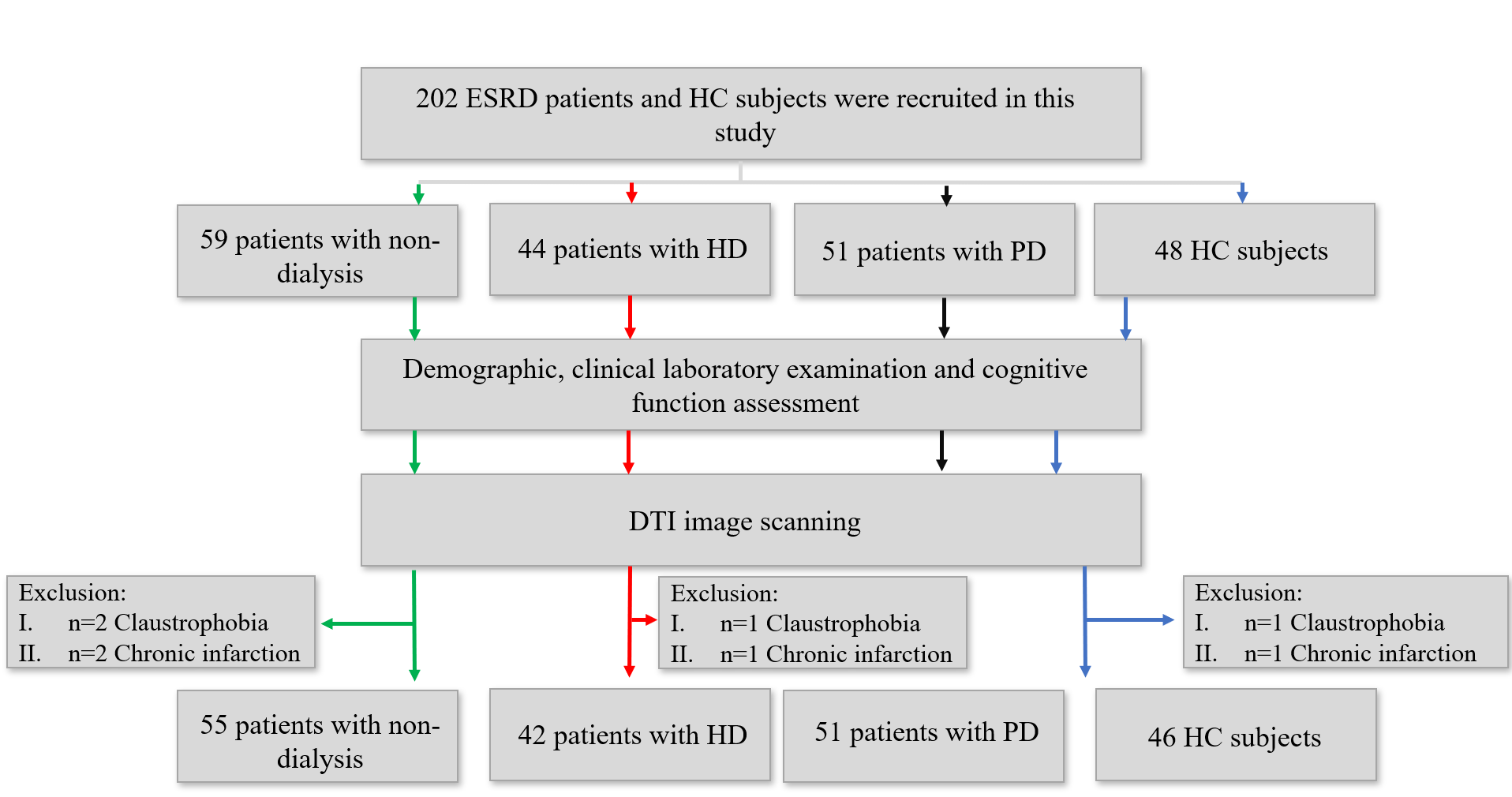
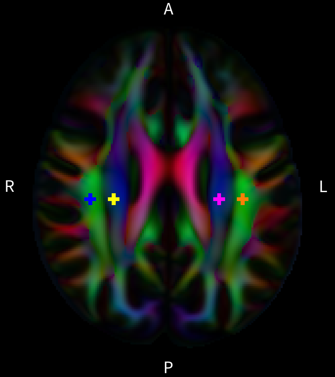
The study was approved by the Ethics Committee of Beijing Friendship Hospital of Capital Medical University and implemented in accordance with the Declaration of Helsinki. A total of 148 patients with CKD, including 55 patients with non-dialysis, 42 patients with HD and 51 patients with PD, were recruited. In addition, 46 sex-, age-, and education-matched healthy controls (HCs) were also enrolled in the study. All participants underwent imaging on a 3T MRI system (Discovery MR750W, General Electric, Milwaukee, Wisconsin, USA) with an eight-channel phased array coil. DTI imaging was performed using a single-shot spin-echo echo planar imaging sequence (TE/TR=97.9ms/8000ms; acquisition matrix=128´128; number of slices=28; voxel size=1.875´1.875´5mm3; b value=0 and 1000 s/mm2 with 64 noncolinear directions). QSIPrep was used to perform the following preprocessing steps: image denoising, motion and eddy current distortion correction, and bias field correcion4. DTI reconstruction was performed using the diffusion toolbox in FSL. Following that, all the results were normalized to the 2 mm isotropic MNI space using linear registration. Eigenvector images were registered using the vecreg command to preserve orientation information. 7-voxel ROIs (56mm3) for left association, left projection, right association, and right projection fiber were located using standard coordiantes in MNI space and manually adjusted their position for each participant (Fig.2). Following previous studies, the ALPS index was calculated as the ratio of the mean of x-axis diffusivity in the area of projection fibers and x-axis diffusivity in the area of association fibers to the mean of the y-axis diffusivity in the area of projection fibers and z-axis diffusivity in the area of association fibers. Intergroup comparisons were accomplished using one sample ANOVA tests. The Pearson correlation test was used to analyze between the ALPS index and MoCA scores.Results
ESRD patients with non-dialysis, HD and PD showed lower DTI-ALPS index compared with HC. Compared with HD and PD, ESRD patients with non-dialysis showed lower DTI-ALPS index (Fig.3). In ESRD patients with non-dialysis group, DTI-ALPS index was positively correlated with MoCA scores (Fig.4).Discussion
Previous studies mainly focused on structure changes in HD patients5-6, to further understand the natural process of structural change in ESRD patients with non-dialysis and the effect of HD, we firstly investigated ESRD patients without HD and PD. The structure changes may be related to the damage of the blood-brain barrier (BBB) and blood-CSF-brain barrier (BCSFB). Penetrating arteriole can be found at the cortical surface, the BBB of penetrating arterio is composed of endothelial cells and joining tight junctions, surrounded by encircling glial end-fee, they act to protect the underlying parenchyma. The choroid plexus (CPs) capillaries contain numerous fenestrae, each of which closed by a thin diaphragm, and the basement membrane overlying the capillary walls lacks astrocyte process input, therefore, the capillaries of CPs are highly permeable to most solutes, however, basement membrane subjacent to the choroidal epithelium is continuous.Conclusion
Our findings reveal abnormal glymphatic function, especially in the regions of brain in ESRD patients with non-dialysis. Regional glymphatic dysfunction may contribute to the pathogenesis of RSRD.Acknowledgements
noReferences
1. Wang, L.; Xu, X.; Zhang, M.; Hu, C.; Zhang, X.; Li, C.; Nie, S.; Huang, Z.; Zhao, Z.; Hou, F. F.; Zhou, M., Prevalence of Chronic Kidney Disease in China: Results From the Sixth China Chronic Disease and Risk Factor Surveillance. JAMA internal medicine 2023, 183 (4), 298-310.
2. Boland B, Yu WH, Corti O, Mollereau B, Henriques A, Bezard E, Pastores GM, Rubinsztein DC, Nixon RA, Duchen MR, Mallucci GR, Kroemer G, Levine B, Eskelinen EL, Mochel F, Spedding M, Louis C, Martin OR, Millan MJ. Promoting the clearance of neurotoxic proteins in neurodegenerative disorders of ageing. Nat Rev Drug Discov. 2018;17(9):660-688.
3. Heo CM, Lee WH, Park BS, Lee YJ, Park S, Kim YW, Lee DA, Yoo BC, Park KM. Glymphatic Dysfunction in Patients With End-Stage Renal Disease. Front Neurol. 2022, 25;12:809438.
4. Cieslak M, Cook PA, He X, Yeh FC, Dhollander T, Adebimpe A, Aguirre GK, Bassett DS, Betzel RF, Bourque J, Cabral LM, Davatzikos C, Detre JA, Earl E, Elliott MA, Fadnavis S, Fair DA, Foran W, Fotiadis P, Garyfallidis E, Giesbrecht B, Gur RC, Gur RE, Kelz MB, Keshavan A, Larsen BS, Luna B, Mackey AP, Milham MP, Oathes DJ, Perrone A, Pines AR, Roalf DR, Richie-Halford A, Rokem A, Sydnor VJ, Tapera TM, Tooley UA, Vettel JM, Yeatman JD, Grafton ST, Satterthwaite TD. QSIPrep: an integrative platform for preprocessing and reconstructing diffusion MRI data. Nat Methods. 2021;18(7):775-778.
5. Guo H, Liu W, Li H, Yang J. Structural and Functional Brain Changes in Hemodialysis Patients with End-Stage Renal Disease: DTI Analysis Results and ALFF Analysis Results. Int J Nephrol Renovasc Dis. 2021;14:77-86.
6. Chen HJ, Qiu J, Xu X, Guo Y, Fu L, Fu Q, Wu Y, Qi Y, Chen F. Abnormal white matter along fibers by automated fiber quantification in patients undergoing hemodialysis. Neurol Sci. 2023 Jul 1.
Figures