2626
Evaluation of glymphatic system and inflammation alteration in patients with brain metastases using DTI-ALPS and NLR1The First Affiliated Hospital of Xi‘an Jiaotong University, Xi'an, China, 2Xi‘an Jiaotong University, Xi'an, China
Synopsis
Keywords: Tumors (Pre-Treatment), Diffusion Tensor Imaging
Motivation: Malignant tumors always metastasize along the blood or meningeal infiltration. We hope to evaluate CSF and blood changes in brain metastases patients through a simple and effective method.
Goal(s): To investigate glymphatic system and inflammation alteration in patients with brain metastases.
Approach: We combined DTI-ALPS and NLR to analyze the differences between healthy controls and patients with brain metastases.
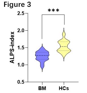
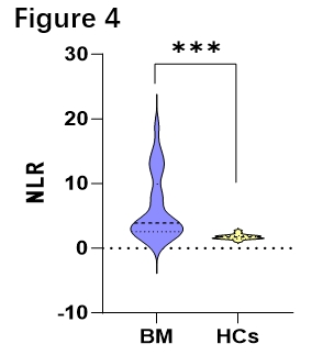
Results: Compared with HCs, the mean ALPS index of patients with brain metastases was significantly decreased, and the NLR was significantly increased.
Impact: ALPS-index and NLR can provide a new perspective for potential assessment of how brain metastasis affects the glymphatic system and inflammatory immune system, and may also support more relevant treatments for patients.
Introduction
Brain metastatic tumor is common intracranial malignancies, it portended the poor prognosis of tumor patients and caused dysfunction and cognitive impairment. The primary tumors usually include lung cancer, breast cancer and kidney cancer and most of these tumors metastasize along the blood or meningeal infiltration[1,2]. Previous study has demonstrated that numerous diseases invade the central nervous system(CNS) through blood-cerebrospinal fluid(CSF)-brain pathway[3]. Blood neutrophil and lymphocyte ratio(NLR) is a key indicator of the progression and prognosis in various malignant tumors[4]. Glymphatic system is a translocation pathway mediating the exchanging of CSF and interstitial fluid (ISF)[5]. Recently, DTI analysis along the perivascular space (DTI-ALPS) as an emerging technology, was used to investigate the activity of the glymphatic system[6]. Studies have revealed glymphatic system dysfunction in both meningeoma and glioma patients, but it is not known whether brain metastases will effect glymphatic system and the blood-CSF-brain pathway. In this study, we took advantage of DTI-ALPS and NLR to evaluate the changes of CSF and blood circulation, in order to provide a new idea for the progress and prognosis of patients with brain metastatic tumor.Methods
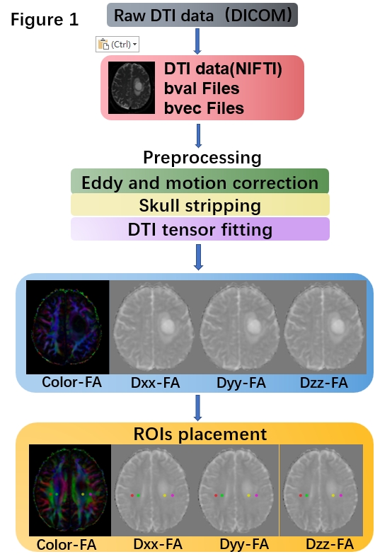
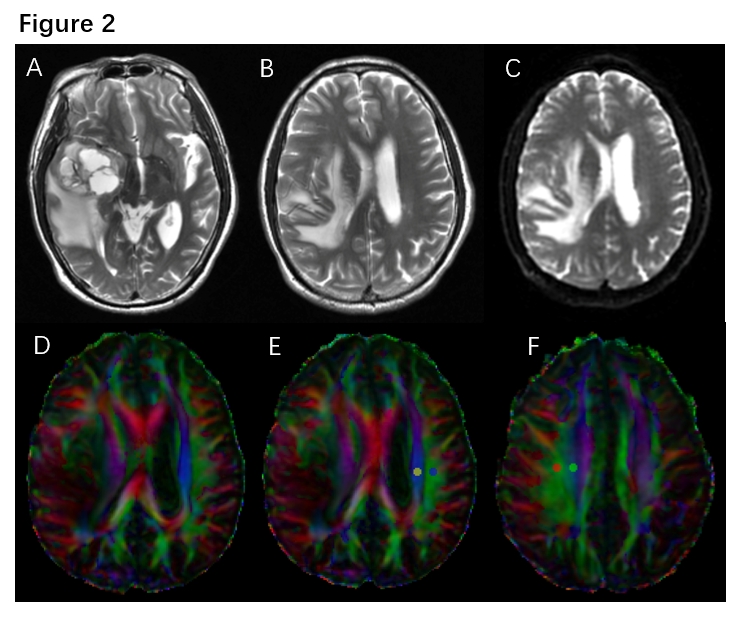
All examinations were performed on GE3.0T MR Scanner (GE Signa HDXT, GE Healthcare) with an 8-channel head receiver coil. DTI scanning parameters are as follows:30diffused sensitive gradient directions, TR=8000ms, TE=83.5ms, b value =1000s/mm2, scanning thickness=5mm, field of view=240mm×240mm, matrix=128×128, interpolation algorithm automatically reconstructed to 256×256, a total of 28 layers were scanned. After converting DTI data to NIFTI format in MRcroGL, data preprocessing was performed using FMRIB Software Library (FSL) (Figure 1). The brief steps included eddy correct and motion correct, skull stripping and tensor fitting. The difusion tensor was calculated, including a colour-coded fractional anisotropy (FA) and Dxx-FA, Dyy-FA, Dzz-FA maps. Then, a 5-mm-diameter region of interest (ROI) was placed in the area of the projection fibers and the association fibers in the bilateral hemispheres(Figure 2). The DTI-ALPS index was calculated in the FA maps using the following formula: DTI-ALPS index=mean (Dxxproj, Dxxassoc)/mean (Dyyproj, Dzzassoc). Blood neutrophil and lymphocyte counts were collected in all patients and HCs, than calculated the neutrophil and lymphocyte ratio(NLR). ALPS index and NLR were expressed as mean and standard deviation (SD). Data normality was tested using Shapiro–Wilk test. The student t test was used to compare ALPS index and NLR between HCs and brain metastases patients. The threshold for the significance level was p< 0.05. All statistical analyses were performed using SPSS version 26.Results
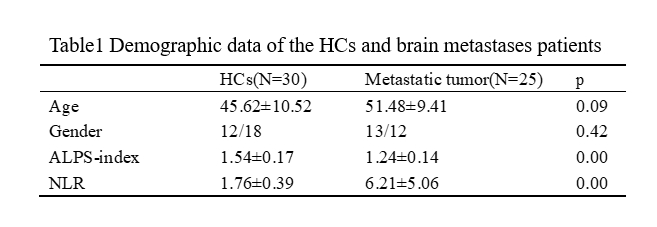
25 patients with brain metastases and 30 HCs were enrolled into the study. Demographic data of the HCs and patients were listed in Table 1. The primary lesion of brain metastases included 13 lung cancer,8breast cancer,3renal carcinoma and 1 malignant melanoma. Compared with HCs, the mean ALPS index of patients with brain metastases was significantly decreased, and the NLR was significantly increased(Figure 3, Figure 4)。Discussion
In this study, the results showed that in patients with brain metastases, ALPS-index was decreased compared to HCs, while NLR was increased. These findings indicated depressed glymphatic system and activated systemic inflammatory state in brain metastases patients. Previous study has suggested that CSF can provide microenvironment for tumor metastasis for tumor cells infiltrating and growing[7]. Glymphatic system dysfunction will cause the accumulation of injurant in CSF. In addition, a blood-CSF-brain pathway was proposed in varieties of neurological diseases, especially in metastases[8]. Our study indicated both ALPS-index and blood NLR alteration in brain metastases, which means peripheral blood and CSF circulation may together aggravate infiltration and progression of metastases. Thus, whether the malignant tumors metastasize through blood or meningeal, the progression of tumors was accompanied by changes in CSF and blood circulation. This may help a better understanding of how tumors metastasize and provide more effective treatments.Conclusion
In this study, our findings showed that DTI-ALPS and NLR can provide detailed information on peripheral blood and CSF circulation alteration in patients with brain metastases. Clinically, this information is valuable for understanding the pathway of brain metastasis from malignant tumors. Therefore, ALPS-index and NLR can provide a new perspective for potential assessment of how brain metastasis affects the glymphatic system and inflammatory immune system, and may also support more relevant treatments for patients.Acknowledgements
No acknowledgement found.References
[1] Sacks P, Rahman M. Epidemiology of Brain Metastases. Neurosurg Clin N Am. 2020 Oct;31(4):481-488.
[2] Gállego Pérez-Larraya J, Hildebrand J. Brain metastases. Handb Clin Neurol. 2014;121:1143-57.
[3] Cousins O, Hodges A, Schubert J, et.al. The blood-CSF-brain route of neurological disease: The indirect pathway into the brain. Neuropathol Appl Neurobiol. 2022 Jun;48(4):e12789. .
[4] Zahorec R. Neutrophil-to-lymphocyte ratio, past, present and future perspectives. Bratisl Lek Listy. 2021;122(7):474-488.
[5] Oshio K. What Is the "Glymphatic System"? Magn Reson Med Sci. 2023 Jan 1;22(1):137-141.
[6] Taoka T, Masutani Y, Kawai H, et.al. Evaluation of glymphatic system activity with the diffusion MR technique: diffusion tensor image analysis along the perivascular space (DTI-ALPS) in Alzheimer's disease cases. Jpn J Radiol. 2017 Apr;35(4):172-178.
[7] Boire A, Zou Y, Shieh J, et.al. Complement Component 3 Adapts the Cerebrospinal Fluid for Leptomeningeal Metastasis. Cell. 2017 Mar 9;168(6):1101-1113.e13.
[8] Toh CH, Siow TY, Castillo M. Peritumoral Brain Edema in Metastases May Be Related to Glymphatic Dysfunction. Front Oncol. 2021 Oct 13;11:725354.
Figures