2625
Association of CSF amyloid-β and tau with diffusion along the perivascular space in Alzheimer’s disease1Research, National Neuroscience Institute; Duke-NUS Medical School, Singapore, Singapore, Singapore, 2National Neuroscience Institute, Singapore, Singapore, 3National Neuroscience Institute; Duke-NUS Medical School, Singapore, Singapore, Singapore, 4Research, National Neuroscience Institute, Singapore, Singapore, 5National Neuroscience Institute; Duke-NUS Medical School, Singapore; Lee Kong Chian School of Medicine, Singapore, Singapore, Singapore
Synopsis
Keywords: Alzheimer's Disease, Alzheimer's Disease
Motivation: Glymphatic clearance of toxic proteins, quantified by the “DTI-along-the-perivascular-space” (ALPS) index, is impaired in Alzheimer’s disease (AD)
Goal(s): We tested the association of the ALPS index to CSF Aβ and tau.
Approach: We used imaging, CSF biomarker, and neuropsychological assessment data from 12 MCI, 21 AD, and 11 other dementia patients. We generated color FA maps and ALPS ROIs, before testing the interaction effects of ALPS × group on each CSF biomarker measure.
Results: In our sample of AD and mild cognitive impairment patients (n=44), we found significant positive association of CSF tau and phosphorylated tau (but not amyloid-β) with the ALPS index.
Impact: Our study establishes a novel link between brain glymphatic function and CSF phosphorylated tau in AD via the ALPS index.
Introduction
The glymphatic system is responsible for clearing toxic proteins from the brain. A hallmark of Alzheimer’s disease (AD) is the accumulation of the toxic proteins, amyloid-β (Aβ) and tau. Accordingly, there is growing interest in the role of glymphatic function in AD aetiology. Diffusion-along-the-perivascular-space (ALPS) can be quantified using DTI, and has been applied widely to study glymphatic function [1]. We tested the association of the ALPS index to Aβ and tau from CSF in a retrospective cross-sectional study of AD patients.Methods
SubjectsAD, mild cognitive impairment (MCI) and other dementia patients (including fronto-temporal dementia and vascular dementia) were recruited from memory clinics at the National Neuroscience Institute, Singapore (2015-2018).
MRI acquisition
We acquired DTI and susceptibility-weighted MRI (SWI) on a 3T Siemens Prisma with the following parameters. DTI: slice thickness 2.3 mm, voxel size 2.30 x 2.30 mm, matrix 96x96x68, TE=0.054s, TR=5.6s, FA=90⁰, 8 b=0 s/mm2 volumes and 61 diffusion-weighted volumes at b=1000 s/mm2. SWI: slice thickness 2.0 mm, voxel size 0.86 x 0.86, matrix 232 x 256, TE=0.020s, TR=0.028s, FA=15⁰.
CSF biomarkers
CSF total tau, phosphorylated tau (p-tau) and Aβ concentrations were determined by INNOTEST ELISAs (Fujirebio). Ratios for total tau:Aβ and p-tau:Aβ were calculated.
Neuropsychological assessment
Global cognition was assessed using the Montreal Cognitive Assessment (MoCA) and Mini-Mental State Exam (MMSE).
Image analysis
DTI data underwent skull-stripping, eddy current and motion correction, denoising, Gibbs’ artefact removal, estimation of diffusion tensors, and generation of colour fractional anisotropy (FA) map using MRTrix3 [2]. Colour FA maps were spatially aligned with SWI and used by two neuroradiologists to place regions of interest (ROIs) according to the scheme in Figure 1. ALPS index calculation was performed according to the conventional formula [1] and then averaged for left and right hemispheres, and for the two raters.
Statistics
CSF biomarkers were right-tailed so we applied a natural log transform. We tested the interaction effect of ALPS × group on each CSF measure. For significant interaction effects, the main outcome was the association of the CSF biomarker as dependent variable with the ALPS index as independent variable whilst controlling for age, which we assessed using linear regression.
Results
We included data for 12 MCI (aged 53.2±8.5 years; 25% female), 21 AD (aged 56.4±5.0 years; 57% female), and 11 with other dementias (aged 61.4±3.3 years; 73% female). Inter-rater reliability for ALPS index was excellent (ICC(2,k)=0.87). One-way ANOVA and post-hoc t-tests revealed significant group differences in total tau, p-tau, p-tau:Aβ ratio, MMSE and MoCA, but not in total tau:Aβ ratio, Aβ or ALPS index (Figure 2). Significant ALPS × group interaction effects (Figure 3) were present for p-tau (F=9.46, p<0.001) and total tau (F=4.84, p=0.014). A weak interaction effect was present for the p-tau:Aβ ratio (F=2.46, p=0.100) and no interaction effect was present for the total tau:Aβ ratio (F=0.84, p=0.441) or Aβ (F=0.35, p=0.710). Linear regression models controlling for age showed that the ALPS index was significantly associated with p-tau in the AD group (β=0.72, p=0.001) but not the MCI (β=-0.72, p=0.090) or other dementias groups (β=0.31, p=0.383). Likewise, the ALPS index was significantly associated with total tau in the AD group (β=0.62, p=0.005) but not the MCI (β=0.02, p=0.975) or other dementias groups (β=-0.44, p=0.292). Cognitive impairment, in both MoCA and MMSE, was significantly associated with all CSF biomarkers in bivariate analyses, but not when accounting for the confounding effect of age (regardless of whole-group or subgroup analyses). Nor were either MoCA or MMSE associated with the ALPS index (whether accounting for age or not, and regardless of which subgroup).Discussion
We present the first study reporting associations of CSF tau and p-tau with the ALPS index in AD, finding positive association of p-tau and ALPS. While there is no direct comparator available, we note one study investigated associations of CSF biomarkers and ALPS index in AD, and found positive association of glymphatic function with Aβ but did not report associations for tau [3]. Our finding contrasts with studies of other neurological conditions [4, 5, 6], which find negative relationships. Our result may be explained by the early age-of-onset of our cohort, preliminary sample size, diurnal variation, sleep quality or ApoE4 status. Tau clearance is complex, and is achieved via multiple different mechanisms. Studies combining CSF biomarkers with MRI and PET imaging are needed to better understand the interplay of glymphatic clearance, tau concentrations in CSF, and their perturbation in AD. We are expanding this study to include 145 participants with healthy control group, and MRI measures of free water, perivascular space fraction, and choroid plexus volume.Acknowledgements
No acknowledgement found.References
1. Taoka T, Masutani Y, Kawai H, Nakane T, Matsuoka K, Yasuno F, Kishimoto T, Naganawa S. Evaluation of glymphatic system activity with the diffusion MR technique: diffusion tensor image analysis along the perivascular space (DTI-ALPS) in Alzheimer's disease cases. Jpn J Radiol. 2017 Apr;35(4):172-178. doi: 10.1007/s11604-017-0617-z. Epub 2017 Feb 14. PMID: 28197821.
2. Tournier JD, Smith R, Raffelt D, Tabbara R, Dhollander T, Pietsch M, Christiaens D, Jeurissen B, Yeh CH, Connelly A. MRtrix3: A fast, flexible and open software framework for medical image processing and visualisation. Neuroimage. 2019 Nov 15;202:116137. doi: 10.1016/j.neuroimage.2019.116137. Epub 2019 Aug 29. PMID: 31473352.
3. Kamagata K, Andica C, Takabayashi K, Saito Y, Taoka T, Nozaki H, Kikuta J, Fujita S, Hagiwara A, Kamiya K, Wada A, Akashi T, Sano K, Nishizawa M, Hori M, Naganawa S, Aoki S; for the Alzheimer's Disease Neuroimaging Initiative. Association of MRI Indices of Glymphatic System With Amyloid Deposition and Cognition in Mild Cognitive Impairment and Alzheimer Disease. Neurology. 2022 Sep 19;99(24):e2648–60. doi: 10.1212/WNL.0000000000201300. Epub ahead of print. PMID: 36123122; PMCID: PMC9757870.
4. Butler T, Zhou L, Ozsahin I, Wang XH, Garetti J, Zetterberg H, Blennow K, Jamison K, de Leon MJ, Li Y, Kuceyeski A, Shah SA. Glymphatic clearance estimated using diffusion tensor imaging along perivascular spaces is reduced after traumatic brain injury and correlates with plasma neurofilament light, a biomarker of injury severity. Brain Commun. 2023 Apr 25;5(3):fcad134. doi: 10.1093/braincomms/fcad134. PMID: 37188222; PMCID: PMC10176239.
5. Qin Y, Li X, Qiao Y, Zou H, Qian Y, Li X, Zhu Y, Huo W, Wang L, Zhang M. DTI-ALPS: An MR biomarker for motor dysfunction in patients with subacute ischemic stroke. Front Neurosci. 2023 Mar 31;17:1132393. doi: 10.3389/fnins.2023.1132393. PMID: 37065921; PMCID: PMC10102345.
6. Ma X, Li S, Li C, Wang R, Chen M, Chen H, Su W. Diffusion Tensor Imaging Along the Perivascular Space Index in Different Stages of Parkinson's Disease. Front Aging Neurosci. 2021 Nov 15;13:773951. doi: 10.3389/fnagi.2021.773951. PMID: 34867300; PMCID: PMC8634754.
Figures
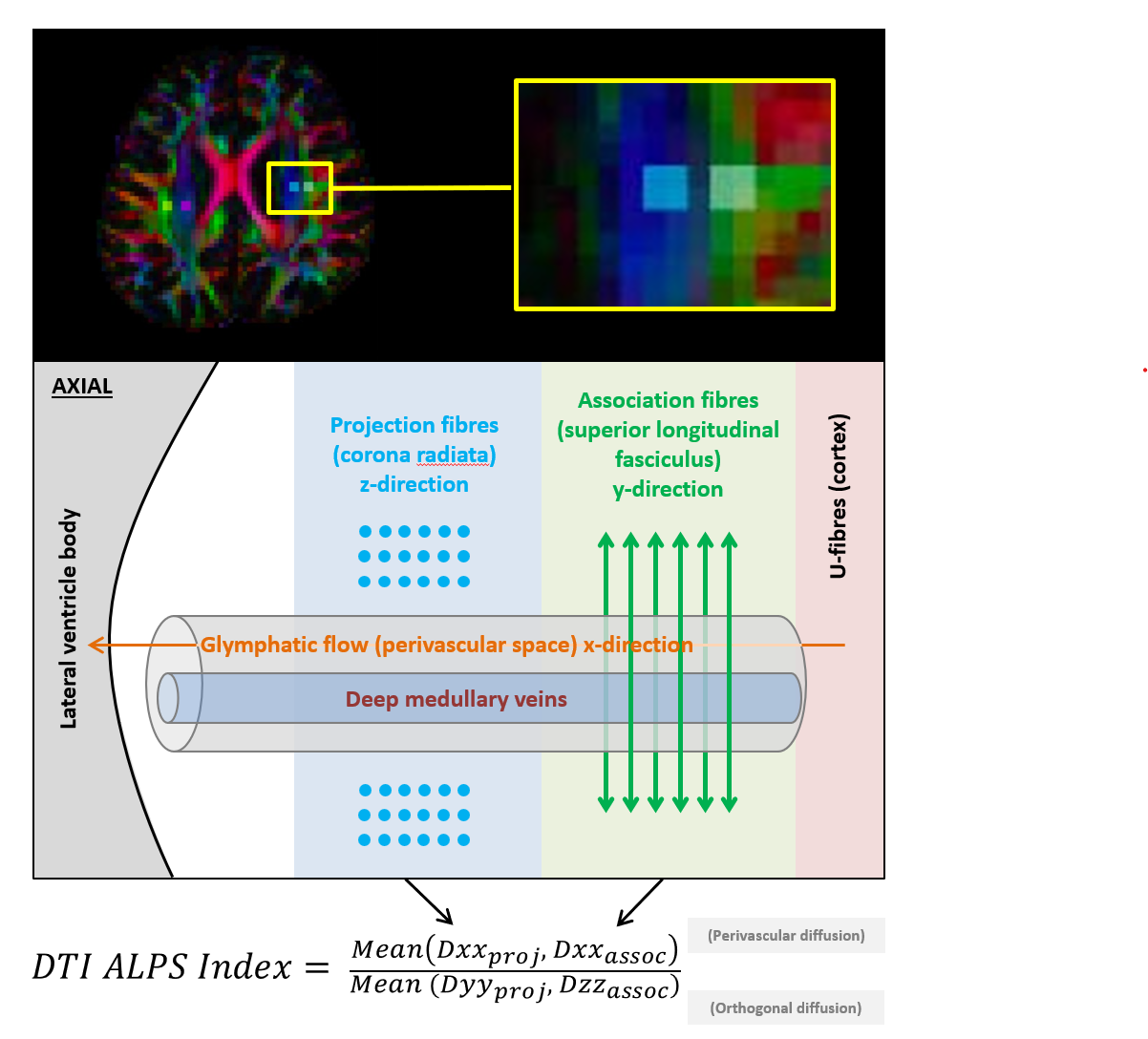
Figure 1. DTI along the perivascular space: concept and measurement.
Regions of interest (ROIs) were manually drawn in a single axial slice at the level of the body of the lateral ventricle. Each ROI comprised 4 voxels placed in a square, constituting 12.17 mm3. Two ROIs were drawn per hemisphere, with the first in the projection fibre bundle and the second in the association fibre bundle (superior longitudinal fasciculus). Susceptibility-weighted MRI was used to avoid large contravening vessels.
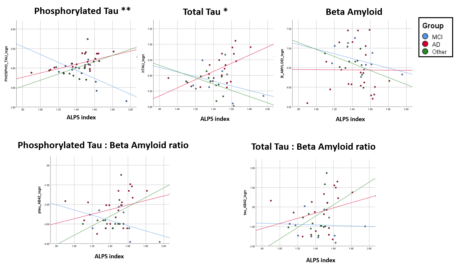
Figure 2. Group differences among CSF biomarkers, cognitive scores and diffusion along the perivascular space.
One-way ANOVA significance is represented by asterisk beside chart title while t-test significance (not corrected for multiple comparisons) is represented by asterisk and line spanning the groups in the significant comparison.
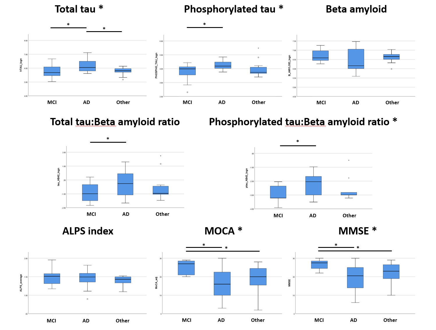
Figure 3. Interaction effects of DTI along the perivascular space and group status for five CSF biomarkers.
The interaction of the average ALPS score and group (Alzheimer’s disease [AD], mild cognitive impairment [MCI] and other dementias) is represented in the line charts by differences in the slope of each group. ** p<0.001. * p<0.05.