2615
Comparing R2* and QSM at 9.4T for the ability to detect increased deoxyhemoglobin (hypoxia) in the mouse brain1Department of Radiology, University of Calgary, Calgary, AB, Canada, 2Department of Neuroscience, University of Calgary, Calgary, AB, Canada, 3Hotchkiss Brain Institute, University of Calgary, Calgary, AB, Canada, 4School of Information Technology and Electrical Engineering, University of Queensland, Queensland, Australia
Synopsis
Keywords: Susceptibility/QSM, Quantitative Imaging, Hypoxia, High-Field MRI
Motivation: Investigating R2* and QSM for deoxyhemoglobin detection is vital for diagnosing and understanding diseases where tissue oxygenation is frequently compromised.
Goal(s): To evaluate R2* against QSM for their ability to detect deoxyhemoglobin changes in a controlled hypoxic environment using a mouse model.
Approach: Employ a graded hypoxia protocol in naïve, female C57Bl/6 mice, capturing 3D MGE images at various oxygen levels (30%, 15% and 10%) to measure R2* and QSM responses.
Results: R2* demonstrated significant sensitivity to hypoxia in brain regions, particularly the hippocampus, unlike QSM, suggesting its potential as a superior hypoxia biomarker.
Impact: This study reveals R2* relaxometry's superior sensitivity to the detection of changes in deoxyhemoglobin over QSM, potentially improving early detection and monitoring of hypoxia-related diseases, such as Multiple Sclerosis, and informing future clinical imaging protocols.
Introduction
Among the various quantitative MRI acquisition techniques, R2* and Quantitative Susceptibility Mapping (QSM) have emerged as promising methods for detecting alterations in paramagnetic substances, a surrogate marker for tissue oxygenation contrasts.1,2 Since R2* is sensitive to changes in magnetic susceptibility and magnetic field inhomogeneities, it serves as a highly effective modality for detecting changes in deoxyhemoglobin. R2* could demonstrate superiority in detecting changes in deoxyhemoglobin compared to QSM, which quantifies magnetic susceptibility of tissues and has been predominantly used for assessing iron content in the brain.3 QSM is increasingly being used to detect iron in conditions such as MS however some of these changes may actually be due to hypoxia.4,5,6 This study aims to compare the efficacy of R2* and QSM in detecting changes in deoxyhemoglobin during a graded hypoxia paradigm in the mouse brain. We aim to determine the sensitivity of these quantitative MRI techniques in capturing the dynamic physiological responses to hypoxia.Methods
Ten naïve, female C57Bl/6 mice were subject to a graded hypoxia paradigm (n=10). A 3D MGE (multi gradient echo) image was acquired at 30%, 15% and 10% inspired oxygen, balanced with nitrogen. There were 5-minute transition periods between each gas manipulation to allow for the respiration rate of the mice to stabilize. Mice were anesthetized with 2% isoflurane. Images were acquired on a Bruker Avance console and 9.4T MRI with a 35 mm volume coil and an MGE sequence: TR= 100ms, 5 echoes (TE= 3.1, 7.1, 11.1, 15.1, 19.1ms), FOV= 1.92x1.59x0.93cm, matrix= 128x106x62, voxel size= 0.15mm isotropic, flip angle= 20°, 2 averages. QSM data were processed by employing phase unwrapping, background field removal using RESHARP and a dipole inversion to acquire susceptibility maps.7 During image acquisition, arterial oxygen saturation (SaO2) was measured with a mouse pulse oximeter at a frequency of 1Hz.Results
The physiological presence of hypoxia was validated by pulse oximetry data, which indicated significant drops in SaO2 when transitioning from 30% to 15% and from 15% to 10% oxygen (p<0.01) (Fig.1). At 10% inspired oxygen, a significant elevation in the R2* was observed in the left and right hippocampus, thalamus, and cortex (p<0.001), when compared to 30% oxygen (Fig.2). Notably, R2* at 10% oxygen demonstrated regional variability, with the thalamus and cortical areas exhibiting a pronounced divergence from the hippocampus (p<0.05 to p<0.001). These differences were not detected in the QSM analysis, which showed no significant changes across the same regions.Discussion
The findings of this study underscore the sensitivity of R2* in detecting variations in deoxyhemoglobin levels during graded hypoxia. The increasing R2* with decreasing oxygen levels aligns with the expected physiological response, as lower oxygenation leads to higher deoxyhemoglobin. The consistency across all stages of oxygenation, reinforces the robustness of R2* as a biomarker for hypoxia. In contrast, QSM did not exhibit significant changes, suggesting its relative insensitivity to the acute fluctuations in deoxyhemoglobin induced by hypoxia. These results may have implications for clinical and research applications, particularly in conditions where rapid assessment of tissue oxygenation is critical. The superior performance of R2* could be attributed to its direct relationship with magnetic field inhomogeneities caused by deoxyhemoglobin, a property that QSM may not capture as effectively due to its reliance on phase information, which could be less sensitive to acute changes in oxygenation. Future research should focus on exploring the mechanistic reasons behind the differential sensitivity of these techniques. Moreover, the integration of R2* into clinical protocols could enhance the diagnostic accuracy for diseases characterized by hypoxic events.Conclusion
This investigation highlights the increased sensitivity of R2* over QSM in detecting brain deoxyhemoglobin changes during hypoxia. The differential response of R2* and QSM to reduced oxygenation offers critical insights into their respective mechanisms of action. R2*’s strong sensitivity to hypoxia, especially in the hippocampus, shows it’s potential for the monitoring of diseases associated with hypoxia. QSM is often used for iron, and iron is also often present in neurological conditions with hypoxia. It will be important to determine which sequence is optimal for clinical neuroimaging. Future studies will be focused on how to detect both iron and hypoxia in models of demyelination and iron overload.Acknowledgements
This work was supported by Canada Institute of Health Research (CIHR), and Natural Sciences and Engineering Research Council (NSERC).
References
1. Dunn J f., Wadghiri YZ, Meyerand M. Regional heterogeneity in the brain’s response to hypoxia measured using BOLD MR imaging. Magn Reson Med. 1999;41(4):850–4.
2. Liu C, Wei H, Gong NJ, Cronin M, Dibb R, Decker K. Quantitative Susceptibility Mapping: Contrast Mechanisms and Clinical Applications. Tomography. 2015 Sep;1(1):3–17.
3. Schweser F, Deistung A, Lehr BW, Reichenbach JR. Quantitative imaging of intrinsic magnetic tissue properties using MRI signal phase: An approach to in vivo brain iron metabolism? NeuroImage. 2011 Feb 14;54(4):2789–807.
4. Rahmanzadeh R, Galbusera R, Lu P, Bahn E, Weigel M, Barakovic M, et al. A New Advanced MRI Biomarker for Remyelinated Lesions in Multiple Sclerosis. Ann Neurol. 2022 Sep;92(3):486–502.
5. Nathoo N, Rogers JA, Yong VW, Dunn JF. Detecting deoxyhemoglobin in spinal cord vasculature of the experimental autoimmune encephalomyelitis mouse model of multiple sclerosis using susceptibility MRI and hyperoxygenation. PloS One. 2015;10(5):e0127033.
6. Adingupu DD, Evans T, Soroush A, Jarvis S, Brown L, Dunn JF. Non-invasive Detection of Persistent Cortical Hypoxia in Multiple Sclerosis Using Frequency Domain Near-Infrared Spectroscopy (fdNIRS). Adv Exp Med Biol. 2022;1395:89-93.
7. Sun H, Ma Y, MacDonald ME, Pike GB. Whole head quantitative susceptibility mapping using a least-norm direct dipole inversion method. NeuroImage. 2018 Oct 1;179:166–75.
Figures
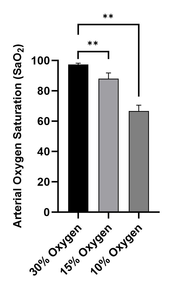
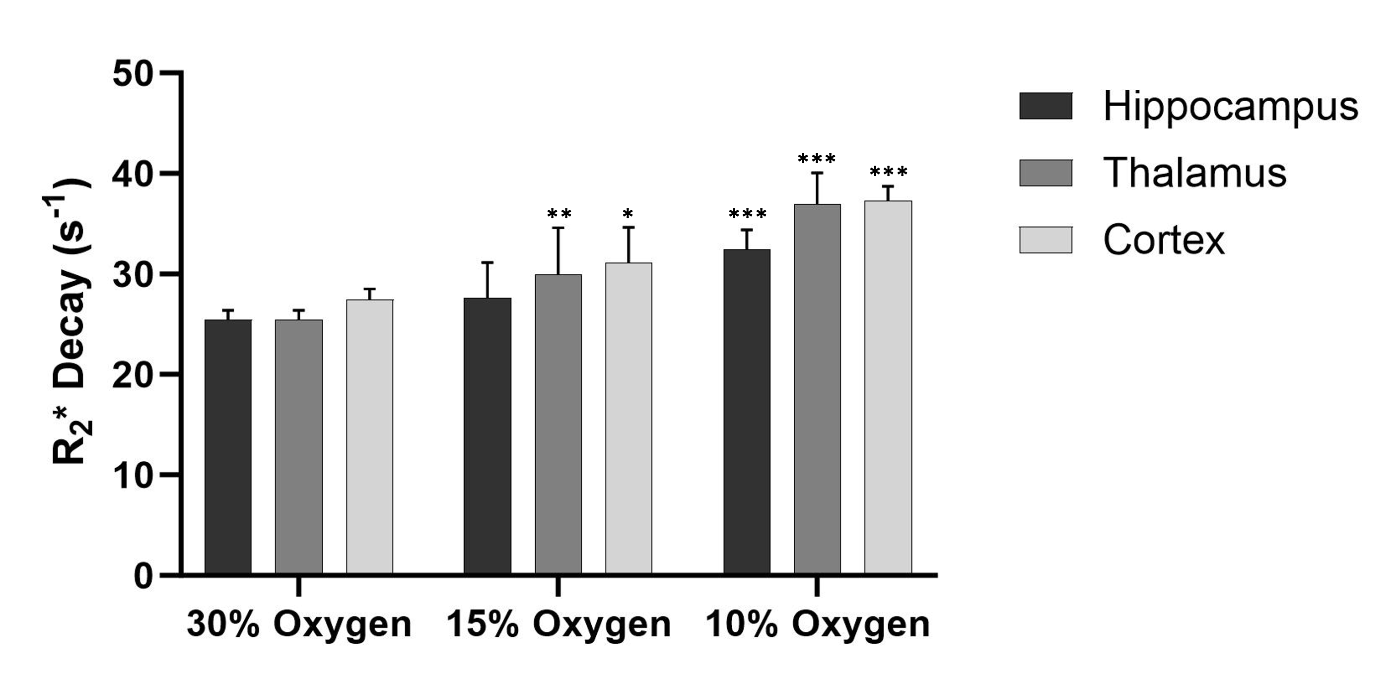
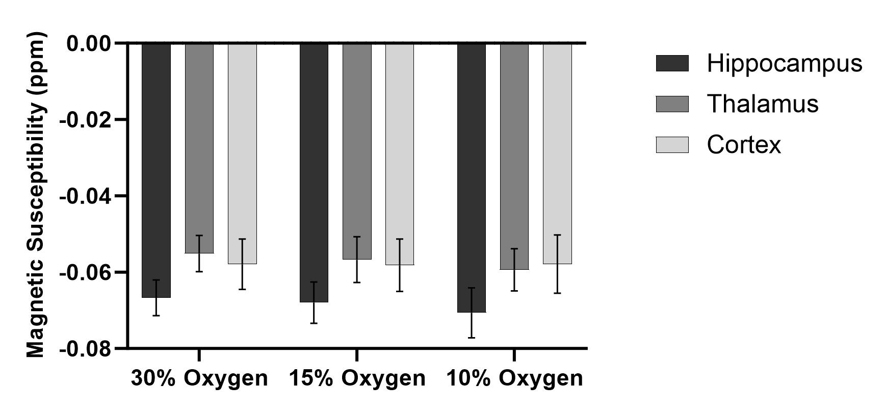
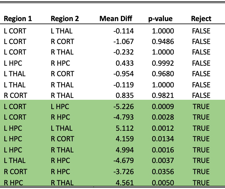
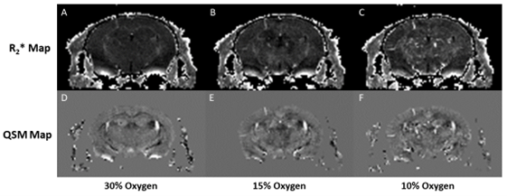
Figure 5: R2* and QSM images of a single slice at varying oxygen concentrations. R2* maps at 30%, 15% and 10% respectively (A-C). QSM maps at 30%, 15% and 10% respectively (D-F).