2606
Animal experimental study of IVIM in assessing obesity-related glomerulopathy1Medical Imaging Center, First Affiliated Hospital of Jinan University, Guangzhou, China, 2MR Research, GE Healthcare, Beijing, China
Synopsis
Keywords: IVIM, Diffusion/other diffusion imaging techniques
Motivation: Early symptoms of obesity-related glomerulopathy (ORG) are atypical and diagnosis depends on invasive kidney biopsy.
Goal(s): This study assessed the potential of intravoxel incoherent motion (IVIM) to detect renal injury induced by ORG and monitor renal function after weight loss therapy.
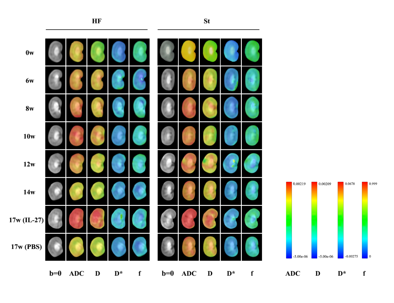
Approach: IVIM were performed in high fat diet (HF) and standard diet (St) rats’ kidney before and after therapy.
Results: Compared with St group, significant decreases in D, D*, f in kidney of HF group were observed, and D was the first to decrease. After therapy, D, D*, f and the renal function were increased simultaneously.
Impact: Obesity with high metabolic demand leads to renal injury and obesity-related glomerulopathy (ORG). Early symptoms of ORG are not obvious, which may increase the risk of chronic kidney diseases. Non-invasively detecting ORG-induced renal injury can prevent poor prognosis.
Introduction
The high metabolic demand caused by obesity will lead to renal injury.1 With the rise of obesity, the prevalence of obesity-related glomerulopathy (ORG) has increased.2.3 The early symptoms of OGR are not obvious which lead to an increased risk of progression to chronic kidney diseases.4 Early identification of OGR are crucial. Renal biopsy is essential for diagnosis, but it is invasive and should not be repeated during therapy. Intravoxel incoherent motion diffusion-weighted imaging (IVIM), as a novel functional magnetic resonance imaging method, can noninvasively and repeatedly obtain in vivo water molecular diffusion and tissue microperfusion information.5 This study aimed to assess the potential of IVIM in detecting early renal injury caused by ORG and monitoring renal function after weight loss therapy.Methods
42 specific pathogen free male Sprague-Dawley rats were randomly divided into two groups: high fat diet (HF) and standard diet (St) (n=21 per group). After 14 weeks of feeding, the ORG model was successfully established. 6 rats were randomly selected from the HF and St groups respectively as HF or St treatment groups and injected intraperitoneally with interleukin-27 (IL-27) for weight loss. 6 rats were randomly selected as HF or St non-treatment group and injected intraperitoneally with phosphate buffered saline (PBS).MRI including IVIM and DWI was performed at 0, 6, 8, 10, 12, 14 and 17 week to obtain ADC and IVIM parameters (f, D, D*) in the right renal cortex (CO), outer stripe of the outer medulla (OOM), inner stripe of the outer medulla (IOM) and inner medulla (IM) in HF and St groups. Single photon emission computed tomography (SPECT) was performed at 6, 8, 10, and 14 week to evaluate glomerular filtration rate (GFR). At 8, 14 and 17 week, serum, urine and kidney samples were collected for follow-up pathological detection and laboratory biochemical analysis. MRI parameters, GFR, urine protein, histopathological changes of kidney were assessed.
Results
1. Before weight loss treatment, at 8 week, D and ADC in IOM in HF group wan firstly decreased compared with St group (P < 0.05). At 12 and 14 week, compared with St group, f, D, D* and ADC in all renal sites in HF group were decreased significantly.2. After weight loss treatment, f, D, D* and ADC in all renal sites in HF treatment group were increased compared with those before weight loss treatment. D and ADC in OOM, IOM and IM in St treatment group were also increased. f, D, D* and ADC in all renal sites in HF treatment group were higher than those in HF non-treatment group.
3. Before weight loss treatment, GFR, IL-1β, adiponectin, triglyceride and total cholesterol concentrations in HF group were higher than those in St group. After weight loss treatment, IL-1β, urinary protein, triglyceride and total cholesterol concentrations in HF treatment group were lower than those in HF non-treatment group.
4. There was no correlation between GFR, urinary protein and f, D, D*, ADC before or after weight loss treatment.
Discussion
The increased metabolic of ORG patient leads to glomerular hyperfiltration, glomerular hypertrophy, glomerular sclerosis,6 reduced intracellular and extracellular space, limiting the diffusion movement of water molecules in the renal parenchyma, resulting in corresponding changes in IVIM parameters. D in HF group was lower than that in St group, indicating that the true diffusion of water molecules in ORG rats’ kidney was limited. The decrease of f in HF group reflected the decrease of microcirculation flow in kidney. In HF treatment group, D and ADC in all renal sites were increased, indicating that the pathologic extracellular space in the kidney was enlarged and the diffusion of water molecules in the tissue was not limited. An increase in f indicated increased blood perfusion in kidney. Changes in IVIM parameters and laboratory indicators showed that renal function improved after weight loss treatment. Rats in this study were in the early stage of ORG while renal function did not deteriorate significantly. This might result in no correlation between GFR, urinary protein and IVIM parameters.Conclusion
IVIM can detect early ORG-related renal injury. D and f have advantages and value in evaluating kidney injury sensitively. The renal function of ORG rats was improved after weight loss treatment while IVIM parameters of kidney were also increased, suggesting that IVIM is a potential tool for dynamic, whole-course and non-invasive detection of renal function improvement after weight loss therapy of ORG.Acknowledgements
No.References
1. Okabayashi Y, Tsuboi N, Sasaki T, et al. Single-Nephron GFR in Patients With Obesity-Related Glomerulopathy. Kidney international reports. 2020;5(8):1218-1227.
2. Tsuboi N, Koike K, Hirano K, et al. Clinical features and long-term renal outcomes of Japanese patients with obesity-related glomerulopathy. Clinical and experimental nephrology. 2013;17(3):379-385.
3. Hu R, Quan S, Wang Y, et al. Spectrum of biopsy proven renal diseases in Central China: a 10-year retrospective study based on 34,630 cases. Scientific reports. 2020;10(1):10994.
4. Hall JE, Mouton AJ, da Silva AA, et al. Obesity, kidney dysfunction, and inflammation: interactions in hypertension. Cardiovascular research. 2021;117(8):1859-1876.
5. Sigmund EE, Vivier PH, Sui D, et al. Intravoxel incoherent motion and diffusion-tensor imaging in renal tissue under hydration and furosemide flow challenges. Radiology. 2012;263(3):758-769.
6. Martínez-Montoro JI, Morales E, Cornejo-Pareja I, et al. Obesity-related glomerulopathy: Current approaches and future perspectives. Obesity reviews: an official journal of the International Association for the Study of Obesity. 2022;23(7):e13450.
Figures