2602
Towards robust myocardial IVIM-DWI: feasibility of single-shot MoCo-DW-EPI with Compressed SENSE AI and elastic registration
Yasuhiro Goto1, Michinobu Nagao2, Masami Yoneyama3, Adam Wu4, Johannes M Peeters5, Isao Shiina1, Kazuo Kodaira1, Yutaka Hamatani1, Takumi Ogawa1, Mana Kato1, Yasuyuki Morita1, and Shuji Sakai2
1Department of Radiological Services, Tokyo Women's Medical University, Tokyo, Japan, 2Department of Diagnostic imaging & Nuclear Medicine, Tokyo Women's Medical University, Tokyo, Japan, 3Philips Japan, Tokyo, Japan, 4Philips Healthcare (Shanghai) Ltd, Shanghai, China, 5Philips Healthcare, Best, Netherlands
1Department of Radiological Services, Tokyo Women's Medical University, Tokyo, Japan, 2Department of Diagnostic imaging & Nuclear Medicine, Tokyo Women's Medical University, Tokyo, Japan, 3Philips Japan, Tokyo, Japan, 4Philips Healthcare (Shanghai) Ltd, Shanghai, China, 5Philips Healthcare, Best, Netherlands
Synopsis
Keywords: Diffusion Analysis & Visualization, Myocardium
Motivation: The purpose of this study was to evaluate the robustness of IVIM mapping using aMoCo-EPICS-AI-DWI-FEIR.
Goal(s): Determine how to achieve better image quality, robustness to motion, and quantitative accuracy in myocardial IVIM-DWI.
Approach: MoCo-TSE-DWI and aMoCo-EPICS-AI-DWI on a 3.0T MR clinical imager for image comparison.
Results: aMoCo-EPICS-AI-DWI-FEIR demonstrated reduced individual and site-specific differences in the parameters obtained from IVIM-DWI imaging, compared with MoCo-TSE-DWI-FEIR.
Impact: aMoCo-EPICS-AI-DWI-FEIR might be the best method for myocardial IVIM-DWI with better image quality, motion robustness and improved quantitative accuracy.
Introduction
Cardiac DWI has a potential to achieve improved diagnosis through novel micro-structural and functional assessment1. The intravoxel incoherent motion (IVIM) theory, proposed by Le Bihan et al.2. enables evaluation of living tissue diffusion movement and micro-vessel perfusion in vivo using multi-b-value DWI, and myocardial IVIM may play an important role assessment of various heart diseases3-6. In previous studies, we have introduced a turbo spin-echo based7 cardiac motion-compensated (MoCo-TSE-DWI)8,9 with fast elastic image registration (FEIR) for myocardial IVIM-DWI9, it demonstrated improved robustness of quality of IVIM mapping without image distortion. However, MoCo-TSE-DWI due to its low signal-to-noise ratio (SNR). Recently, it has been reported the usefulness of EPI-DWI with deep-learning constrained Compressed SENSE (EPICS-AI) to enhance the SNR while reducing the image distortion by using higher reduction factor10-12. In this study, we attempted to combine all promising techniques towards robust cardiac IVIM-DWI, including second order (acceleration) motion-compensated MPG scheme13,14 (aMoCo), EPICS-AI and FEIR. The purpose of this study was to evaluate the robustness of IVIM mapping using aMoCo-EPICS-AI-DWI-FEIR.Methods
Ten healthy volunteers (age range: 22-48 years) underwent MoCo-TSE-DWI and aMoCo-EPICS-AI-DWI (Fig.1) on a 3.0T MR clinical imager (Ingenia Elition X, Philips Healthcare) for image comparison. Imaging parameters for aMoCo-EPICS-AI-DWI-IVIM: FOV=300x300mm2, actual matrix size = 1.38 x 1.38 mm, AI Compressed SENSE-factor = 4.0, TE = 72ms, TR = 2beats, slice thickness = 8 mm, acquisition time = 1m53s, and three directions with b-values 0, 20, 40, 60, 80, 100, 200, 300, 400 s/mm2. Free-breathing acquisition with navigator respiratory gating and tracking is applied. The actual trigger delay (TD) and data acquisition window were visually determined by TD scout scan15. For quantitative image evaluation, three slices of left ventricular short-axis images were divided into 16 segments. The ADC, D*, D and f values of each segment were measured. These four parameters were defined by the following formula. The bi-exponential IVIM model is expressed as: SDWI /S0 = f*exp ( -bD*) + (1-f) *exp (-bD) where S0 is SDWI at b = 0 seconds/mm2, SDWI the signal intensity at a given b-value, D the self-diffusion coefficient, f the self-diffusion fraction, and D* the pseudo-diffusion coefficient. The coefficient of variation (CV), mean, and standard deviations for a total 96 of four parameters were calculated and compared among the two IVIM-DWI imaging. Statistical analysis was performed with Wilcoxon signed-rank test and judged the difference as significant at p<0.05.Results and Discussion
Representative DWI source images and corresponding IVIM maps of MoCo-TSE-DWI-FEIR (Fig.2) and aMoCo-EPICS-AI-DWI-FEIR (Fig.3) are shown. Table 1 shows the comparison of IVIM quantitative values. There was significant difference in ADC, D*, D and f values CVs between MoCo-TSE-DWI-FEIR and aMoCo-EPICS-AI-DWI-FEIR. CVs for all four parameters were significantly smaller in aMoCo-EPICS-AI-DWI-FEIR than in MoCo-TSE-DWI. aMoCo-EPICS-AI-DWI-FEIR demonstrated reduced individual and site-specific differences in the parameters obtained from IVIM-DWI imaging, compared with MoCo-TSE-DWI-FEIR. ADC values of aMoCo-EPICS-AI-DWI-FEIR approximated the D value, while MoCo-TSE-DWI-FEIR showed a discrepancy between ADC and D values. Figure 4 shows the representative ADC maps of MoCo-TSE-DWI-FEIR and aMoCo-EPICS-AI-DWI-FEIR. The signal uniformity of the cardiac wall was improved with aMoCo-EPICS-AI-DWI-FEIR compared with MoCo-TSE-DWI-FEIR. These results suggest that IVIM parameters obtained from aMoCo-EPICS-AI-DWI-FEIR could be more accurate.Conclusion
aMoCo-EPICS-AI-DWI-FEIR might be the best method for myocardial IVIM-DWI with better image quality, motion robustness and improved quantitative accuracy.Acknowledgements
No acknowledgement found.References
1. Nielles-Vallespin S, et.al, Cardiac Diffusion: Technique and Practical Application. J Magn Reson Imaging. 2020 Aug;52(2):348-368. doi: 10.1002/jmri.26912. 2. Le Bihan, D. et al. Separation of diffusion and perfusion in intravoxel incoherent motion MR imaging. Radiology 1988;168, 497–505, https://doi.org/10.1148/radiology.168.2.3393671. 3. Spinner GR, et al. Bayesian intravoxel incoherent motion parameter mapping in the human heart. J Cardiovasc Magn Reson. 2017 Nov 6;19(1):85. doi: 10.1186/s12968-017-0391-1. 4. Xiang SF, et al. STROBE-A preliminary investigation of IVIM-DWI in cardiac imaging. Medicine (Baltimore). 2018 Sep;97(36):e11902. doi: 10.1097/MD.0000000000011902. 5. Spinner GR, et al. On probing intravoxel incoherent motion in the heart-spin-echo versus stimulated-echo DWI. Magn Reson Med. 2019 Sep;82(3):1150-1163. doi: 10.1002/mrm.27777. 6. Xiang SF, et al. Intravoxel Incoherent Motion Magnetic Resonance Imaging with Integrated Slice-specific Shimming for old myocardial infarction: A Pilot Study. Scientific Reports 2019:9:19766. 7. Goto Y, et.al, Simple and robust cardiac diffusion weighted imaging using single-shot Turbo Spin-Echo with peripheral pulse gating. Proc. ISMRM:2016.3476). 8. Goto Y, et al, Improvement of distortion-free cardiac diffusion weighted imaging (DWI) using motion-compensated single-shot turbo spin-echo (MoCo-TSE) DWI. Proc. ISMRM:2020.2077. 9. Goto Y, et al, Improvement of distortion-free cardiac IVIM-DWI using motion-compensated single-shot turbo spin echo DWI with fast elastic image registration. Proc. ISMRM:2022.4562. 10. Yoneyama M, et al, SNR boost in whole-body DWIBS utilizing deep learning constrained Compressed SENSE reconstruction. Proc. ISMRM:2022.3655. 11. Yoneyama M, et al, SNR enhancement in rapid high b-value prostate single-shot DW-EPI utilizing deep learning constrained Compressed SENSE reconstruction. Proc. ISMRM:2022.3712. 12. Yoneyama M, et al, Pseudo-3D whole-brain ultra-thin-slice diffusion-weighted imaging of the brain utilizing deep learning constrained Compressed SENSE. Proc. ISMRM:2022.4732. 13. Chen R, et al, Second-order motion compensated single-shot spin echo planar imaging sequence using Compressed SENSE: a pilot study. Proc. ISMRM:2023.4290. 14. Luo W, et al, Computed DWI with 2nd Order motion Compensated DWI for Diagnosis of the Myocardial Infarction without Contrast Agents. Proc. ISMRM:2023.4291. 15. Moulin K, et al. Probing cardiomyocyte mobility with multi-phase cardiac diffusion tensor MRI. PLoS One. 2020 Nov 12;15(11):e0241996. doi: 10.1371/journal.pone.0241996.Figures
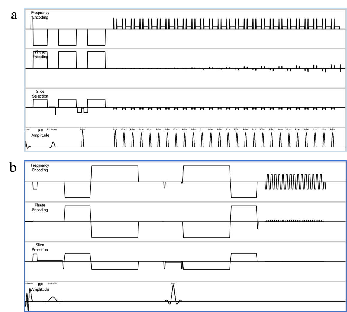
Figure 1. Sequence diagram of MoCo-TSE-DWI (a) and aMoCo-EPICS-AI-DWI (b).
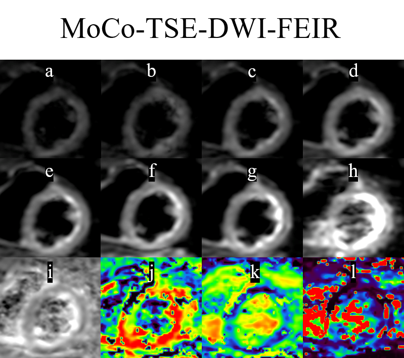
Figure 2. Representative IVIM-DWI using
MoCo-TSE-DWI raw images of the mid-ventricular short axis slices: b-value =
400, 300, 200, 100, 60, 40, 20, 0 s/mm2 (a-h) and calculated parametric maps:
ADC ( i ), D ( j ), f ( k ), and D* ( l ) maps.
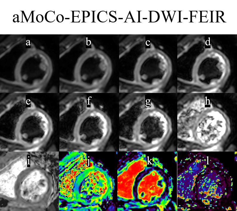
Figure 3. Representative IVIM-DWI using aMoCo-AI-EPICS-DWI raw images of the
mid-ventricular short axis slices: b-value = 400, 300, 200, 100, 60, 40, 20, 0 s/mm2 (a-h) and calculated
parametric maps: ADC ( i ), D ( j ), f ( k ), and D* ( l ) maps.
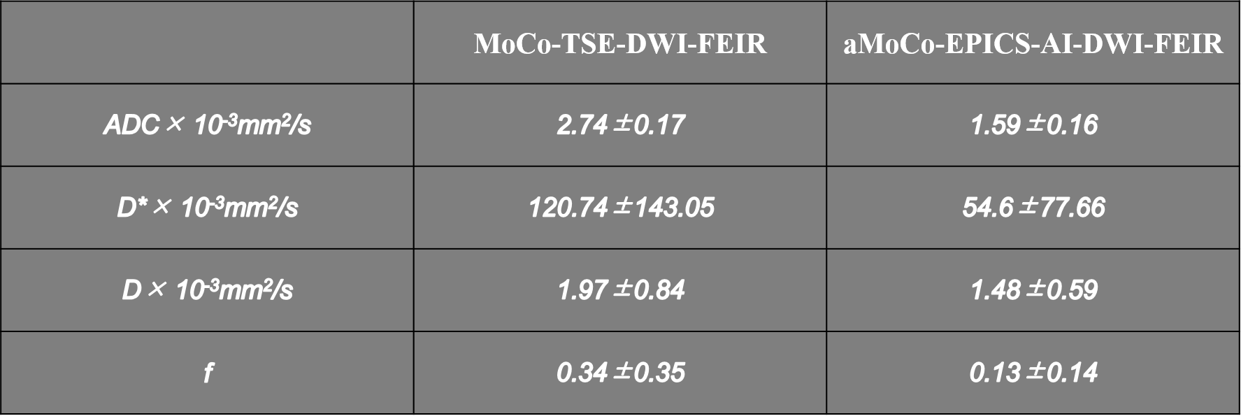
Table 1. IVIM parameters measured on 16 myocardial
segments of ten volunteers.
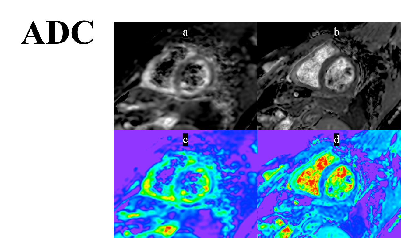
Figure 4. Representative ADC maps of MoCo-TSE-DWI (a,c) and aMoCo-AI-EPICS-DWI (b,d). The signal uniformity of the cardiac wall was improved with aMoCo-AI-EPICS-DWI compared with MoCo-TSE-DWI.
DOI: https://doi.org/10.58530/2024/2602