2600
Spatial profiling of parameters derived from diffusion weighted magnetic resonance imaging in the healthy human kidney1Center for Advanced Imaging Innovation and Research, Department of Radiology, NYU Langone Health, New York, NY, United States, 2Siemens Healthcare GmbH, Erlangen, Germany, 3The Institute of Cancer Research and The Royal Marsden NHS Foundation Trust, London, United Kingdom, 4MR Application Predevelopment, Siemens Healthcare GmbH, Erlangen, Germany
Synopsis
Keywords: Microstructure, Microstructure, IVIM, DTI, Renal
Motivation: Kidney microstructure has received relatively less attention compared to other organs due to EPI artifacts convolved with motion and cardiac pulsatility. Recently, mitigations of these artifacts have enabled more microstructural explorations of the kidney.
Goal(s): The study aimed at deriving estimates of microstructure and microcirculation from the perspective of concentric layers segmentations.
Approach: We scrutinized the dependence of renal DWI parameters from conventional representations (DTI, IVIM), an advanced hybrid DTI-IVIM approach and a multiple encoded FC-IVIM model on concentric layers.
Results: The most significant layer dependence was observed for pseudodiffusion parameters and structural fractional anisotropy, with weaker dependences observed for structural diffusivity parameters.
Impact: Potential extrapolations to kidney microstructure using in vivo MRI could be highly impactful in the study of renal dysfunction.
Introduction
Diffusion-weighted MRI is a versatile technique that can infer many microstructural and microcirculatory features from biological tissue, with particular application to renal tissue. There is an extensive literature on diffusion tensor imaging (DTI)1 of anisotropy in renal medulla, intravoxel incoherent motion (IVIM)2 measurements separating microstructural from microcirculation effects, and combinations of the two3. However, interpretation of these features and adaptation of more specific models remains an ongoing challenge. There is a well-developed literature on preclinical murine and excised human kidney microstructure using in vivo or ex vivo modalities and their combinations4. Transferring this knowledge to in vivo human kidney imaging and extrapolations to its microstructure could be highly impactful in the study of renal dysfunction. However, the diffusion-weighted MR parameters are generally not specific5 due to the dependency of the diffusion signal on different physiological parameters and restriction, and the fact that inter-species microstructural features are substantially different.Macroscopic segmentation strategies are evolving to better assess intra-organ functional variations. The twelve-layer concentric object method (TLCO)6 has enabled better differentiation of CKD kidneys from controls compared to the classical cortex and medulla segmentations. Recently, TLCO was applied to analysis of apparent diffusion coefficient (ADC)7 and arterial spin labelling (ASL) parameters8.
In this work, we probe the spatial dependence of diffusion MRI metrics with concentrically layered segmentation in healthy kidneys. The metrics include those from DTI, IVIM, a combined approach (REFMAP)3, and a multiply encoded model (FC-IVIM)9 providing estimates of fluid velocity and branching length.
Methods
In this HIPAA-compliant and IRB-approved prospective study, 7 healthy volunteers (3 male, age 28±5 years) provided written informed consent and had abdominal imaging performed in a 3 T MRI system (MAGNETOM Prisma; Siemens Healthcare, Erlangen, Germany) in supine position with posterior spine array and anterior body array RF coils and chest leads for ECG gating. Coronal oblique T2-weighted HASTE images were collected for anatomical reference. Sagittal phase contrast (PC) MRI images through the left renal artery were collected at multiple cardiac phases to estimate the systolic cardiac phase for kidney tissue. With a research application single shot echo-planar imaging DWI sequence with dynamic field correction, cardiac triggered oblique coronal DWI, aligned to the prior HASTE imaging, at multiple echo times (bipolar and flow compensated pulse sequence, TR/TE1/TE2 2800/81/120 ms, matrix 192/192/1, resolution 2.2/2.2/5 mm, GRAPPA acceleration factor 2) were collected at 10-12 b-values between 0-800 s/mm2 and 12 directions. Acquisition time for a given echo time and encoding was approximately 6 minutes. Additionally, to correct for motion and field inhomogeneity, 16 right-to-left and 16 left-to-right phase-encoding b=0 images were acquired sampling the full range of motion. 3 left kidneys were excluded due to excessive noise 10.The 4 DWI acquisitions (2 echo times for bipolar and flow-compensated acquisitions) were registered together and underwent a 6-layer segmentation implemented as multiple zones concentric objects (MZCO) generation on the freely available software FireVoxel (build 421, https://firevoxel.org/).
First, average IVIM maps (Dt, fp, Dp) were generated from segmented biexponential fits of the directionally averaged bipolar gradient diffusion-weighted image sets at TE = 81 ms. Secondly, the directional DWI signals from the bipolar acquisition at TE=81 ms was processed analogously to the Renal Flow and Microstructure AnisotroPy (REFMAP) [9] to extract DTI, IVIM, and directional IVIM parameters simultaneously (i.e. MD, FA, D∥, D⊥, f, D* , D*∥, D*⊥). Finally, both bipolar and flow compensated diffusion signals at both echo times, were averaged over all directions and used to generate signal intensity curves from each layer for input into the multiple encoded FC-IVIM method to estimate flow velocity v, vessel segment length l, and perfusion fraction f for each layer.
Results
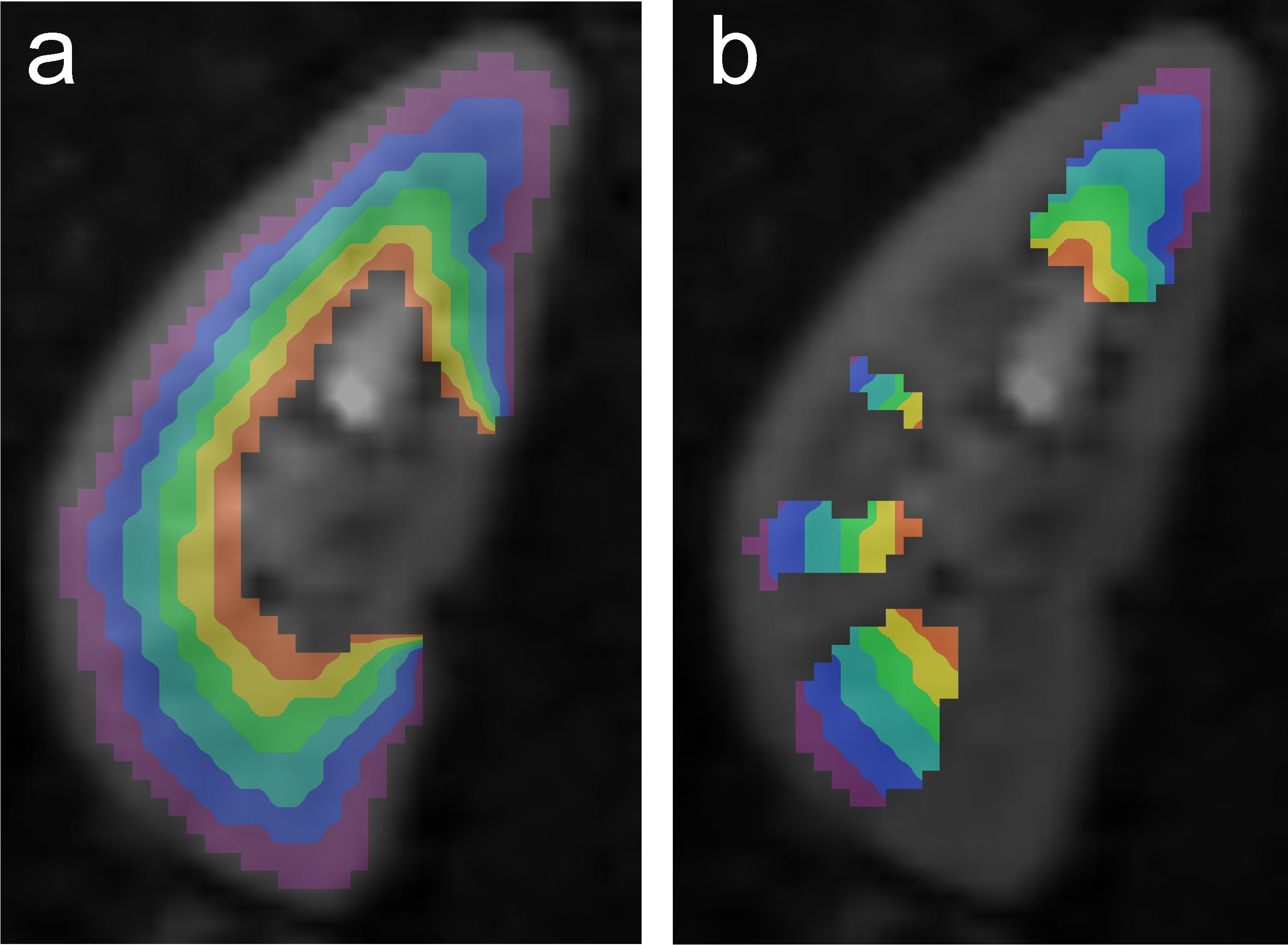
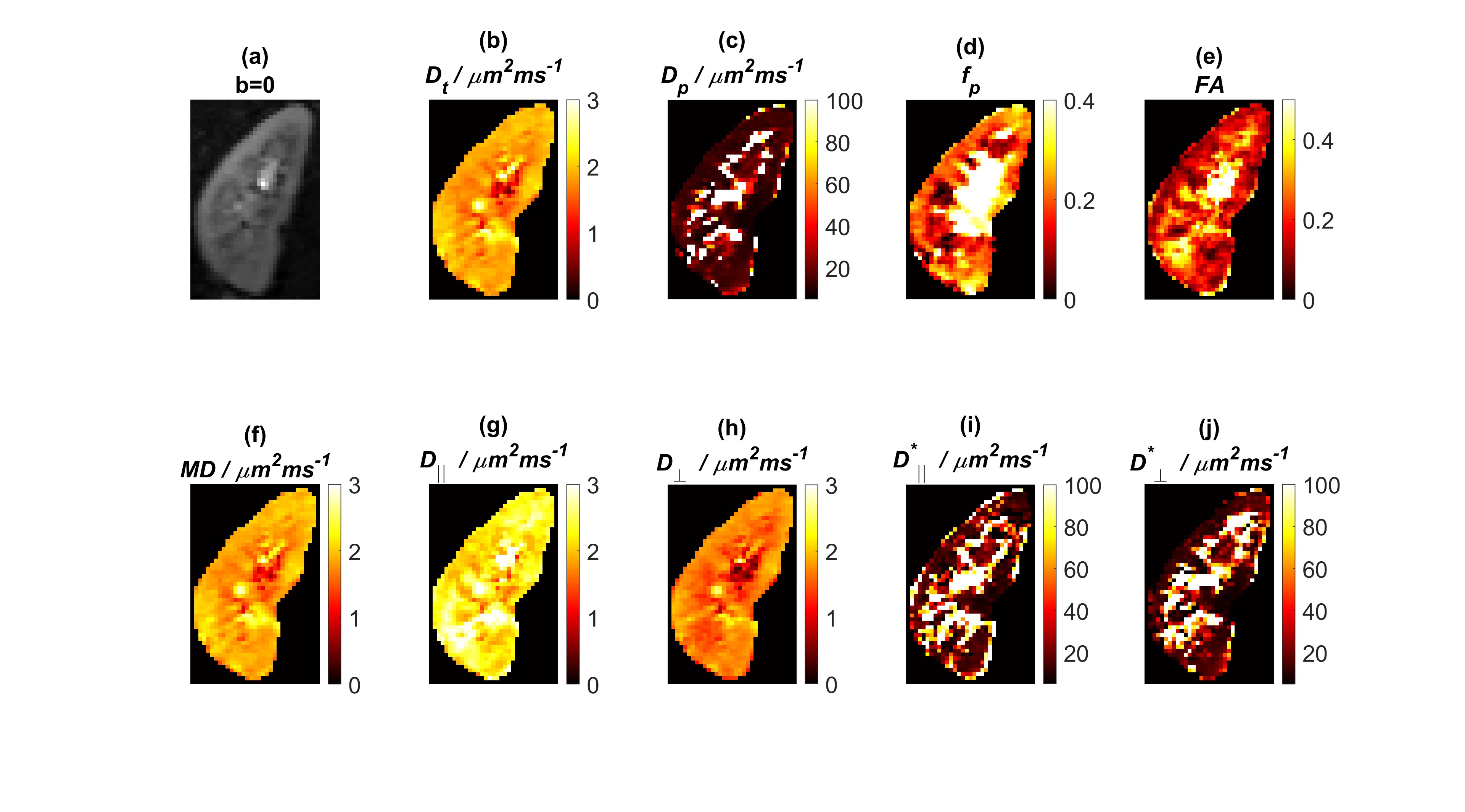
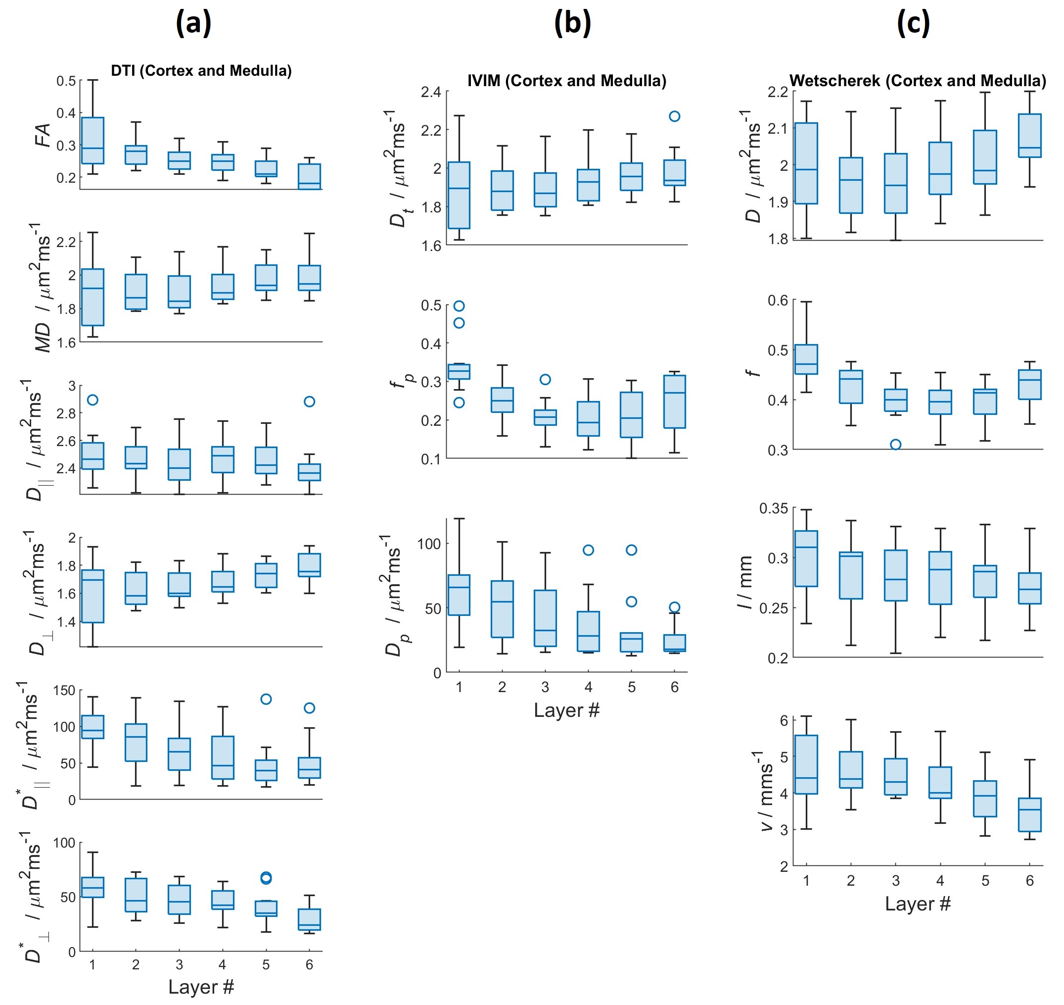
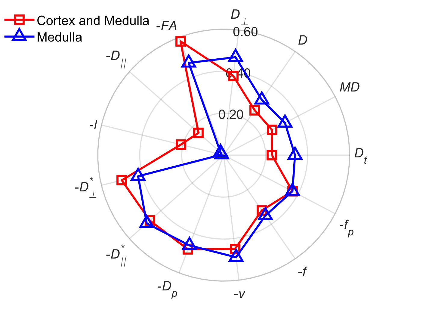
Fig. 1 is a sample kidney divided into 6 layers using the multiple zones concentric objects.Fig. 2 is sample maps derived from each of the three diffusion models for this case. Fig. 3 shows the variation of these parameters vs. kidney layers for all of the subjects in the study. Fig. 4 illustrates Spearman correlation coefficients for each parameter vs. kidney layer.The results indicate significant inner to outer increases in structural diffusivities, and significant inner to outer decreases in pseudodiffusivities and velocities.
Discussion
We studied the dependence of a set of conventional to more advanced DWI derived parameters on concentric layers in healthy human kidney. The most significant layer dependence was observed for pseudodiffusion parameters and structural fractional anisotropy, with weaker dependences observed for structural diffusivity parameters. Further validation of these trends in comparison with histologic reference, as well as correlation with measures of renal function, is required for maximal interpretation and utility.Acknowledgements
This work was supported by the National Institutes of Health (NIH).References
1. Basser, P.J. and C. Pierpaoli, Microstructural and physiological features of tissues elucidated by quantitative-diffusion-tensor MRI. Journal of Magnetic Resonance Series B, 1996. 111(3): p. 209- 219.
2. Le Bihan, D., et al., Separation of diffusion and perfusion in intravoxel incoherent motion MR imaging. Radiology, 1988. 168(2): p. 497-505.
3. Sigmund, E.E., et al., Cardiac Phase and Flow Compensation Effects on REnal Flow and Microstructure AnisotroPy MRI in Healthy Human Kidney. J Magn Reson Imaging, 2023. 58(1): p. 210-220.
4. Xie, L., et al., MRI tools for assessment of microstructure and nephron function of the kidney. Am J Physiol Renal Physiol, 2016. 311(6): p. F1109-F1124.
5. Afzali, M., et al., The sensitivity of diffusion MRI to microstructural properties and experimental factors. J Neurosci Methods, 2021. 347: p. 108951.
6. Milani, B., et al., Reduction of cortical oxygenation in chronic kidney disease: evidence obtained with a new analysis method of blood oxygenation level-dependent magnetic resonance imaging. Nephrol Dial Transplant, 2017. 32(12): p. 2097-2105
7. Zhao, K., et al., Diagnostic and prognostic performance of renal compartment volume and the apparent diffusion coefficient obtained from magnetic resonance imaging in mild, moderate and severe diabetic kidney disease. Quant Imaging Med Surg, 2023. 13(6): p. 3973-3987.
8. Sanmiguel Serpa, L.C., et al. A NEW METHOD TO ANALYSE RENAL PERFUSION: A PROOF OF CONCEPT. in Proc. Intl. Soc. Mag. Reson. 2023. Toronto, Canada.
9. Wetscherek, A., B. Stieltjes, and F.B. Laun, Flow-compensated intravoxel incoherent motion diffusion imaging. Magn Reson Med, 2015. 74(2): p. 410-9.
10. Führes T., et al. Impact of velocity- and acceleration-compensated encodings on signal dropout and black-blood state in diffusion-weighted magnetic resonance liver imaging at clinical TEs. PLoS One. 2023. Oct 5;18(10):e0291273.
Figures