2597
An Optimized Analytical Segmented (opAS) Approach to Extract Intravoxel Incoherent Motion (IVIM) Parameters1Department of Veterinary Clinical Sciences, University of Minnesota, Saint Paul, MN, United States, 2Center for Magnetic Resonance Research, University of Minnesota, Minneapolis, MN, United States
Synopsis
Keywords: IVIM, Perfusion, IVIM post processing
Motivation: Despite the IVIM model’s ability to noninvasively provide insight into tissue microstructure and perfusion, estimation of IVIM parameters in low-perfused tissues such as bone marrow remains a challenge.
Goal(s): In this work, we propose an optimized analytical segmented (opAS) approach for extracting IVIM diffusion (Ds) and perfusion (Df and f).
Approach: We compare the performance of this new approach against the analytical segmented (AS) method previously proposed for low-perfused tissues using numerical simulations and a piglet model.
Results: Our findings indicate that opAS outperforms AS in the estimation of all IVIM parameters (Ds, Df, and f), particularly in the low perfusion regime.
Impact: The proposed opAS method outperforms the prior AS method in estimating IVIM diffusion (Ds) and perfusion (Df and f) parameters, bringing IVIM a step closer to being a clinically useful non-contrast-enhanced technique to assess bone marrow perfusion.
Introduction
Intravoxel incoherent motion (IVIM) is a multi-b-value diffusion-weighted imaging (DWI) technique that uses a two-compartment model to simultaneously measure tissue diffusion coefficient (Ds), perfusion coefficient (Df), and perfusion fraction (f).1 IVIM has provided insight into various pathological conditions, mostly in highly perfused tissues and organs.2, 3 However, in low-perfused tissues, such as bone marrow, obtaining reliable IVIM measurements can be challenging due to the inherently weak perfusion signals,4 necessitating robust post-processing algorithms. The Analytical Segmented (AS) fitting approach has been shown to outperform other partial segmentation approaches in the low perfusion regime5, 6 and has been utilized to distinguish ischemic from perfused femoral heads in a piglet model of osteonecrosis.7 Nevertheless, AS performance relies on the choice of b threshold (bt) used to calculate Ds (i.e., b-value beyond which perfusion effects can be ignored), as error associated with the measurement of Ds propagates into the estimated perfusion parameters (Df and f). This work aims to propose an optimized analytical segmented (opAS) method to extract IVIM parameters and to compare its performance against the AS approach using numerical simulations and in vivo imaging of femoral head ischemia in the piglet model.Methods
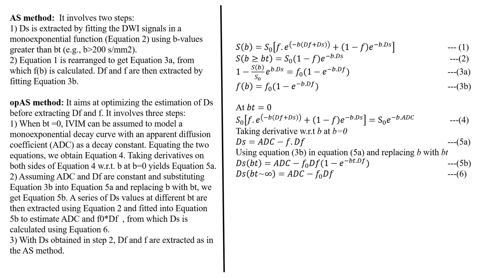
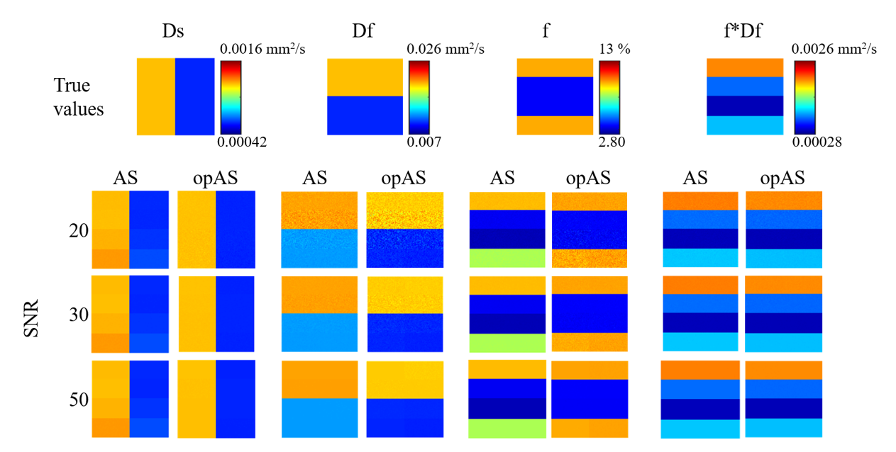
Theory: IVIM models signal attenuation due to the application of diffusion weightings (b-values) as a two-compartment model (Equation 1, Figure 1). The AS and proposed opAS approaches are described in Figure 1.Numerical Simulations: MRI signals were generated using Equation 1 for tissues with different combinations of low and high IVIM parameters. Using IVIM parameters previously reported for ischemic and perfused-control femoral heads of a piglet model,7 the following IVIM parameters were chosen: Ds=0.0006 and 0.0012 mm2/s; Df=0.01 and 0.02 mm2/s; and f=4 and 10%. Each IVIM parameter was defined in a 100x100 matrix resulting in 1250 numerical tissue samplings for each of eight low and high parameter combinations. 18 b-values were used, ranging from 0 to 500 s/mm2. Gaussian noise was added to generate SNR levels of 20, 30, and 50 at each sample.
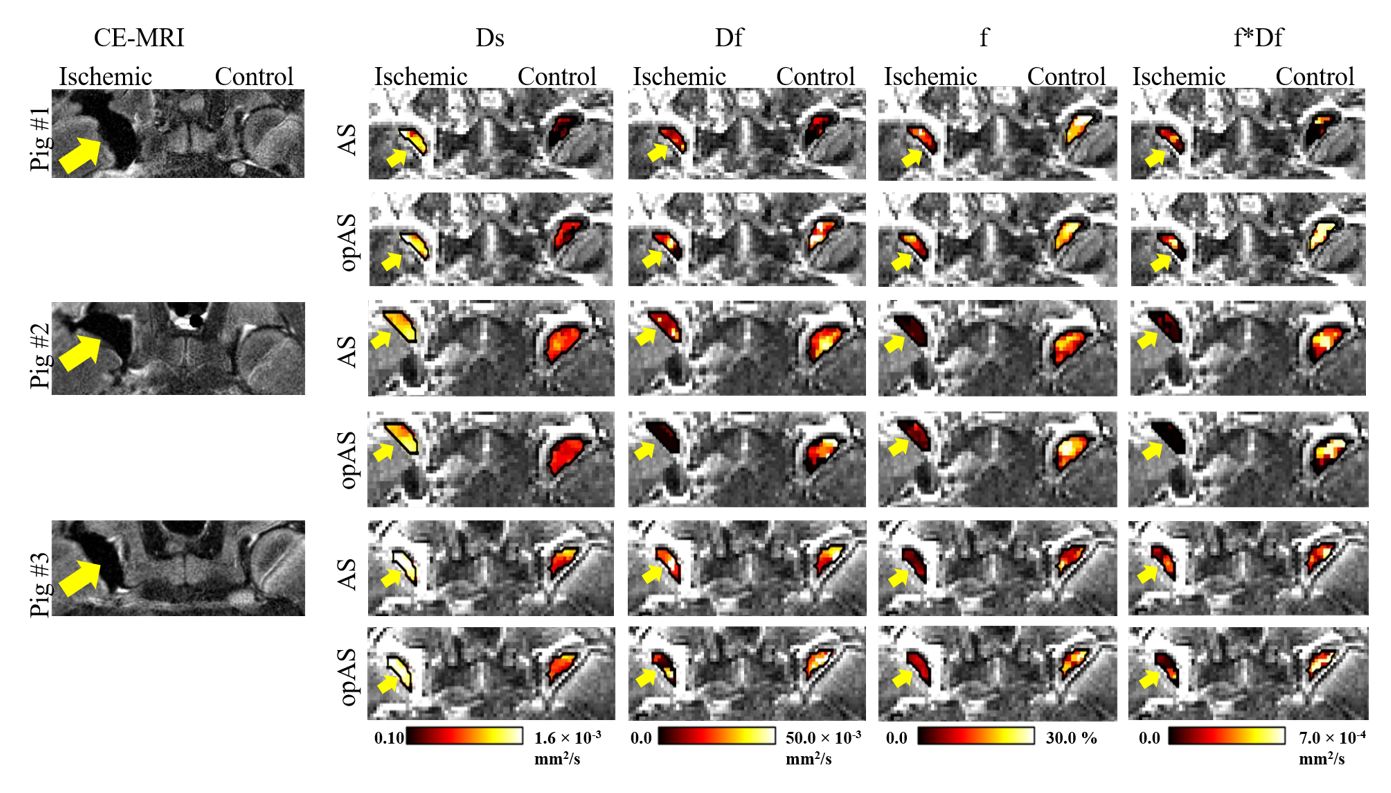
In Vivo 3T MRI of Piglet Model: We retrospectively analyzed IVIM data from a piglet model study of femoral head ischemia7 to compare the AS and opAS approaches. Data were included from 10 piglets that underwent surgery at six weeks of age to induce global ischemia in one of their femoral heads.8 The unoperated, contralateral femoral head served as a perfused control. One week after surgery, the bilateral hips of each piglet were imaged in vivo at 3T MRI using RESOLVE DWI with a subset 13 b-values used in the simulations (b=0,20,30,40,50,60,70,80,90,100,200,300,500 s/mm2), and lack of perfusion (ischemia) in the operated femoral heads was confirmed using gadolinium contrast-enhanced MRI (CE-MRI).
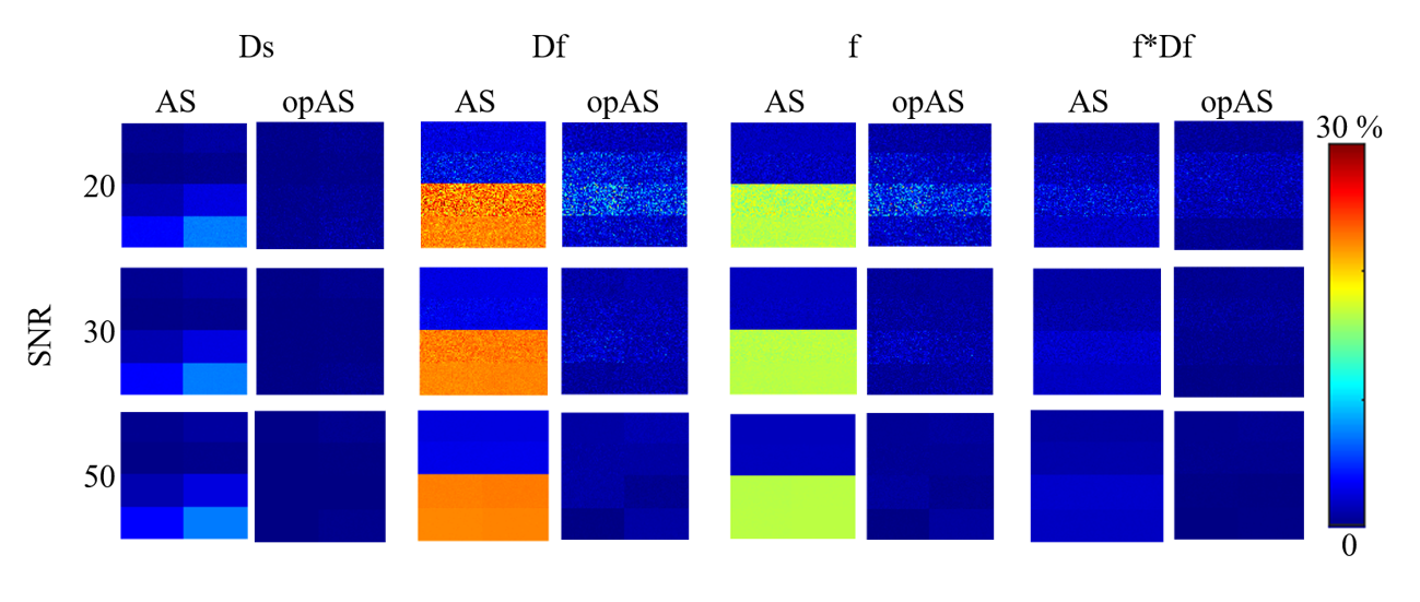
Data Analysis: IVIM parameters estimated from simulated data were observed against the true values, and absolute percentage error was calculated for each parameter. For the in vivo data, regions of interest constituting the bone and bone marrow of the operated and control femoral heads were manually drawn using the b=0 diffusion-weighted image. Measures of IVIM parameters in ischemic vs. control femoral heads were compared using the AS and opAS approaches.
Results
True and estimated IVIM parameters are shown in Figure 2, and their corresponding absolute percentage errors in Figure 3. opAS outperformed AS in estimating all IVIM parameters across the SNR range, with the greatest improvement occurring at low Df. opAS slightly improved the estimation of Ds. The estimated f*Df using either approach was comparable to true values. Both opAS and AS consistently showed an increase in Ds and a decrease in f in the ischemic vs. control femoral heads. For Df and f*Df, opAS showed a consistent decrease in ischemic vs. control femoral heads, while AS showed mixed outcomes (Figure 4).Discussion and Conclusion
Our simulation findings demonstrate that the opAS approach improves the estimation of all IVIM parameters at all SNR levels compared to the AS method. The results indicate that more accurate estimation of Ds significantly improves estimation of the perfusion parameters (Df, f, and f*Df). The opAS method was particularly effective at low Df values, which suggests that lower Df has an impact at relatively high b-values, leading to an overestimation of Ds and poor measurements of the perfusion parameters. opAS accounts for this by estimating Ds independent of bt. Our retrospective analysis of the piglet model data shows that more consistent IVIM parameter measurements can be achieved using the opAS vs. AS approach, further validating its superiority. This improvement further motivates clinical translation of IVIM mapping to simultaneously assess bone marrow diffusion (Ds) and perfusion (Df, f, and f*Df).Acknowledgements
This project was supported by NIH grants R01AR081877 and R56AR078315. The piglet model data were acquired with support from NIH grants K01AR070894, UL1TR002494, and P41EB027061.References
1. Le Bihan D, Breton E, Lallemand D, Aubin ML, Vignaud J, Laval-Jeantet M. Separation of diffusion and perfusion in intravoxel incoherent motion MR imaging. Radiology. 1988 Aug; 168(2):497-505.
2. Barbieri S, Donati OF, Froehlich JM, Thoeny HC. Impact of the calculation algorithm on biexponential fitting of diffusion‐weighted MRI in upper abdominal organs. Magnetic resonance in medicine. 2016 May; 75(5):2175-84.
3. Federau C. Measuring Perfusion: Intravoxel Incoherent Motion MR Imaging. Magn Reson Imaging Clin N Am. 2021 May; 29(2):233-242. doi: 10.1016/j.mric.2021.01.003. PMID: 33902905.
4. Meeus EM, Novak J, Withey SB, Zarinabad N, Dehghani H, Peet AC. Evaluation of intravoxel incoherent motion fitting methods in low‐perfused tissue. Journal of magnetic resonance imaging. 2017 May; 45(5):1325-34.
5. Buko E, Zhang J, Ajala A, Hor PH, and Muthupillai R. An analytical segmented (AS) approach for extracting intravoxel incoherent motion (IVIM) model parameters. Proc. 26th ISMRM Annual Meeting 2018; No. 5361.
6. Buko EO, Ajala A, Zhang J, Hor PH, and Muthupillai R. An analytical segmented approach for extracting TE independent perfusion fraction in intravoxel incoherent motion (IVIM) MRI. In: Proceedings of the 28th Annual Meeting of ISMRM, Virtual. Abstract 4393.
7. Buko EO, Bhave S, Tóth F, Johnson CP. IVIM Detects Bone Ischemia in a Piglet Model of Legg-Calvé-Perthes Disease. Proc. 31st Joint ISMRM-ESMRMB & ISMRT Annual Meeting 2022; No. 1598.
8. Kim HK, Su PH. Development of flattening and apparent fragmentation following ischemic necrosis of the capital femoral epiphysis in a piglet model. J Bone Joint Surg Am 2002; 84-A (8):1329-34.
Figures