2596
Effect of simultaneous multislice imaging and repetition time on biexponential liver intravoxel incoherent motion1Uniklinikum Erlangen, Erlangen, Germany, 2Siemens Healthineers AG, Erlangen, Germany
Synopsis
Keywords: IVIM, Diffusion/other diffusion imaging techniques
Motivation: Intravoxel incoherent motion (IVIM) measurements are often performed with various acquisition parameters among different studies. Previous studies showed differences in f during the application of simultaneous multislice (SMS).
Goal(s): The goal is to investigate the influence of SMS and the concomitant T1-effects due to the reduction of TR.
Approach: Diffusion-weighted images of the liver were acquired for conventional and SMS excitation with long and short TR each. Results were verified with a biophysical model.
Results: The evaluation indicates no dependence of IVIM parameters on SMS. Yet, the reduction of TR appears to be relevant for small gaps between slices in the simulations.
Impact: This study indicates that while comparing different IVIM studies, TR and gaps between slices must be taken into account, as they may substantially alter the measured f. IVIM parameters for SMS excitation are well comparable with conventional excitation, though.
Introduction
Intravoxel incoherent motion (IVIM) describes the initial signal decay at small b-values due to perfusion in diffusion-weighted imaging (DWI).1,2 The signal decay is often described as$$S(b)/S(b=0)=(1-f)\exp{(-bD)}+f\exp{(-bD^*)}$$
with the measured signal $$$S(b)$$$, the perfusion fraction $$$f$$$, the diffusion coefficient $$$D$$$, and the pseudodiffusion coefficient $$$D^*$$$.
The simultaneous multislice imaging (SMS) technique3 significantly shortens scan time4-7 by enabling the excitation of multiple slices. Recently, several studies employed SMS for IVIM imaging applications.8-11 Some of them observed a significant decrease of $$$f$$$ from conventional to SMS accelerated acquisitions.8,9,11 So far, it is ambiguous whether this effect is intrinsically caused by the SMS technique itself or due to T1-effects. The mentioned studies made use of the scan time acceleration and drastically reduced TR for the SMS acquisition. With T1 times of blood between 1,300 ms12 and 1,900 ms13, large reductions of TR may have a major impact on $$$f$$$.14
For this reason, the influence of both SMS and TR on biexponential IVIM parameters in the liver were investigated.
Methods
Diffusion-weighted images of the liver were acquired at 3T (MAGNETOM Prisma, Siemens Healthineers, Erlangen, Germany) of ten healthy volunteers. Image acquisition was performed with a short TR (1,300 ms, prefix s) for conventional slice excitation (sAF1) and SMS excitation with acceleration factor three (sAF3). Similarly, a long TR (4,500 ms, prefix l) was used for both modalities (lAF1 and lAF3). Nine 5 mm thick transversal slices were acquired at 19 b-values between 0 and 800 s/mm² and at three orthogonal diffusion directions each.Regions of interest (ROI) were defined in the parenchyma of the liver on the $$$b=0$$$ images and transferred to the remaining diffusion-weighted images in MITK v2021.10 (Medical Imaging Interaction Toolkit).
The subsequent evaluation was performed in Python 3.9. Median values of the ROIs were computed over all voxels for each slice, b-value, and diffusion direction and were normalized to the median at $$$b=0$$$. These values were arithmetically averaged over diffusion directions and the same nominal b-values. The biexponential IVIM equation was fitted in a segmented manner14-17 to the median signal at all b-values. Data points were weighted in the fit according to the number of averages.
Statistical analysis was performed with the Kruskal-Wallis test. In order to validate the measurements and compare it to the literature, a biophysical model was developed accounting for in- and outflow effects of blood magnetization and the resulting T1-effects depending on the gap between slices, TR and blood flow velocities, similarly to a previous study.14 In this way, $$$f$$$ can be modeled depending on the blood flow velocity $$$v_0$$$. Computations were performed with 5 mm gaps between slices and zero gaps between slices. T1 of blood was set to 1,300 ms.12
Results
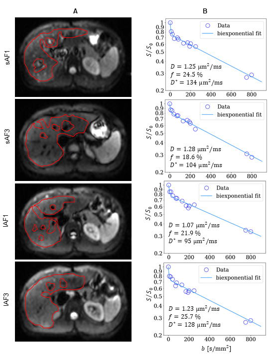
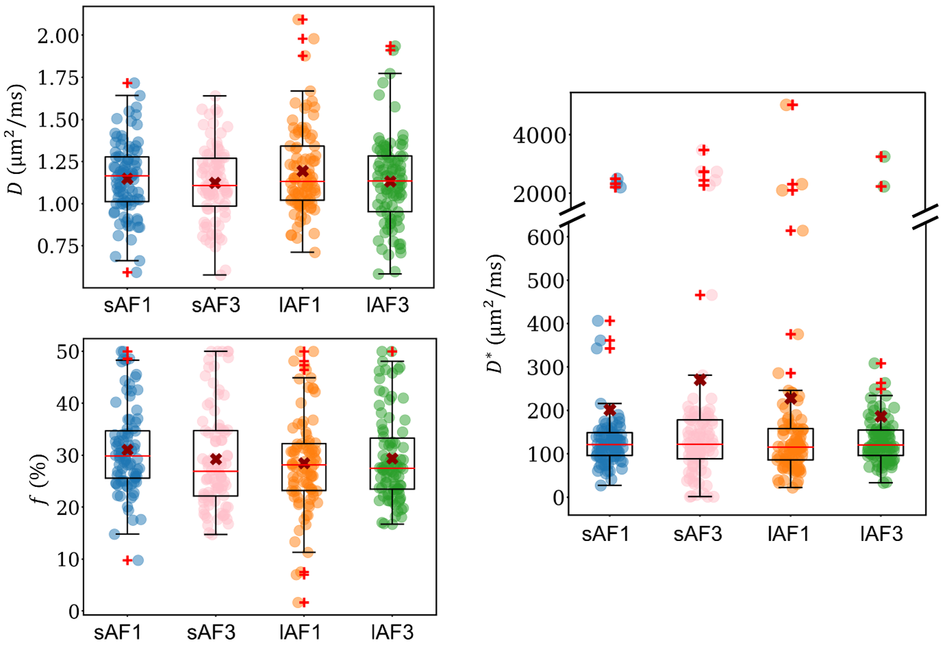
Figure 1 illustrates representative diffusion-weighted images for 130 s/mm² with respective segmentation and IVIM curves.Figure 2 shows the boxplots of the biexponential IVIM parameters. No significant differences between the four modalities were observed. The p-values were 0.553 ($$$D$$$), 0.122 ($$$f$$$) and 0.856 ($$$D^*$$$).
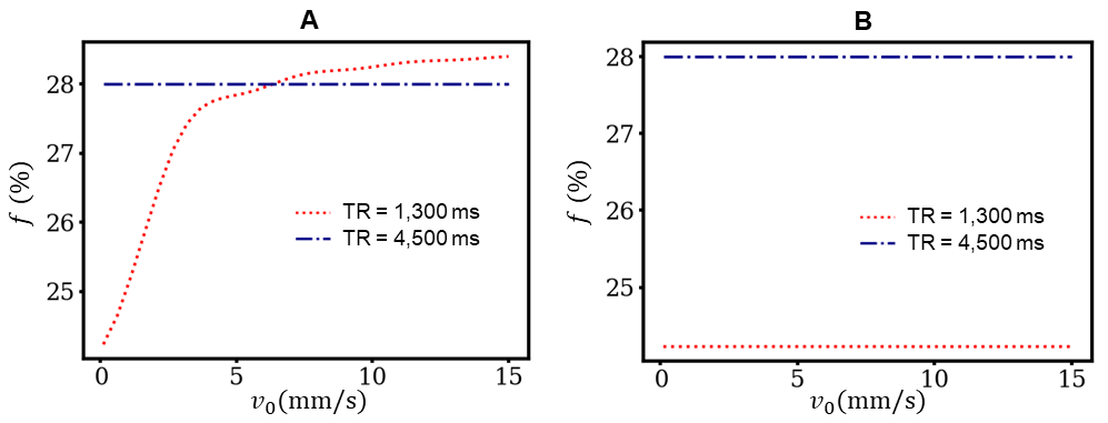
Figure 3 shows the results from the biophysical simulations. For 5 mm gaps between slices (Figure 3A), $$$f$$$ remains constant for the given $$$v_0$$$ for TR = 4,500 ms, while it is monotonically increasing for TR = 1,300 ms with increasing $$$v_0$$$. From $$$v_0=2~\mathrm{mm/s}$$$ upwards, the measured values for $$$f$$$ are comparable for long and short TR. With no gaps in between slices (Figure 3B), both computed $$$f$$$ values remain constant over $$$v_0$$$, but with a noteworthy difference (28 % vs 24 %).
Discussion
Our study neither showed a dependency on SMS excitation nor on TR. This is in contradiction to previous studies.In this work, we used much fewer slices and larger gaps between slices than commonly used in the literature. In this way, we reduced the effect of in- and outflowing blood magnetization.
However, pre-saturated blood magnetization flowing from neighboring slices may have a substantial influence at small slice gaps and small TR as confirmed by the biophysical model. Large enough slice gaps contain fully relaxed blood magnetization that can flow into the measured slice for appropriate $$$v_0$$$. In the case of no gaps between slices, a considerable amount of blood is already pre-saturated when being measured again.
This likely explains the mismatch between previous studies and the importance of considering TR and the distance between slices.
Conclusion
The biexponential IVIM parameters were not found to have any significant dependency on the slice excitation mode and on TR for the given measurement protocol. However, small distances between slices might considerably influence $$$f$$$ at small TR affirmed by biophysical computations.Acknowledgements
Funding by the Deutsche Forschungsgemeinschaft is gratefully acknowledged (DFG project 446875476).References
1. Le Bihan D, Breton E, Lallemand D, Grenier P, Cabanis E, Laval-Jeantet M. MR imaging of intravoxel incoherent motions: application to diffusion and perfusion in neurologic disorders. Radiology. Nov 1986;161(2):401-7. doi:10.1148/radiology.161.2.3763909
2. Le Bihan D, Breton E, Lallemand D, Aubin ML, Vignaud J, Laval-Jeantet M. Separation of diffusion and perfusion in intravoxel incoherent motion MR imaging. Radiology. Aug 1988;168(2):497-505. doi:10.1148/radiology.168.2.3393671
3. Larkman DJ, Hajnal JV, Herlihy AH, Coutts GA, Young IR, Ehnholm G. Use of multicoil arrays for separation of signal from multiple slices simultaneously excited. J Magn Reson Imaging. Feb 2001;13(2):313-7. doi:10.1002/1522-2586(200102)13:2<313::aid-jmri1045>3.0.co;2-w
4. Ohlmeyer S, Laun FB, Palm T, et al. Simultaneous multislice echo planar imaging for accelerated diffusion-weighted imaging of malignant and benign breast lesions. Investigative Radiology. 2019;54(8):524-530.
5. Manoliu A, Ho M, Piccirelli M, et al. Simultaneous multislice readout‐segmented echo planar imaging for accelerated diffusion tensor imaging of the mandibular nerve: a feasibility study. Journal of Magnetic Resonance Imaging. 2017;46(3):663-677.
6. Sotiropoulos SN, Jbabdi S, Xu J, et al. Advances in diffusion MRI acquisition and processing in the Human Connectome Project. Neuroimage. 2013;80:125-143.
7. Barth M, Breuer F, Koopmans PJ, Norris DG, Poser BA. Simultaneous multislice (SMS) imaging techniques. Magn Reson Med. Jan 2016;75(1):63-81. doi:10.1002/mrm.25897
8. Tang C, Qin Y, Hu Q, Ai T. Diagnostic value of multi-model high-resolution diffusion-weighted MR imaging in breast lesions: Based on simultaneous multi-slice readout-segmented echo-planar imaging. Eur J Radiol. Sep 2022;154:110439. doi:10.1016/j.ejrad.2022.110439
9. Phi Van VD, Becker AS, Ciritsis A, Reiner CS, Boss A. Intravoxel Incoherent Motion Analysis of Abdominal Organs: Application of Simultaneous Multislice Acquisition. Invest Radiol. Mar 2018;53(3):179-185. doi:10.1097/RLI.0000000000000426
10. Xu H, Zhang N, Yang DW, et al. Scan Time Reduction in Intravoxel Incoherent Motion Diffusion-Weighted Imaging and Diffusion Kurtosis Imaging of the Abdominal Organs: Using a Simultaneous Multislice Technique With Different Acceleration Factors. J Comput Assist Tomogr. Jul-Aug 01 2021;45(4):507-515. doi:10.1097/RCT.0000000000001189
11. Boss A, Barth B, Filli L, et al. Simultaneous multi-slice echo planar diffusion weighted imaging of the liver and the pancreas: Optimization of signal-to-noise ratio and acquisition time and application to intravoxel incoherent motion analysis. Eur J Radiol. Nov 2016;85(11):1948-1955. doi:10.1016/j.ejrad.2016.09.002
12. van Leeuwen FHP, Lena B, Zwanenburg JJM, et al. Detecting low blood concentrations in joints using T1 and T2 mapping at 1.5, 3, and 7 T: an in vitro study. Eur Radiol Exp. Dec 2 2021;5(1):51. doi:10.1186/s41747-021-00251-z
13. Stanisz GJ, Odrobina EE, Pun J, et al. T1, T2 relaxation and magnetization transfer in tissue at 3T. Magn Reson Med. Sep 2005;54(3):507-12. doi:10.1002/mrm.20605
14. Loh M, Führes T, Stuprich C, Uder M, Saake M, Laun FB. Influence of saturation effects on biexponential liver intravoxel incoherent motion. Magn Reson Med. Mar 2 2023;doi:10.1002/mrm.29622
15. Führes T, Riexinger AJ, Loh M, et al. Echo time dependence of biexponential and triexponential intravoxel incoherent motion parameters in the liver. Magn Reson Med. Feb 2022;87(2):859-871. doi:10.1002/mrm.28996
16. Patel J, Sigmund EE, Rusinek H, Oei M, Babb JS, Taouli B. Diagnosis of cirrhosis with intravoxel incoherent motion diffusion MRI and dynamic contrast-enhanced MRI alone and in combination: preliminary experience. J Magn Reson Imaging. Mar 2010;31(3):589-600. doi:10.1002/jmri.22081
17. Sigmund EE, Vivier PH, Sui D, et al. Intravoxel incoherent motion and diffusion-tensor imaging in renal tissue under hydration and furosemide flow challenges. Radiology. Jun 2012;263(3):758-69. doi:10.1148/radiol.12111327
Figures