2589
Comparison of MUSE-DWI and Conventional DWI in the Application of Invasive Breast Cancer and Malignancy Grade Prediction: A Comparative Study1Weifang medical university, Weifang, China, 2Weifang People‘s Hosptial, Weifang, China, 3GE Healthcare China, beijing, China
Synopsis
Keywords: DWI/DTI/DKI, Cancer
Motivation: MUSE-DWI was used to better display breast cancer and histological grade prediction.
Goal(s): This study aim to compare MUSE-DWI and conventional DWI for evaluating invasive breast cancer lesions, focusing on ADC values for preoperative histological grading.
Approach: It included 63 confirmed lesions, assessed for qualitative parameters like sharpness, artifacts, and distortion, and quantitative measures including SNR and ADC. The results demonstrated that MUSE-DWI significantly reduces artifacts and distortions, enhancing image quality. Moreover, it showed higher diagnostic efficacy in the preoperative histological grading of breast cancer.
Results: MUSE-DWI was superior to SS-EPI-DWI in image display and histological grade prediction.
Impact: Compared with SS-EPI-DWI, MUSE-DWI can display the lesions of invasive breast cancer more intuitively, and it has higher robustness to the histological grade of invasive breast cancer.
Introduction
Breast cancer is the leading fatal cancer among women, comprising 30% of all new cancer diagnoses and 15% of female cancer deaths. The variation in clinical treatment and prognosis is significant among breast cancer patients [1]. Histological grading, recognized as a key surrogate marker in various malignancies, is crucial in invasive breast cancer [2]. Traditional histopathological grading relies on invasive methods like surgery or biopsy [3]. Diffusion-weighted imaging (DWI) offers a non-contrast MRI technique for breast cancer detection [4]. It quantitatively assesses lesions by calculating Apparent Diffusion Coefficient (ADC) values [5]. Traditional DWI, however, is limited to morphological lesion analysis, often challenged by artifacts and distortions. This affects radiologists’ ability to precisely analyze lesion morphology. Multiple Sensitivity Encoding DWI (MUSE-DWI) is a groundbreaking technique, extending existing sensitivity encoding. It utilizes interleaved trajectory k-space, enhancing signal-to-noise ratio (SNR) and spatial resolution without navigational pulses [6]. This study explores MUSE-DWI's role in breast cancer, particularly in preoperative histological grading. It aims to assess whether MUSE-DWI can outperform traditional DWI in diagnostic efficacy for histological grading of invasive breast cancer. This innovative approach could revolutionize breast cancer diagnosis and treatment strategies, offering more accurate and non-invasive alternatives to current methods.Materials and Methods
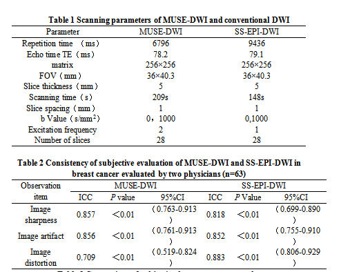
SubjectsA total of 63 lesions confirmed as invasive breast cancer by surgical or biopsy pathology were recruited. Patients' ages ranged from 26 to 73 years, with an average of 51.56 ± 10.54 years. Diagnoses included Grade I (n=2), Grade II (n=33), Grade III (n=28). Due to few grade I cases and no significant survival or recurrence differences between grades I and II, these were combined into a low-grade group, while grade III was high-grade [7]. MRI Imaging MRI experiments used a 3.0T scanner (SIGNATM Architect, GE) with an 8-channel breast coil. Patients, positioned prone with breasts in the coil and arms raised, underwent feet-first imaging. Image reconstruction for b=0s/mm² and b=1000s/mm² was automatically completed. Specific parameters are in Table 1. Data Analysis Two physicians with 5-10 years of breast MR diagnosis experience, respectively, analyzed images.Employing a 4-point scale, the two physicians independently assessed the image clarity, distortion, and artifacts across all DWI sequences, while remaining blind to each other's evaluations. ADC maps were generated on a GE ADW4.7 workstation. In each patient, ROIs on lesions in ADC maps were identified by both radiologists. SNR was calculated as average signal intensity / standard deviation of noise.Statistical Analysis Statistical analysis was performed using SPSS version 26.0 software. Normally distributed continuous data were expressed as "x±s," and non-normally distributed data were expressed as interquartile range. Paired t-tests were used for parametric tests, and Wilcoxon signed-rank tests were used for non-parametric tests.The intraclass correlation coefficient between the two radiologists was evaluated according to the following interpretation: a value of 0.75–1.00 was considered excellent agreement, 0.60–0.74 was considered good agreement, 0.40–0.59 was considered fair agreement, and less than 0.40 was considered poor agreement [8].Receiver Operating Characteristic (ROC) curves were used to evaluate the diagnostic performance of ADC values derived from MUSE-DWI and conventional DWI in stratifying the level of invasive breast cancer. The area under the curve (AUC) was calculated. A p-value of less than 0.05 was considered statistically significant.Results
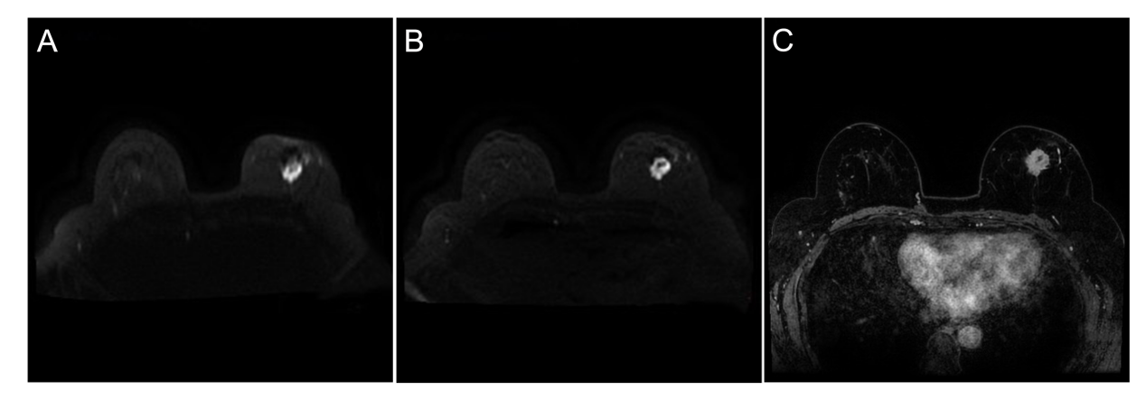
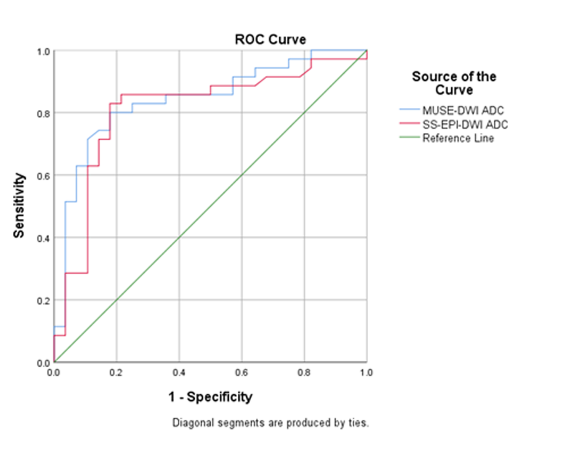
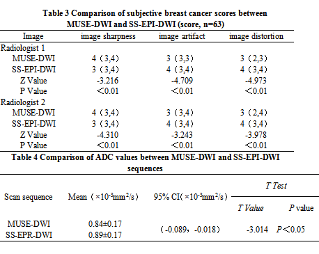
The interobserver agreement between the two experienced radiologists was robust.These results are summarized in Table 2. Qualitative Results Regarding image clarity, both radiologists gave higher ratings to MUSE-DWI compared to SS-EPI-DWI (z = -3.216, P < 0.01; z = -4.310, P < 0.01). Conversely, MUSE-DWI received lower scores for image artifacts (z = -4.709, P < 0.01; z = -3.243, P < 0.01) and distortion (z = -4.973, P < 0.01; z = -3.978, P < 0.01) compared to SS-EPI-DWI. These results are summarized in Table 3 and illustrated in Figure 1. Quantitative Results All the lesions demonstrated solid-enhancement morphology. Notably, the ADC values derived from MUSE-DWI (0.84 ± 0.17) were significantly lower than those from SS-EPI-DWI (0.89 ± 0.17) (P < 0.05), as presented in Table 4. Additionally, the ROC curve analysis revealed that the AUC values for predicting the grade grouping of invasive breast cancer were 0.841 for MUSE-DWI ADC values and 0.809 for SS-EPI-DWI ADC values, indicating a statistically significant difference (P < 0.05). This data is visually represented in Figure 2.Discussion and Conclusions
The results demonstrate that MUSE-DWI images have higher resolution, less distortion, and fewer artifacts. Both qualitative and quantitative results from this study revealed that the performance of MUSE-DWI in terms of image deformation was superior to that of SS-EPI-DWI. In conclusion, MUSE-DWI is feasible and can be seamlessly integrated into routine clinical breast MRI protocols.Acknowledgements
No Acknowledgements fundReferences
1. Veronesi U, Boyle P, Goldhirsch A, Orecchia R, Viale G. Breast cancer. Lancet. 2005;365(9472):1727-1741. 1. Rakha EA, El-Sayed ME, Lee AH, et al. Prognostic significance of Nottingham histologic grade in invasive breast carcinoma. J Clin Oncol. 2008;26(19):3153-3158.3.Dalton LW, Page DL, Dupont WD. Histologic grading of breast carcinoma. A reproducibility study. Cancer. 1994;73(11):2765-2770.4.Partridge SC, Nissan N, Rahbar H, Kitsch AE, Sigmund EE. Diffusion-weighted breast MRI: Clinical applications and emerging techniques. J Magn Reson Imaging. 2017;45(2):337-355.5.Spick C, Bickel H, Pinker K, et al. Diffusion-weighted MRI of breast lesions: a prospective clinical investigation of the quantitative imaging biomarker characteristics of reproducibility, repeatability, and diagnostic accuracy. NMR Biomed. 2016;29(10):1445-1453.6.Baxter GC, Patterson AJ, Woitek R, Allajbeu I, Graves MJ, Gilbert F. Improving the image quality of DWI in breast cancer: comparison of multi-shot DWI using multiplexed sensitivity encoding to conventional single-shot echo-planar imaging DWI. Br J Radiol.2021;94(1119):20200427.7.Takahashi H, Oshi M, Asaoka M, Yan L, Endo I, Takabe K. Molecular Biological Features of Nottingham Histological Grade 3 Breast Cancers. Ann Surg Oncol. 2020;27(11):4475-4485.8.Cicchetti D V.Guidelines,Criteria,and Rules of Thumb for Evaluating Normed and Standardized Assessment Instruments in Psychology[J].Psychological Assessment,1994,6(4).Figures