2586
Application of 5 diffusion models in bladder cancer staging based on the date of diffusion spectrum imaging1Affiliated Cancer Hospital of Zhengzhou University; Henan Cancer Hospital, Zhengzhou, China, 2Radiology, Affiliated Cancer Hospital of Zhengzhou University; Henan Cancer Hospital, Zhengzhou, China, 3Siemens Healthcare, Shanghai ,China, Shanghai, China
Synopsis
Keywords: Diffusion Modeling, Diffusion/other diffusion imaging techniques
Motivation: The pathological grade of bladder cancer is closely related to the choice of treatment and the prognosis1. Diffusion spectrum imaging (DSI) is a newly developed model, which is helpful in distinguish benign and malignant tumors2.
Goal(s): This study was to explore the quantitative parameter from DSI in differentiating pathology grade of bladder cancer.
Approach: The differences of DSI quantitative parameter between high-grade and low-grade lesions and the differentiation performance of the indices were evaluated .
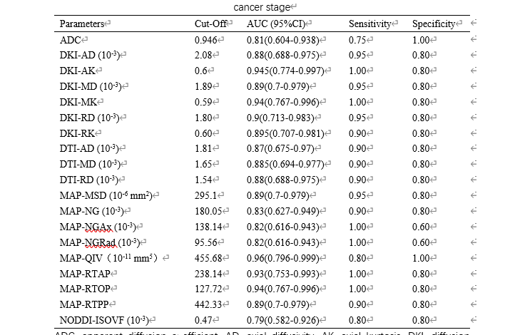
Results: Quantitative parameters of DSI could effectively distinguish pathology grade of bladder cancer, and AUC ranged from 0.790 to 0.960.
Impact: The quantitative parameters obtained from Diffusion spectrum imaging are more stable and can predict the pathological grade of bladder cancer better than traditional ADC.
Introduction
Prognosis and treatment strategies differs between high-grade and low-grade bladder cancer, especially non-muscle-invasive bladder cancer3-4. Therefore, it is necessary to judge the pathological grade of bladder cancer in patients before surgery to help clinicians develop individualized treatment plans for patients. DSI is a model that uses multiple b-values and gradient directions to sample the diffusion signal of water molecules in the entire q-space, and quantitatively estimates it through a probability density function, which is mathematically and physically superior to other diffusion MRI2. It has been used in the diagnosis of central nervous system and breast diseases, but has not been reported in the bladder. To prospectively investigate the value of quantitative parameters derived from diffusion spectrum imaging (DSI) in preoperatively predicting the staging in patients with bladder cancer.Methods and Materials
A total of 25 patients with bladder cancer were included in this study, and each patient was categorized as having either High-grade (20 cases) or low-grade (5 cases) urothelial cancer based on the pathologic result. All patients underwent DSI and conventional MRI including diffusion weighted imaging (DWI). The MR scanning were performed on a 3T MR scanner (Magnetom Prisma, Siemens Healthineers, Erlangen, Germany). The parametric results of 5 different DWI models were calculated using an in-house prototype software developed by MRStation. The tumor size, apparent diffusion coefficient (ADC), and quantitative parameters derived from DSI, such as diffusion tensor imaging (DTI), diffusion kurtosis imaging (DKI), mean apparent propagator (MAP), and neurite orientation dispersion and density imaging (NODDI), were measured and compared between the two groups using T test or rank sum test. The discriminative ability of these quantitative parameters was assessed using receiver operating characteristic (ROC) curve analyses.Results
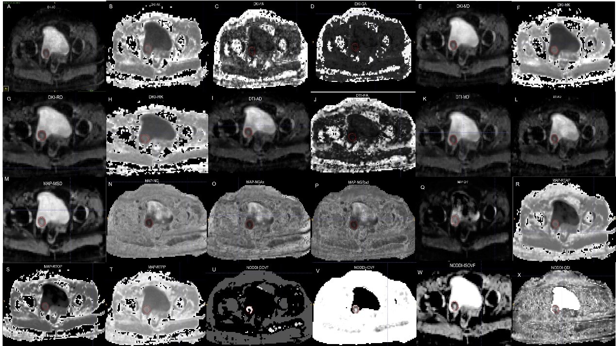
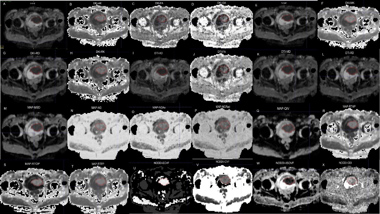
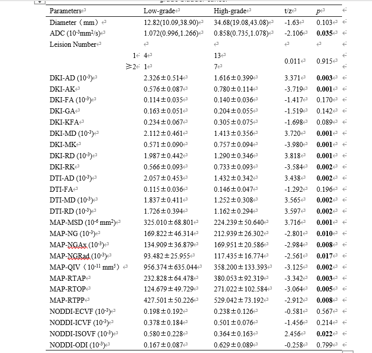
ADC showed a significant difference between the two groups, while the tumor size and lesion number did not. Several DSI quantitative parameters, including axial kurtosis (DKI_AK), mean kurtosis of DKI(DKI-MK), non-Gaussianity (MAP_NG), axial non-Gaussianity (MAP_NGAx), radial non-Gaussianity (MAP_NGRad), return-to-origin probability (MAP_RTOP), return-to-plane probability (MAP_RTPP), return-to-axis probability of MAP (MAP_RTAP) were lower in the low-grade group than in the high-grade group (p ≤ 0.05). In contrast, other DSI quantitative parameters, including axial diffusivity (DKI_AD), mean diffusivity (DKI_MD), radial diffusivity of DKI (DKI_RD), axial diffusivity (DTI_AD), mean diffusivity (DTI_MD), radial diffusivity of DTI (DTI_RD), mean squared diffusion (MAP_MSD), and q-space inverse variance (MAP_QIV), volume fraction of the isotropic compartment of NODDI (NODDI-ISOVF) were higher in the low-grade group than in the high-grade group (p ≤ 0.05). The AUC values of DSI quantitative parameters ranged from 0.790 to 0.960, and most of these values were higher than that of ADC(AUC=0.81).Discussion
The kurtosis parameter is related to the complexity of organizational structures within the region of interest. The more complex the biological tissue structure within the imaging voxel, the greater deviation from a Gaussian distribution in water molecule diffusion, resulting in a higher kurtosis value. The apparent diffusion coefficient (ADC) value, which has been corrected for non-Gaussian distribution, represents the displacement distance of water molecules during unit time and reflects both the overall diffusion level and resistance within tissue. DKI-AD, DKI-MD, DKI-RD were higher in low-grade group than in high-grade group, and the difference between the two groups was statistically significant. DKI-AK, DKI-MK, DKI-RK were higher in high-grade group, the disparity between the two groups demonstrated a statistically significant difference. The results of this study are consistent with previous research findings. Compared to low-grade bladder cancer, high-grade bladder cancer cells exhibit increased proliferation, significant nuclear atypia, reduced extracellular space, restricted diffusion of water molecules between cells, and some high-grade lesions may present necrosis, resulting in a more complex tissue composition.Conclusion
Quantitative parameters from 5 different diffusion models can be helpful for preoperative prediction of bladder cancer grade, which could lead to improved patient outcomes and management.Acknowledgements
No acknowledgement found.References
1. Wang F, Xu Y, Xiang Y, et al. The feasibility of amide proton transfer imaging at 3 T for bladder cancer: a preliminary study. Clin Radiol. 2022 Oct;77(10):776-783. doi: 10.1016/j.crad.2022.07.002. Epub 2022 Aug 16. PMID: 35985845.
2. Mao C, Jiang W, Huang J, et al. Quantitative Parameters of Diffusion Spectrum Imaging: HER2 Status Prediction in Patients With Breast Cancer. Front Oncol. 2022 Feb 3;12:817070. doi: 10.3389/fonc.2022.817070. PMID: 35186753; PMCID: PMC8850631.
3. Cai Q, Wen Z, Huang Y, et al. Investigation of Synthetic Magnetic Resonance Imaging Applied in the Evaluation of the Tumor Grade of Bladder Cancer. J Magn Reson Imaging. 2021 Dec;54(6):1989-1997. doi: 10.1002/jmri.27770. Epub 2021 Jun 3. PMID: 34080268.
4. Wang HJ, Cai Q, Huang YP, et al. Amide Proton Transfer-weighted MRI in Predicting Histologic Grade of Bladder Cancer. Radiology. 2022 Oct;305(1):127-134. doi: 10.1148/radiol.211804. Epub 2022 Jun 28. Erratum in: Radiology. 2022 Oct;305(1):E59. PMID: 35762886.
Figures