2582
Diffusion Weighted Imaging for The Pathological Classification of Parotid Gland Tumors1Department of Medical Imaging, Affiliated Hospital of Guizhou Medical University, Guiyang, China, 2Department of Medical Imaging, Tongren City People's Hospital, Tongren, China, 3Philips Healthcare, Shanghai, China
Synopsis
Keywords: DWI/DTI/DKI, Tumor
Motivation: Parotid gland tumors require various treatments, and it is essential to assess malignancy before surgery.
Goal(s): To evaluate the effectiveness of diffusion-weighted imaging in diagnosing salivary gland tumors.
Approach: In a retrospective study, Conventional MRI (cMRI) and DWI data from 85 cases were analyzed. ADC values were measured and compared among different tumor types. Salivary gland tumors are classified into three categories: pleomorphic adenoma (PA), Warthin tumor, and malignant tumors.
Results: ADC values successfully distinguish PA from other tumors, but they do not differentiate between benign and malignant tumors. Lower ADC values are associated with higher grades of malignancy.
Impact: DWI, a non-invasive imaging technique, provides critical insights into the cellular structure of tissues, aiding in the differentiation of benign and malignant thyroid nodules. It enhances diagnostic accuracy, reduces unnecessary biopsies, and contributes to the optimization of treatment strategies.
Introduction
Salivary gland tumors, which make up 3–6% of head and neck neoplasms, require various treatment approaches. The most common type is pleomorphic adenoma (PA), which is primarily treated with surgical removal [1, 2]. Warthin's tumor (WT), the second most common type, often undergoes conservative management due to its low potential for malignancy. In cases of deep-seated or highly malignant tumors (MT), total parotidectomy and neck dissection may be necessary. Accurate assessment of malignancy before surgery is crucial. Conventional MRI (cMRI) provides information about the shape, while diffusion-weighted imaging (DWI) is excellent for evaluating the cellular structure and determining malignancy[3-5]. Ultrasound has limited reproducibility and concerns about diagnostic performance. This study aims to predict the pathology of salivary gland tumors before surgery by analyzing the apparent diffusion coefficient (ADC) values, with the goal of improving diagnostic accuracy.Methods
After obtaining approval from the Ethics Committee, we conducted a retrospective analysis of cases involving salivary gland tumors that underwent preoperative cMRI and DWI at our institution from November 2015 to September 2021. Pathological results were obtained for a total of 85 cases, which were then divided into three groups (PA, WT, and MT) based on specific criteria. The MRI scans were performed using 1.5T (Philips Achieva) and 3.0T (Philips Ingenia CX ) scanners equipped with head and neck array coils. The imaging sequences included T1WI, T2WI, fat-suppressed T2WI, coronal T2WI, contrast-enhanced T1WI, and DWI with b-values of 0 and 1000 mm2/s. ADC maps were automatically generated, and ADC values were independently obtained from non-necrotic areas using a double-blind approach. The signal intensities and structural features of T1WI and T2WI were documented. For data analysis, we utilized SPSS software. Continuous variables such as ages and ADC values underwent Kruskal-Wallis analysis with post-hoc comparisons. Receiver Operating Characteristic (ROC) curve analysis was employed to evaluate the imaging sensitivity and specificity in predicting histological characteristics. A significance level of P<0.05 was considered statistically significant.Results
The study included 54 male and 31 female patients, with an average age of 53.0±15.1 years. Among them, 35 cases were diagnosed as PA, 31 as WT, and the remaining 19 patients had seven different types of malignant tumors. It is worth noting that the PA group mainly consisted of younger female patients, while the malignant tumor group mainly consisted of elderly individuals. The mean ADC values on DWI for the PA, WT, and MT groups were 1.53±0.29, 0.77±0.20, and 1.06±0.19 (×10-3 mm2/s), respectively (figure 1). The difference between the PA group and the other two groups was statistically significant (P < 0.001). In the ROC analysis, the area under the curve (AUC) values for PA and WT were 0.987 and 0.935, respectively(figure 2). While there was a statistically significant difference in average ADC values between WT and MT, there was a significant overlap in the range, resulting in an AUC of only 0.156.Discussion
This study reveals that ADC values are highly valuable for distinguishing the most common salivary gland tumor, PA, from other histopathological conditions, rather than for discriminating between benign and malignant lesions. The use of ADC values proves effective in distinguishing PA from both WT and MT, with excellent diagnostic performance. However, ADC values are not effective in differentiating between WT and MT. In general, low-grade malignant tumors tend to have relatively higher ADC values, while high-grade malignant tumors have lower ADC values, reflecting the structural characteristics of tumor cells [6, 7]. It is worth noting that lymphomas typically exhibit lower ADC values compared to other malignant tumors [8]. These features can be clinically valuable in assisting the differentiation of malignancies based on their grades and types, aiding in the development of more precise treatment plans. The boundary characteristics on cMRI and signal intensity on T2WI images also contribute to the differentiation of benign and malignant lesions [9]. However, the results of cMRI examinations are not very satisfactory, with sensitivity and specificity being only 40% and 88%, respectively[10]. The dynamic enhancement patterns are advantageous for differentiation, with WT showing the highest accuracy in being of the exophytic type, followed by MT and PA.Conclusions
DWI can assess the histopathological characteristics of salivary gland tumors. When used in conjunction with cMRI, it provides benefits in terms of visualizing the tumor's position, structure, boundaries, neighboring structures, and lymph node participation. This combination enables the distinction between PA, WT, and MT. Nevertheless, there is a significant overlap in ADC values between WT and MT, which requires the inclusion of MRI dynamic contrast enhancement or other sequences to achieve a more precise evaluation.Acknowledgements
I would like to express my heartfelt gratitude to Professor Gao Bo, my Ph.D. supervisor, for his careful guidance, and I am thankful to Dr. Yongjun Cheng from Philips Healthcare for his assistance.
References
[1] Patella F, Franceschelli G, Petrillo M, et al. A multiparametric analysis combining DCE-MRI- and IVIM -derived parameters to improve differentiation of parotid tumors: a pilot study[J]. Future Oncol, 2018,14(28):2893-2903.DOI:10.2217/fon-2017-0655.
[2] Karaman C Z, Tanyeri A, Ozgur R, et al. Parotid gland tumors: comparison of conventional and diffusion-weighted MRI findings with histopathological results[J]. Dentomaxillofac Radiol, 2021,50(4):20200391.DOI:10.1259/dmfr.20200391.
[3] Huang N, Chen Y, She D, et al. Diffusion kurtosis imaging and dynamic contrast-enhanced MRI for the differentiation of parotid gland tumors[J]. Eur Radiol, 2022,32(4):2748-2759.DOI:10.1007/s00330-021-08312-y.
[4] Paris P, Fath L, Schultz P, et al. Diffusion-weighted and gadolinium-enhanced dynamic MRI in parotid gland tumors[J]. Eur Arch Otorhinolaryngol, 2023,280(1):391-398.DOI:10.1007/s00405-022-07590-6.
[5] Kato H, Kawaguchi M, Ando T, et al. Current status of diffusion-weighted imaging in differentiating parotid tumors[J]. Auris Nasus Larynx, 2023,50(2):187-195.DOI:10.1016/j.anl.2022.07.002. [6] Elmokadem A H, Abdel K A, Abdel W R, et al. Diagnostic Accuracy of Multiparametric Magnetic Resonance Imaging for Differentiation Between Parotid Neoplasms[J]. Can Assoc Radiol J, 2019,70(3):264-272.DOI:10.1016/j.carj.2018.10.010.
[7] Wen B, Zhang Z, Fu K, et al. Value of pre-/post-contrast-enhanced T1 mapping and readout segmentation of long variable echo-train diffusion-weighted imaging in differentiating parotid gland tumors[J]. Eur J Radiol, 2023,162:110748.DOI:10.1016/j.ejrad.2023.110748.
[8] Munhoz L, Ramos E, Im D C, et al. Application of diffusion-weighted magnetic resonance imaging in the diagnosis of salivary gland diseases: a systematic review[J]. Oral Surg Oral Med Oral Pathol Oral Radiol, 2019,128(3):280-310.DOI:10.1016/j.oooo.2019.02.020.
[9] Mantsopoulos K, Sievert M, Iro A K, et al. Histopathological comparison of pleomorphic adenomas of the parotid and submandibular gland[J]. Oral Dis, 2021.DOI:10.1111/odi.13858.
[10] Fassnacht W, Schmitz S, Weynand B, et al. Pitfalls in preoperative work-up of parotid gland tumours: 10-year series[J]. B-ENT, 2013,9(2):83-88.DOI:10.1159/000350246.
Figures
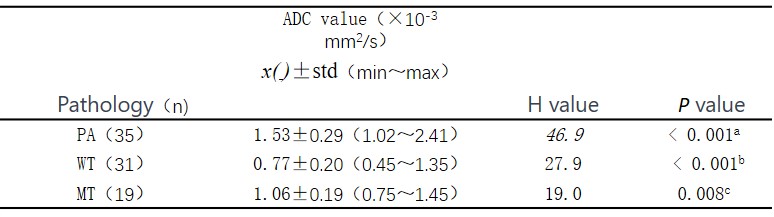
Statistical table of ADC values by pathological subgroup
Using the Kruskal-Wallis test, the P-value represents the comparison of ADC values among PA, WT, and MT. In the top left corner labels: "a" indicates a comparison between PA and WT, "b" represents a comparison between PA and MT, and "c" signifies a comparison between MT and WT.
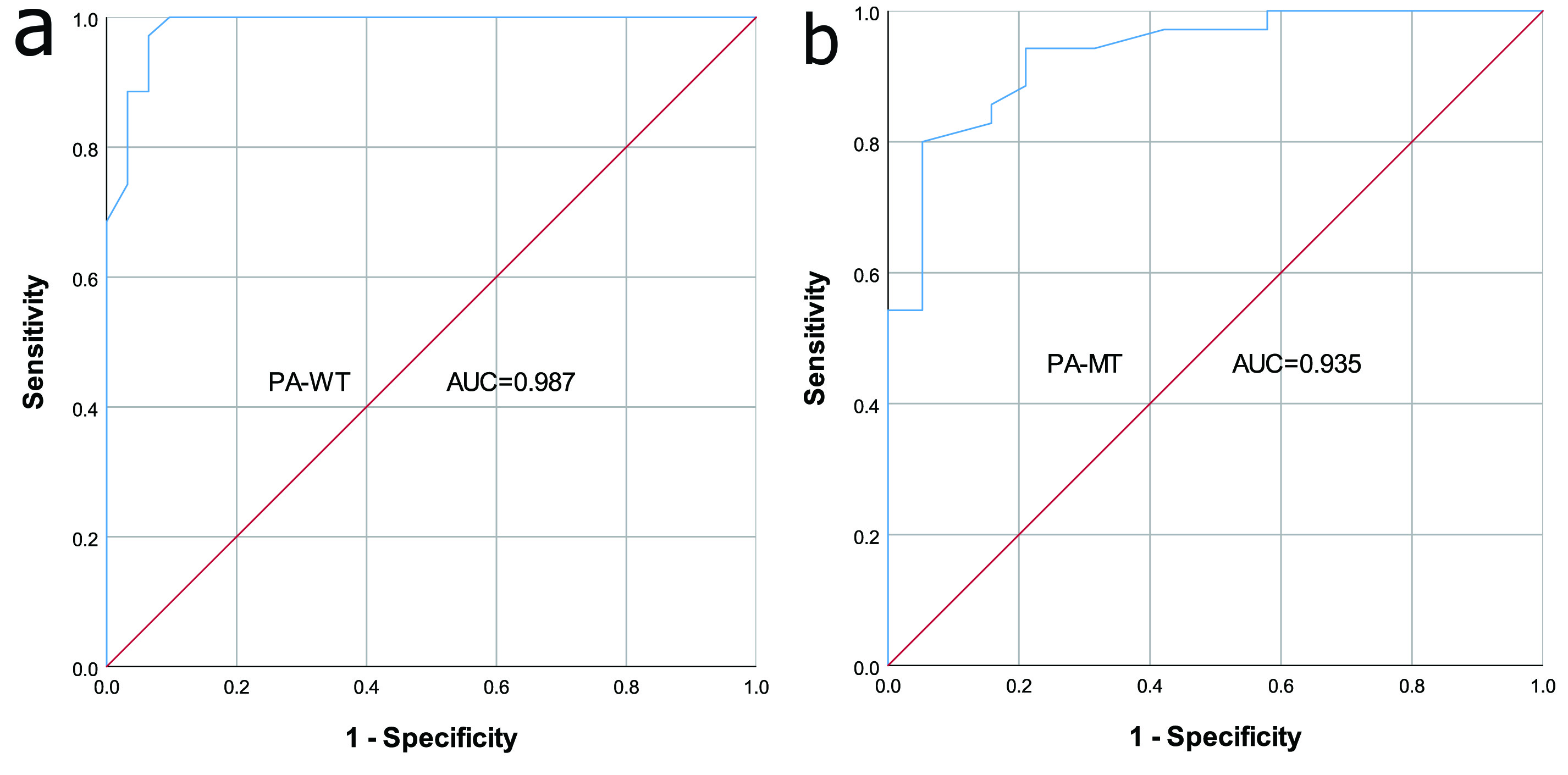
ROC Curve for Discriminating PA from WT and MT based on ADC
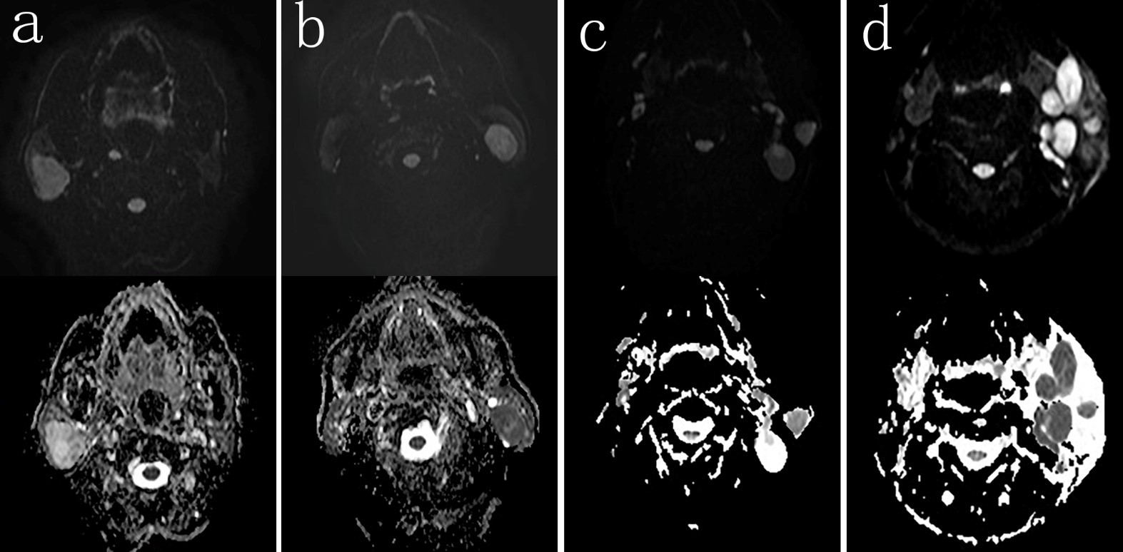
Examples of Three Types of Salivary Gland Tumors
DWI images in the upper row and ADC maps in the lower row. Three cases are presented: pleomorphic adenoma (a), Warthin's tumor (b) and mucoepidermoid carcinoma (c). The ADC values are as follows: 1.64×10⁻³mm2/s, 0.89×10⁻³mm2/s, and 0.94×10⁻³mm2/s, respectively.