2581
Correction of B0-related distortions in diffusion-weighted images of malignant and normal cervical tissue1Department of Radiology, University of California San Diego, La Jolla, CA, United States, 2Department of Surgical Sciences, Uppsala University, Uppsala, Sweden, 3Center for Medical Imaging, Uppsala University Hospital, Uppsala, Sweden, 4Department of Obstetrics, Gynecology and Reproductive Sciences, University of California San Diego, La Jolla, CA, United States, 5Department of Women's and Children's Health, Uppsala University, Uppsala, Sweden, 6Department of Radiation Medicine, University of California San Diego, La Jolla, CA, United States, 7Department of Bioengineering, University of California San Diego, La Jolla, CA, United States, 8Department of Immunology, Genetics and Pathology, Uppsala University, Uppsala, Sweden, 9Department of Oncology, Uppsala University Hospital, Uppsala, Sweden, 10Department of Neuroscience, University of California San Diego, La Jolla, CA, United States
Synopsis
Keywords: Diffusion Software, Data Processing, distortion correction, cervical cancer, cervix
Motivation: DWI in cervical cancer evaluation suffers from distortions, especially pronounced by intestinal gas, potentially impeding lesion detection/delineation.
Goal(s): To evaluate a combined prospective-retrospective distortion reduction approach, based on distortion correction of female pelvic DWI acquired with reduced-FOV.
Approach: Two correction methods, RPG and Topup, were applied on images from cervical cancer patients and volunteers. Distortion correction performance was evaluated by the concordance between tumour/cervix borders in images before and after correction.
Results: Topup outperformed RPG, with statistically significant but modest improvements of initially small distortions resulting from the reduced-FOV acquisition. Occasional correction failures and limitations in the proton density correction warrant alternative approaches.
Impact: Topup distortion correction, commonly used for brain studies, shows statistically significant but modest improvements in female pelvic reduced-FOV DWI. Despite indications of clinical utility, limited improvements and occasional correction failures suggest alternative approaches and prospective distortion reduction as potential strategies.
Introduction
Cervical cancer staging and radiation treatment planning are highly dependent on the accuracy of evaluating parametrial invasion and extent of disease with MRI. B0-inhomogeneity distortion artifacts in the phase encoding (PE) direction limit the evaluation and staging of cervical cancer with DWI. In the female pelvis, distortion reduction has been targeted by prospective approaches1-3. A combined prospective-retrospective approach, based on reduced-FOV (rFOV) acquisition and reverse gradient distortion correction, might further improve geometric accuracy.Aim
To evaluate the performance of two reverse gradient methods for distortion correction of rFOV-DWI, intended for studies of cervical tissue and tumours at 3T.Methods
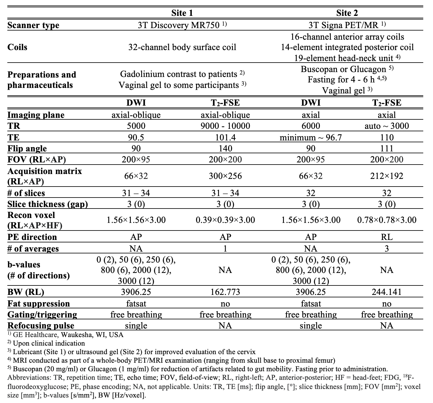
Cervical cancer patients and healthy volunteers underwent MRI at two sites (Site 1: 16 patients/35 volunteers; Site 2: 3 patients/2 volunteers). Patients were examined after diagnostic biopsy and before definite therapy. MRI included a multi-shell rFOV-DWI and a T2-weighted fast spin-echo (T2-FSE), the latter used as anatomical reference to the DWI (details in Table 1). Within the DWI, two volumes with b-value 0 were sequentially acquired, the first (b0,rvs) using a reverse PE gradient polarity compared to the forward polarity used for the second (b0,fwd) and remaining volumes with non-zero b-values. Reverse gradient methods accomplish distortion correction on DWI with bipolar volumes of b-value 0, according to:4,5- The b0,fwd and b0,rvs are used for estimating a 3D deformation field, specifying the voxel-wise displacements in the PE direction that minimizes distortions. The algorithm uses consecutively decreasing Gaussian smoothing kernels (K) and can be optimized in terms of the maximum K (Kmax) and deformation field regularization parameters (λ).
- The deformation field is applied for geometric correction of the whole DWI dataset of multiple b-values.
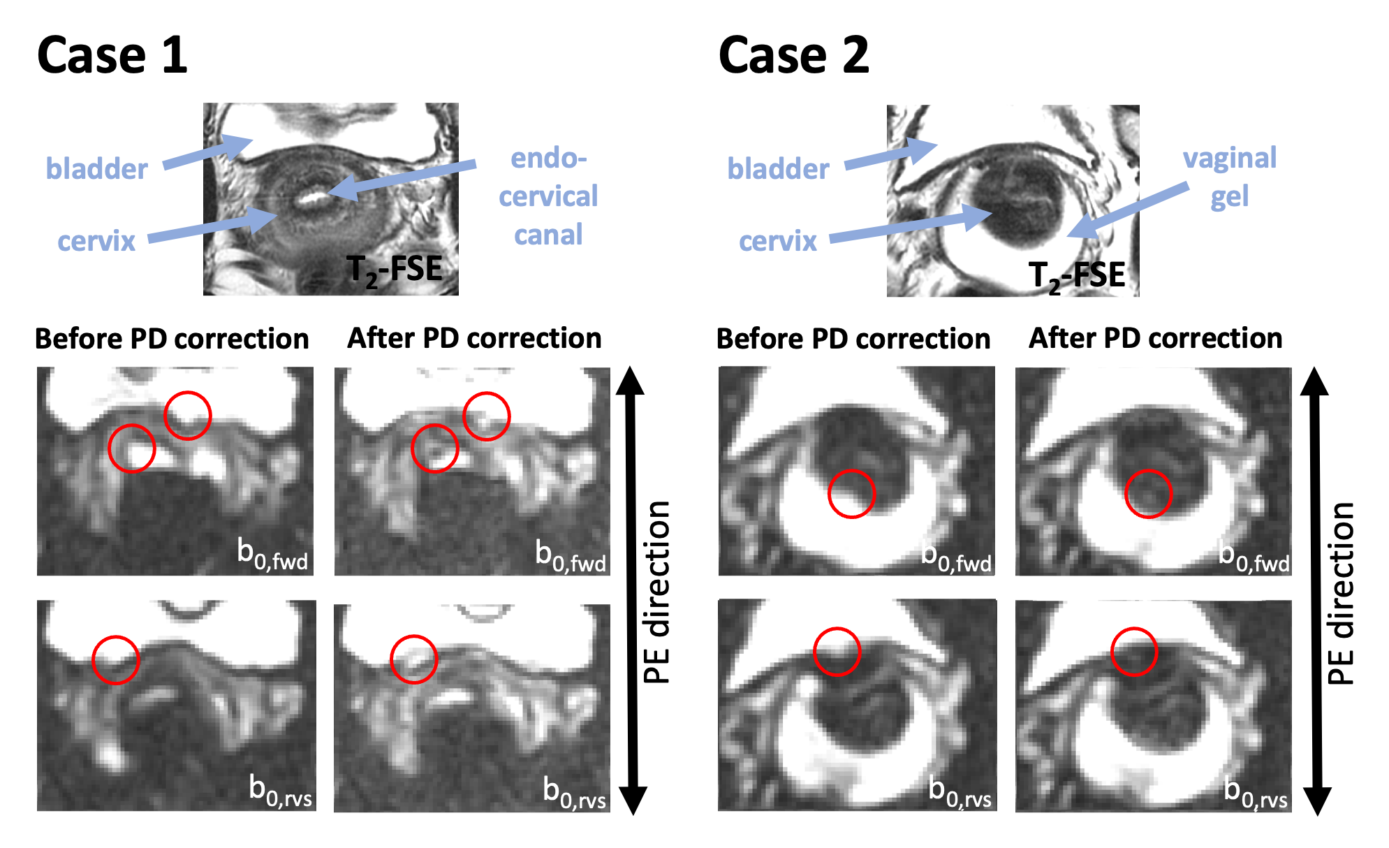
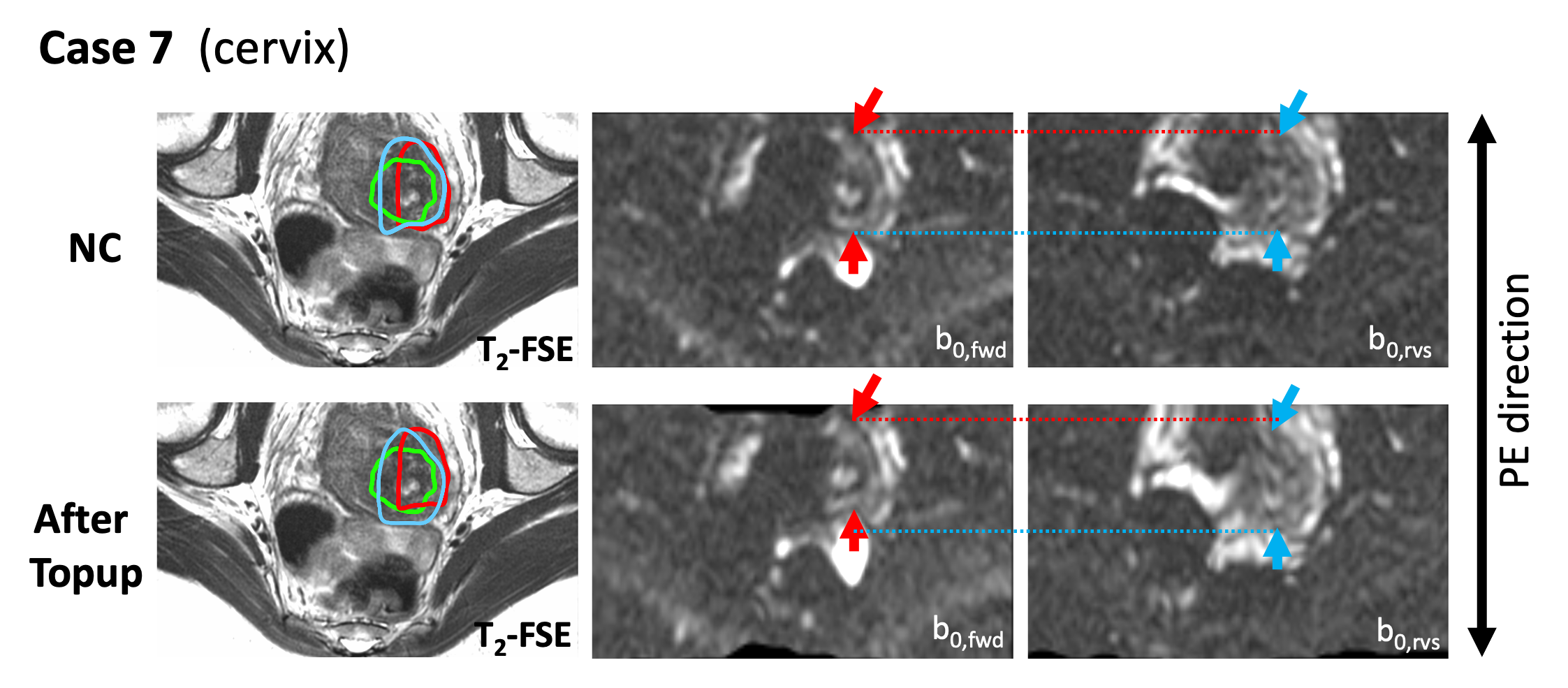
- Proton density (PD) correction, using the Jacobian determinant of the deformation field, is conducted. As previously reported,6 this step can introduce a false appearance of accurate correction not supported by Step 2. This phenomenon was observed also in this study (Fig. 1), whereby Step 3 was omitted.
- RPG.5 The regularization parameters λ1=0 and λ2= 500, 1500, 2500 were combined with Kmax= 5, 10, 15, 20, 25, 30, 35 voxels.
- Topup.4,7 The default configuration file was used as basis for subsequent modification into five configuration files with Kmax= 8, 16, 24, 32 and 40 mm. λ=0.5 for large K (≥16–20 mm) and thereafter decreasing with decreasing K. As PD correction cannot be disabled in Topup, in-house Matlab software was used for Step 2.
The tumour circumference (in patients with macroscopic tumour, stage≥IB1) and normal cervix circumference (in volunteers and patients without macroscopic tumour) were manually delineated in a single slice using OsirixMD (Pixmeo SARL, Switzerland, v.10.0.2). Delineation was performed in b0,fwd and b0,rvs, before and after distortion correction, and in T2-FSE images. The average distance in the PE direction between edge voxels of the b0,fwd and b0,rvs segmentations, and between edge voxels of the b0,fwd and T2-FSE segmentations, was used to assess the degree of distortion. Larger distance implied more severe distortions and reduced distance after correction (Δdistance<0) reflected improved distortions. Comparisons were conducted with Wilcoxon sign-rank test and Spearman rank correlation. P<0.05 represented statistical significance.
Results
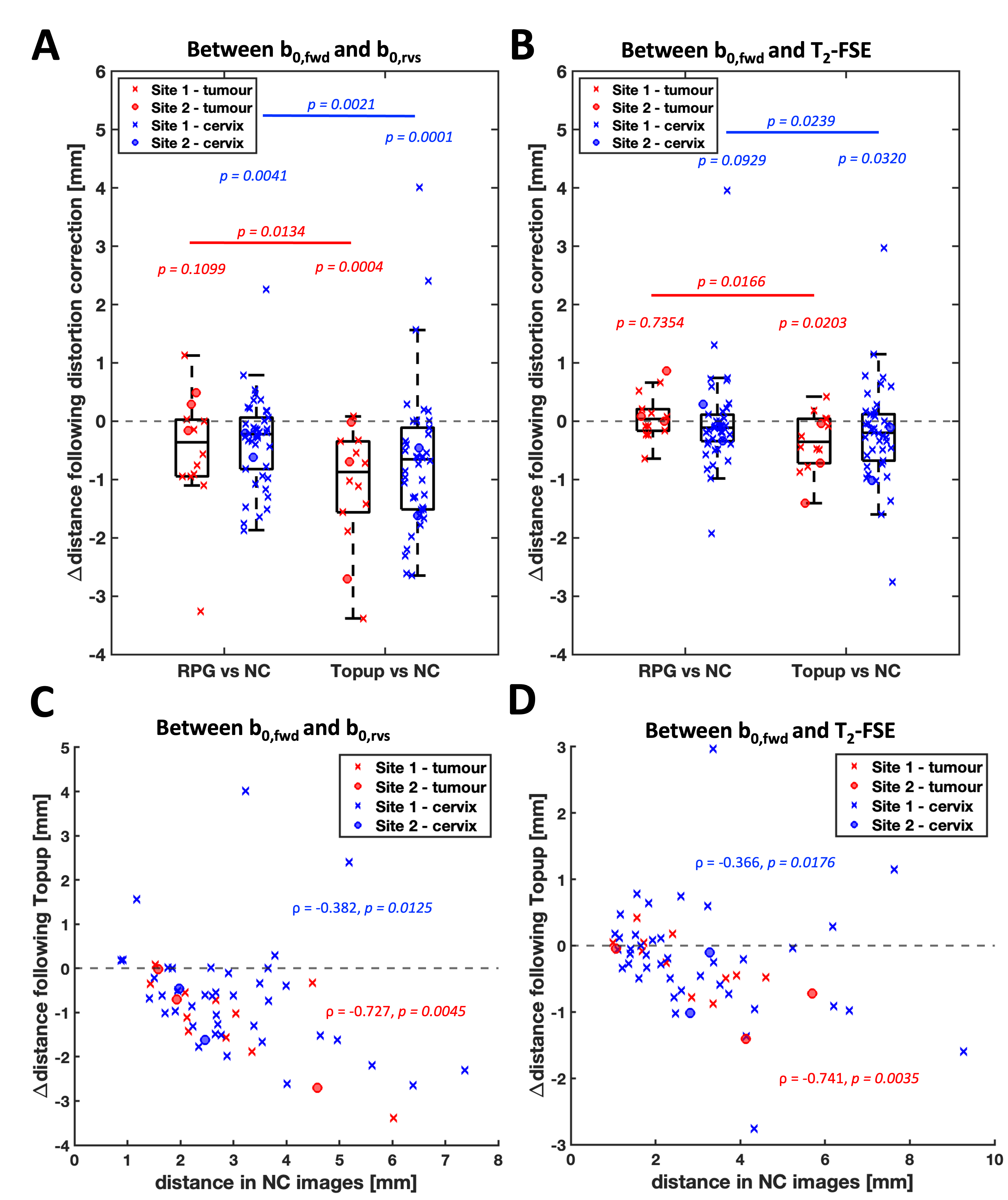
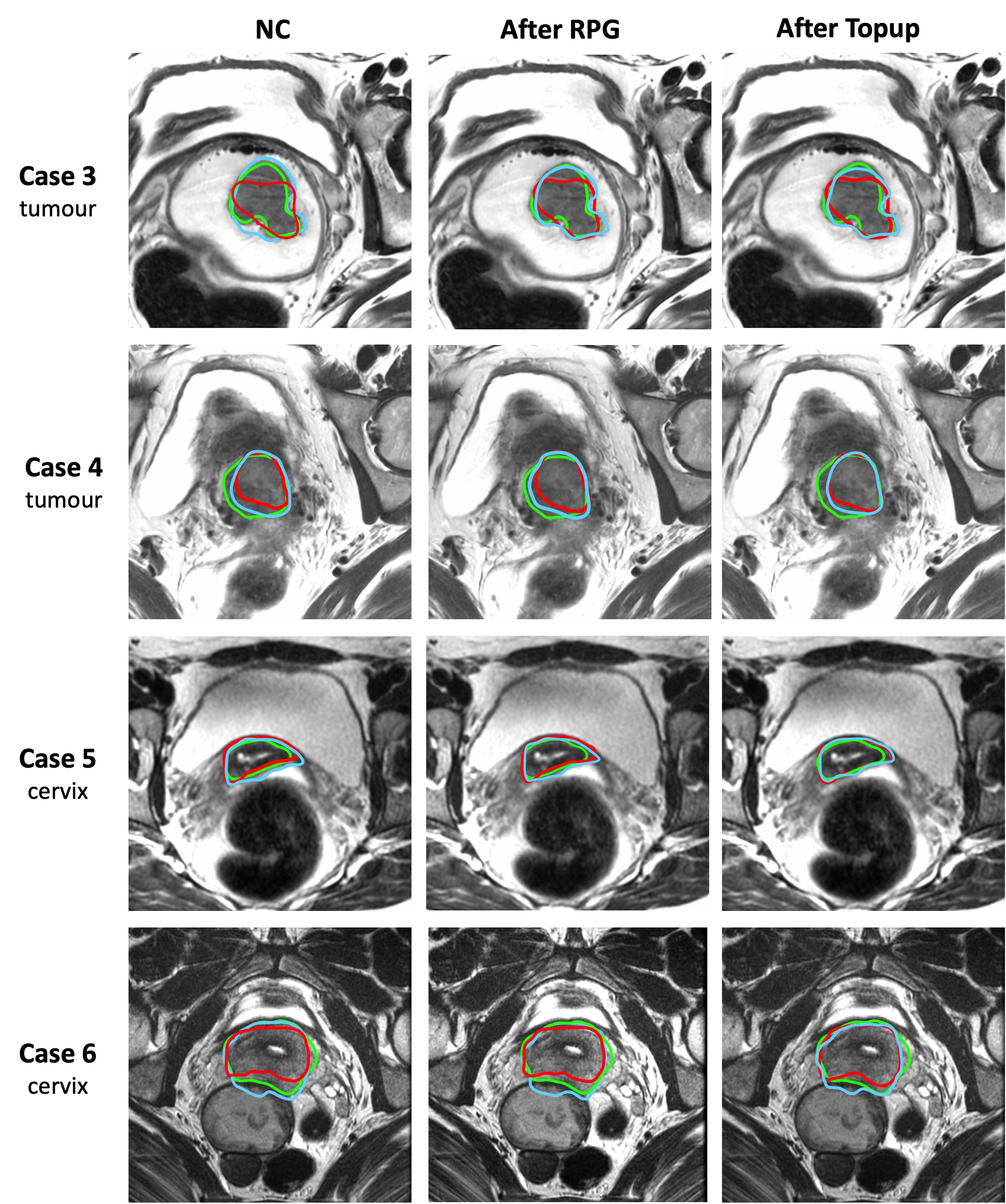
Topup showed superior distortion correction results for tumour and cervix segmentations compared to RPG, both between b0,fwd and b0,rvs and between b0,fwd and T2-FSE (Fig. 2A–B). The distortion reduction by Topup was related to the initial distortion before correction (Fig. 2C–D). The RPG and Topup correction performances are demonstrated by image examples in Fig. 3. Outliers corresponding to Topup correction failures were also observed (image example in Fig. 4).Discussion and Conclusion
Topup showed superior distortion correction performance and provided higher correspondence between DWI and T2-FSE compared to RPG in most situations with minor distortions. However, Topup was limited with major distortions where RPG may prove more useful. Small but statistically significant improvements in geometric accuracy after distortion correction demonstrate clinical utility of Topup, particularly for radiotherapy planning, where anatomic accuracy is critical for targeting tumour and sparing adjacent healthy tissue. Modest initial distortions (improved by utilizing rFOV) and motion-related misalignments between T2-FSE and DWI limited the benefit of Topup, although larger initial distortions led to greater improvements after correction. These results indicate that prospective distortion reduction, such as rFOV, is an appropriate primary approach. However, subsequent correction of full-FOV DWI (suffering from more initial distortion but benefitting from higher signal-to-noise ratio9), is an alternative technique. The Jacobian determinant-based PD correction was omitted due to cases of misleading correction results.Acknowledgements
This study was supported by the Swedish Research Council (dnr 2021-00427), NIH R37CA249659, Lions Cancer Fund Mellansverige Uppsala-Örebro, Makarna Eriksson Foundation and a research grant from GE Healthcare.References
1. Thian YL, Xie W, Porter DA, Weileng Ang B. Readout-segmented echo-planar imaging for diffusion-weighted imaging in the pelvis at 3T-A feasibility study. Acad Radiol 2014;21(4):531-537.
2. An H, Ma X, Pan Z, Guo H, Lee EYP. Qualitative and quantitative comparison of image quality between single-shot echo-planar and interleaved multi-shot echo-planar diffusion-weighted imaging in female pelvis. Eur Radiol 2020;30(4):1876-1884.
3. Ghosh A, Singh T, Singla V, Bagga R, Srinivasan R, Khandelwal N. Read-out segmented echo planar diffusion imaging of the female pelvis-utility in endometrial carcinoma-a preliminary experience. Br J Radiol 2018;91(1090):20180018.
4. Andersson JL, Skare S, Ashburner J. How to correct susceptibility distortions in spin-echo echo-planar images: application to diffusion tensor imaging. Neuroimage 2003;20(2):870-888.
5. Holland D, Kuperman JM, Dale AM. Efficient correction of inhomogeneous static magnetic field-induced distortion in Echo Planar Imaging. Neuroimage 2010;50(1):175-183.
6. Rodriguez-Soto AE, Fang LK, Holland D, et al. Correction of Artifacts Induced by B0 Inhomogeneities in Breast MRI Using Reduced-Field-of-View Echo-Planar Imaging and Enhanced Reversed Polarity Gradient Method. Journal of magnetic resonance imaging : JMRI 2021;53(5):1581-1591.
7. Smith SM, Jenkinson M, Woolrich MW, et al. Advances in functional and structural MR image analysis and implementation as FSL. Neuroimage 2004;23 Suppl 1:S208-219.
8. Cover T, Thomas J. Elements of information theory. Hoboken, NJ, USA: Wiley Interscience: 2006.
9. Jeong EK, Kim SE, Guo J, Kholmovski EG, Parker DL. High-resolution DTI with 2D interleaved multislice reduced FOV single-shot diffusion-weighted EPI (2D ss-rFOV-DWEPI). Magnetic resonance in medicine : official journal of the Society of Magnetic Resonance in Medicine / Society of Magnetic Resonance in Medicine 2005;54(6):1575-1579.
Figures