2580
Time-dependent diffusion-weighted MRI for pathological grade of clear cell renal cell carcinoma1Tongji Hospital, Tongji Medical College, Huazhong University of Science and Technology, Wuhan, China, 2MR Research Collaboration Team, Siemens Healthineers Ltd., Wuhan, China
Synopsis
Keywords: Diffusion Modeling, Tumor
Motivation: Accurate grading of clear cell renal cell carcinoma (ccRCC) is essential for treatment decisions, particularly in patients with comorbidities.
Goal(s): This study aims to investigate time-dependent diffusion MRI to noninvasively characterize the microstructural properties of ccRCC and distinguish between different pathological grades.
Approach: A total of 66 patients with histologically confirmed ccRCC underwent MRI scans, and microstructural parameters were estimated using time-dependent diffusion MRI techniques.
Results: The study revealed distinct microstructural differences between low-grade and high-grade ccRCC, showing the potential of time-dependent diffusion MRI for preoperative pathological grade characterization.
Impact: This study has the potential to reshape the landscape of preoperative ccRCC grading, promote objectivity in treatment planning.
Introduction
Clear Cell Renal Cell Carcinoma (ccRCC) is the most common malignant tumor of the urinary system, and its incidence has been steadily increasing over the past few decades [1]. The aggressiveness of ccRCC is graded according to the WHO/ISUP classification. However, pathological grading is based on postoperative pathology rather than preoperative biopsy, making the preoperative prediction of ccRCC pathological grading crucial for treatment guidance and clinical outcome prediction. Diffusion-weighted imaging (DWI) serves as a noninvasive modality enabling the quantification of water molecule movement driven by diffusion. Nevertheless, it is essential to recognize that the DWI-derived apparent diffusion coefficient (ADC) is only a comprehensive measure of water diffusivity that is determined by multiple microstructural features, such as the intra- and extracellular space, cell size, permeability, and intrinsic diffusivity. The recent development of time-dependent diffusion MRI has demonstrated unique advantages in depicting cellular microstructures by characterizing the diffusion time dependence of restricted water diffusion and relating the diffusion time dependence to specific microstructural parameters [2]. Therefore, the purpose of this study was to investigate the time-dependent diffusion MRI to non-invasively characterize the microstructural properties of ccRCC and differentiate between different pathological grades of ccRCC.Methods
In total of 66 patients with histologically confirmed ccRCC (50 low-grade and 16 how-grade) were recruited in this study. All patients underwent axial MRI scans at 3T (Magnetom Skyra, Siemens Healthcare, Erlangen, Germany) with an 18-element body matrix coil. OGSE data were acquired at oscillating frequencies of 33 Hz (effective diffusion time = 7.5 msec, two cycles, b = 300 and 600 sec/mm2) and 17 Hz (effective diffusion time = 15 msec, one cycle, b = 400, 800, and 1200 sec/mm2), and pulsed gradient spin-echo at diffusion duration and separation of 10 and 30 msec, respectively (b = 400, 800, and 1200 sec/mm2). The following parameters were used for both sequences: three diffusion directions; TR/TE, 5000/82; FOV, 380 × 322.5 mm; and section thickness, 5 mm. Microstructural parameters including cell diameter d, fin, and Dex were estimated using a nonlinear least squares algorithm in Matlab R2018a (Mathworks, Natick, MA) with a fixed intracellular diffusion coefficient at 1.0 µm2/ms. The fitting was repeated 100 times with randomized initializations to avoid local minimums, under the following physiological constraints: 0.01< fin <1, 0.5< Dex <3.5 µm2/ms. Cellularity was defined as fin/d*100 for simplicity. ADC maps were calculated at each td according to the log-linear fitting S/S0 = e-bD to obtain D0Hz (PGSE), D17Hz, and D33Hz.Results
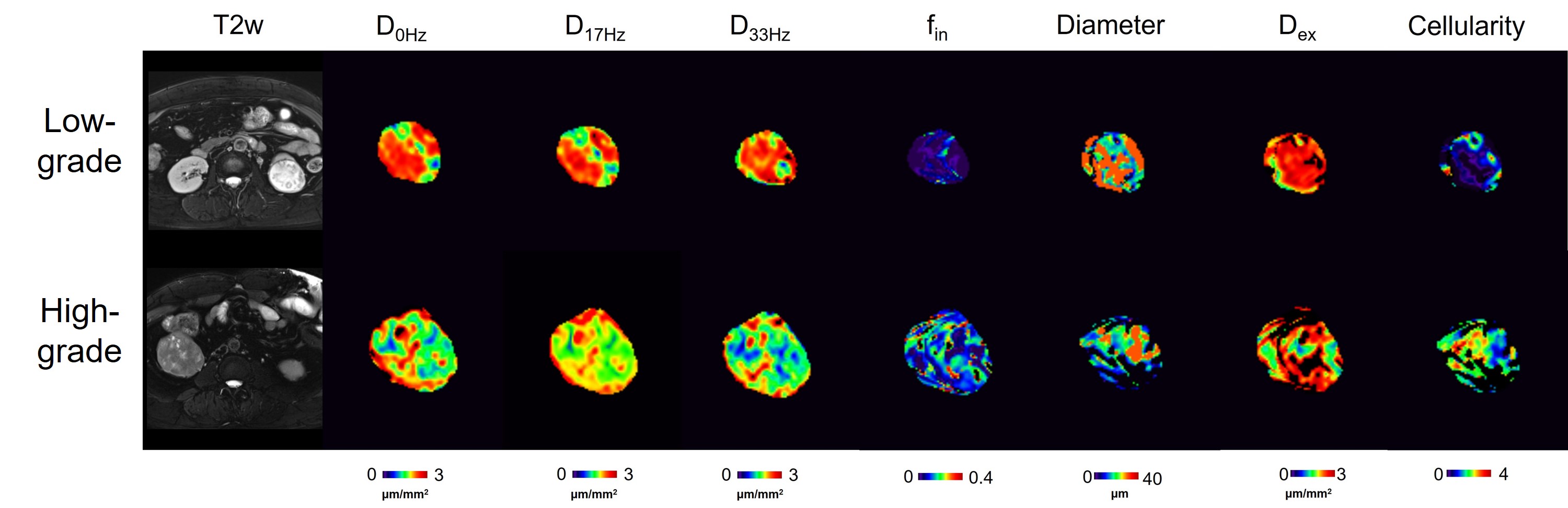
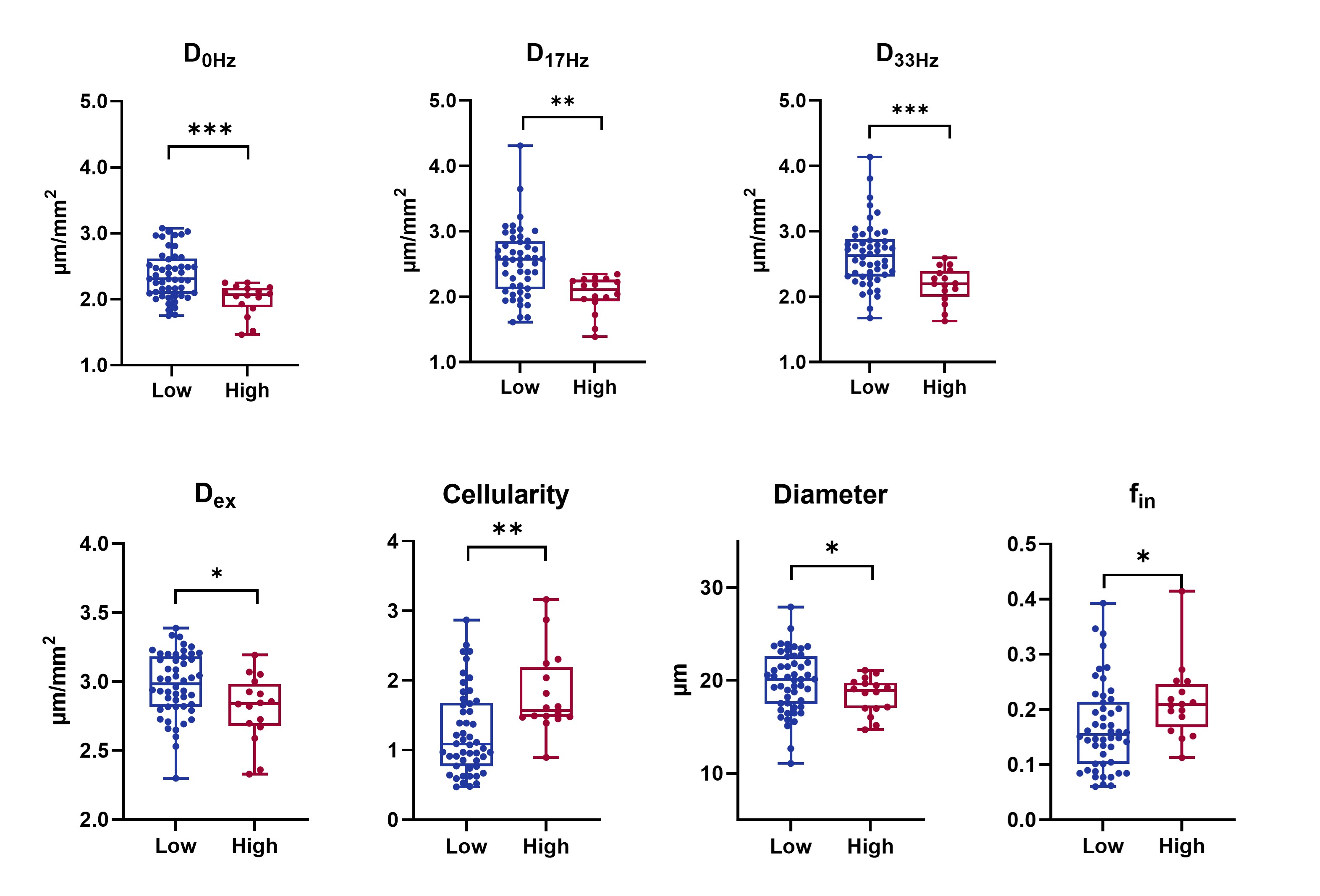
Figure 1 shows the IMPULSED-fitted microstructural parameter maps for low- and high-grade ccRCC, along with the ADC maps at different oscillating frequencies and T2-weighted images. Intracellular volume fraction and cellularity in the cancerous tissues were higher with a higher grade, while the diffusivity measurements (at 0 Hz, 17 Hz, and 33 Hz) were lower with a higher grade. There were differences in several time-dependent diffusion MRI–derived parameters across low- and high-grade tumors. The low-grade lesion exhibited significantly high values in D0Hz, D17Hz, D33Hz, Dex, and Diameter. While Cellularity and fin have typically been found to be higher in the high-grade lesion.Discussion
In summary, this study utilizes time-dependent diffusion MRI to noninvasively characterize the microstructural properties of clear cell renal cell carcinoma (ccRCC) and differentiate between different pathologic grades. Our results reveal distinct microstructural differences between low-grade and high-grade ccRCC. High-grade tumors exhibit elevated intracellular volume fraction and cellularity, indicating increased cellular density. This study reveals the great potential of time-dependent diffusion MRI for preoperative characterization of tumor pathological grade.Acknowledgements
No acknowledgement found.References
1 Pedrosa I, Cadeddu JA (2022) How We Do It: Managing the Indeterminate Renal Mass with the MRI Clear Cell Likelihood Score. Radiology 302:256-269
2 Wu D, Jiang K, Li H et al (2022) Time-Dependent Diffusion MRI for Quantitative Microstructural Mapping of Prostate Cancer. Radiology 303:578-587
Figures