2579
Dynamic Changes in the White Matter Microstructure of Breast Cancer Patients During Neoadjuvant Chemotherapy1School of Medicine, Chongqing University, Chongqing, China, 2Department of Radiology, Chongqing University Cancer Hospital, Chongqing, China, 3School of Biological Engineering, Chongqing University, Chongqing, China, 4MR Research Collaborations, Siemens Healthineers Ltd, Chengdu, China
Synopsis
Keywords: Microstructure, Diffusion/other diffusion imaging techniques, chemotherapy; breast cancer.
Motivation: Chemotherapy can cause cognitive impairment in breast cancer patients. However, the microstructural changes in white matter during chemotherapy remain unknown.
Goal(s): To explore the patterns of white matter microstructural changes during chemotherapy.
Approach: By using three diffusion models (DTI, DKI and NODDI), white matter microstructure of 72 female breast cancer patients was detected at baseline (TP1), at the completion of the first cycle of neoadjuvant chemotherapy (TP2) and at the completion of neoadjuvant chemotherapy but before surgery (TP3).
Results: Different diffusion MRI metrics reflected dynamic changes in white matter microstructure in breast cancer patients receiving neoadjuvant chemotherapy.
Impact: By combining DTI, DKI and NODDI metrics, alterations in white matter microstructure among breast cancer patients were identified from baseline to two subsequent follow-up time points during neoadjuvant chemotherapy. These findings could potentially help the early diagnosis and treatment of chemo-brain.
Introduction
Although chemotherapy has markedly improved the survival rates of breast cancer patients, one of its side effects is chemotherapy-related cognitive impairment (CRCI), significantly impacting patients’ quality of life1. Chemotherapy agents have the potential to induce central neurotoxicity2, resulting in damage to the white matter microstructure. This damage reduced the efficiency of information transmission among neural systems, subsequently leading to impaired cognitive processes. Diffusion MRI provides the capability to characterize various microstructural properties of brain white matter. Studies have discovered reduced fractional anisotropy (FA) in white matter tracts associated with cognitive function in breast cancer patients who underwent chemotherapy, observed at approximately several months3, one year4, and even nine years5 after completion of postoperative chemotherapy. It is worth noting that alternations in white matter microstructure evaluated through DKI and NODDI models have not yet been reported in the field of CRCI. Investigating these changes in white matter microstructure might offer valuable non-invasive imaging markers for understanding the underlying mechanism of CRCI. Therefore, in this study, we aim to explore the patterns of white matter microstructural changes during chemotherapy using diffusion models.Methods
1. ParticipantsThe study was approved by the Ethics Committee of our institute. In total, 72 breast cancer patients were recruited at our institute from 2021 to 2022, with written informed consent. Pre-treatment MRI examination occurred before the neoadjuvant chemotherapy (TP1; baseline). Follow-up measurements were conducted after completion of the first cycle of neoadjuvant chemotherapy (TP2) and after completion of all cycles of neoadjuvant chemotherapy but before surgery (TP3).
2. MRI data acquisition
All MRI data were acquired on a 3T scanner (Magnetom Prisma; Siemens Healthcare, Erlangen, Germany) equipped with a 64-channel head-neck coil. Detailed scan parameters are summarized in the Table 1.
3. Diffusion data processing and analysis
Diffusion data processing included TOPUP and EDDY correction in FSL (the FMRIB Software Library)6. DTI metrics (FA, AD, RD and MD) and DKI metrics (AK, RK, and MK) were generated using FDT toolbox7, while NODDI metrics (NDI, ODI, and ISOVF) were calculated via AMICO toolbox8. Specific diffusion metrics and their abbreviations were list in the Table 2. The tract-based spatial statistics method (FSL, TBSS)9 was then used for voxel-wise statistical analysis. A voxel-wise general linear model analysis with paired permutation test was utilized to compare differences among the parameter values between every two time points. Correction for multiple comparisons used threshold-free cluster enhancement9 and a familywise error rate–corrected value of p < 0.05. White matter tracts were considered statistically significant when the surviving white matter voxels numbered more than 100.
Results
The study included 72 female breast cancer patients who underwent three MRI scans, with demographic and clinical characteristics detailed in Table 3.Longitudinal changes in white matter microstructure in breast cancer patients during neoadjuvant chemotherapy were observed (Figure 1). Patients showed decreased FA in specific brain regions TP2 and TP3, such as the corpus callosum, bilateral anterior corona radiata, and left superior corona radiata. Reductions in FA were also noticed in the corpus callosum genu, body, and bilateral anterior corona radiata from TP1 to TP3. No significant FA differences were noted between TP1 and TP2. Additionally, reductions in AD, RD, MD, AK, RK, and MK occurred at TP2 and TP3, but not between TP2 and TP3. ISOVF decreased between TP1 and TP3 at the splenium of the corpus callosum, left retro-lenticular part of the internal capsule, left posterior thalamic radiation, and left sagittal stratum, but remained unchanged between TP1 and TP2 or TP2 and TP3. NDI and ODI showed no significant changes across the three time points.
Discussion
Our study monitors white matter microstructure changes in breast cancer patients during neoadjuvant chemotherapy. Reduced FA aligns with previous findings3-5, suggesting axonal membrane and myelin sheath alterations10. Chemotherapy revealed decreased AD, RD, and MD primarily in initial stages, contrasting prior studies showing increased values3, 11. Our findings suggest evolving diffusivity changes at different stages of chemotherapy-induced cognitive impairment (CRCI).The alterations in AK, RK, and MK nearly overlapped with changes in AD, RD, and MD, indicating the enhanced sensitivity of DKI metrics over traditional DTI metrics12.
Reduced ISOVF possibly signifies swelling in these tracts and surrounding brain tissue, due to increased ISOVF associated with brain atrophy in prior studies13. However, no significant differences were detected in NDI or ODI during chemotherapy, implying that neurite density and orientation might not show acute changes during this treatment phase.
Conclusion
The acute response of white matter microstructure during neoadjuvant chemotherapy found in this study could provide a basis for early intervention of CRCI in the future.Acknowledgements
No acknowledgement found.References
1. Wefel JS, Kesler SR, Noll KR, et al. Clinical characteristics, pathophysiology, and management of noncentral nervous system cancer-related cognitive impairment in adults[J]. CA Cancer J Clin, 2015, 65: 123-138.2. Gibson EM, Nagaraja S, Ocampo A, et al. Methotrexate Chemotherapy Induces Persistent Tri-glial Dysregulation that Underlies Chemotherapy-Related Cognitive Impairment[J]. Cell, 2019, 176: 43-55.e13.
3. Deprez S, Amant F, Yigit R, et al. Chemotherapy-induced structural changes in cerebral white matter and its correlation with impaired cognitive functioning in breast cancer patients[J]. Hum Brain Mapp, 2011, 32: 480-493.
4. Bukkieva T, Pospelova M, Efimtsev A, et al. Microstructural Properties of Brain White Matter Tracts in Breast Cancer Survivors: A Diffusion Tensor Imaging Study[J]. Pathophysiology, 2022, 29: 595-609.
5. de Ruiter MB, Reneman L, Boogerd W, et al. Late effects of high-dose adjuvant chemotherapy on white and gray matter in breast cancer survivors: converging results from multimodal magnetic resonance imaging[J]. Hum Brain Mapp, 2012, 33: 2971-2983.
6. Woolrich MW, Jbabdi S, Patenaude B, et al. Bayesian analysis of neuroimaging data in FSL[J]. Neuroimage, 2009, 45: S173-186.
7. Jbabdi S, Sotiropoulos SN, Savio AM, et al. Model-based analysis of multishell diffusion MR data for tractography: how to get over fitting problems[J]. Magn Reson Med, 2012, 68: 1846-1855.
8. Daducci A, Canales-Rodríguez EJ, Zhang H, et al. Accelerated Microstructure Imaging via Convex Optimization (AMICO) from diffusion MRI data[J]. Neuroimage, 2015, 105: 32-44.
9. Smith SM, Jenkinson M, Johansen-Berg H, et al. Tract-based spatial statistics: voxelwise analysis of multi-subject diffusion data[J]. Neuroimage, 2006, 31: 1487-1505.
10. Deprez S, Billiet T, Sunaert S, et al. Diffusion tensor MRI of chemotherapy-induced cognitive impairment in non-CNS cancer patients: a review[J]. Brain Imaging Behav, 2013, 7: 409-435.
11. Chen BT, Ye N, Wong CW, et al. Effects of chemotherapy on aging white matter microstructure: A longitudinal diffusion tensor imaging study[J]. J Geriatr Oncol, 2020, 11: 290-296.
12. Konieczny MJ, Dewenter A, Ter Telgte A, et al. Multi-shell Diffusion MRI Models for White Matter Characterization in Cerebral Small Vessel Disease[J]. Neurology, 2021, 96: e698-e708.
13. Colgan N, Siow B, O'Callaghan JM, et al. Application of neurite orientation dispersion and density imaging (NODDI) to a tau pathology model of Alzheimer's disease[J]. Neuroimage, 2016, 125: 739-744.
Figures
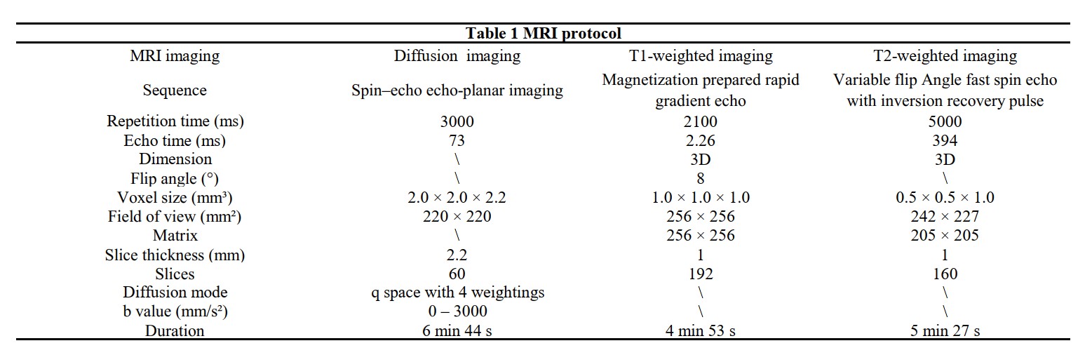
Table 1 MRI protocol

Table 2 Diffusion metrics and their abbreviations
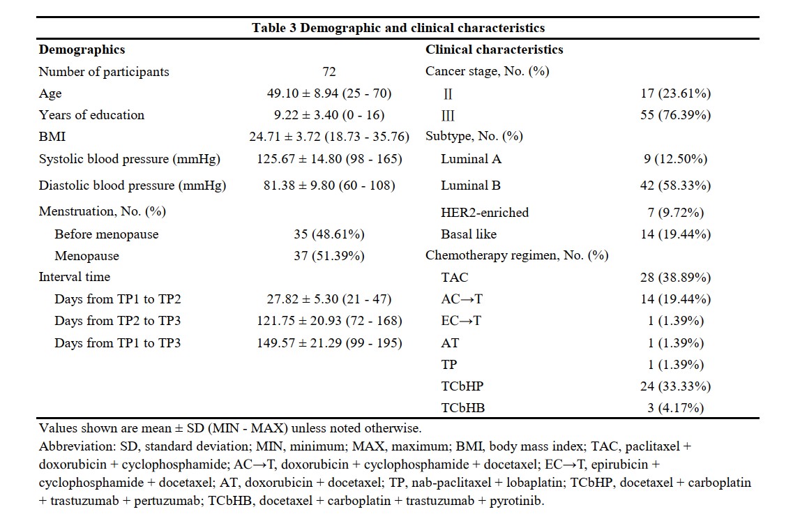
Table 3 Demographic and clinical characteristics
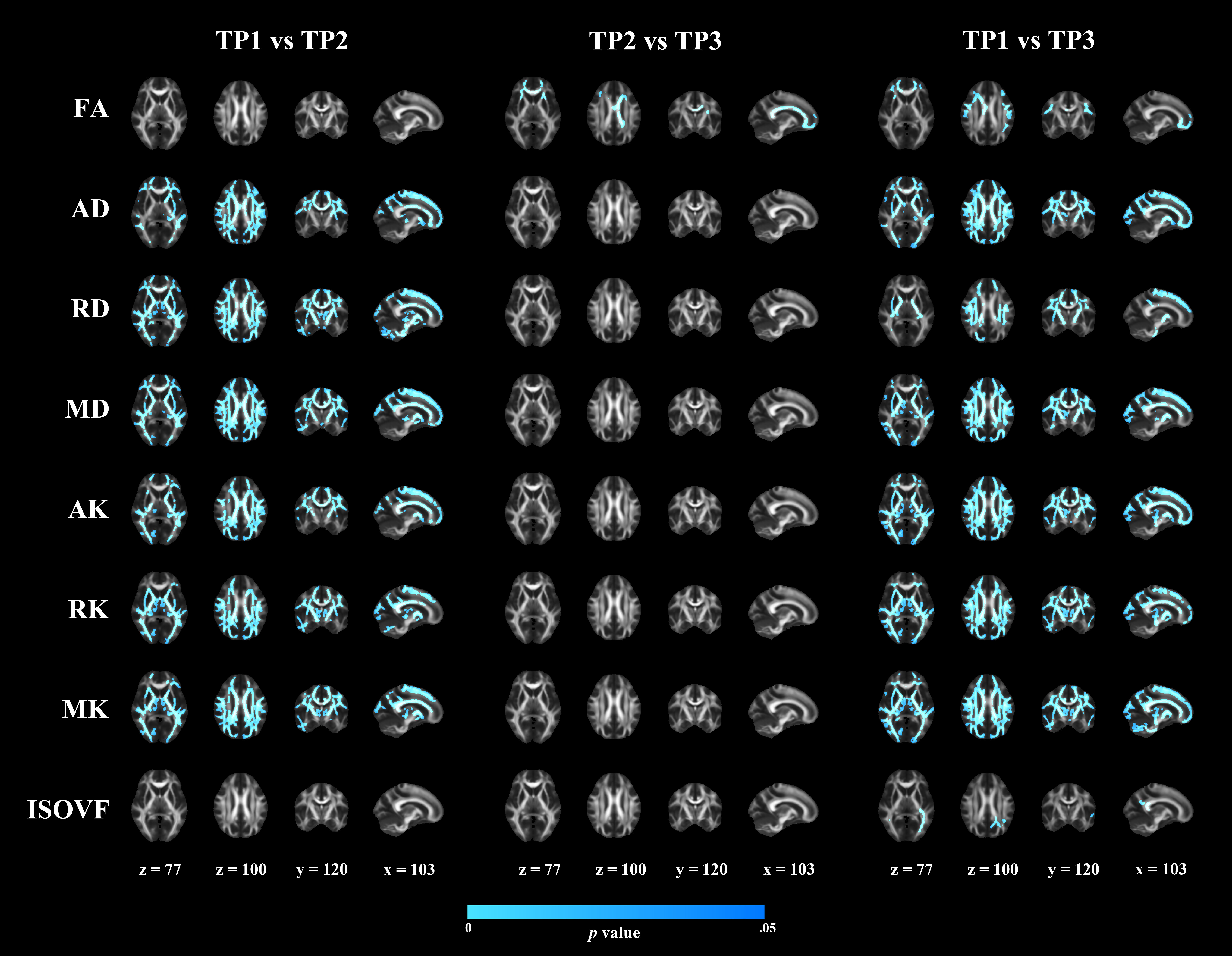
Figure 1 Results of longitudinal diffusion metric comparison.
The significant white matter regions in each diffusion metric are shown. Regions in blue had significant lower values of each diffusion metric over time. TP1, baseline; TP2, after completion of the first cycle of neoadjuvant chemotherapy; TP3, after completion of all cycles of neoadjuvant chemotherapy but before surgery; FA, fractional anisotropy; AD, axial diffusivity; RD, radial diffusivity; MD, mean diffusivity; AK, axial kurtosis; RK, radial kurtosis; MK, mean kurtosis; ISOVF, isotropic volume fraction.