2578
Impact of Extracellular Matrix Protein Decorin on Diffusion MR in the 4T1 Breast Cancer Model
Santosh K Yadav1, Balaji Krishnamachary2, Derek Liu3, Yelena Mironchik2, Sridhar Nimmagadda4, and Zaver Bhujwalla3
1Department of Radiology, Johns Hopkins School of Medicine, Baltimore, USA, Baltimore, MD, United States, 2Johns Hopkins School of Medicine, Baltimore, USA, Baltimore, MD, United States, 3Johns Hopkins School of Medicine, Baltimore, Baltimore, MD, United States, 4Division of Nuclear Medicine and Molecular Imaging, Johns Hopkins School of Medicine, Baltimore, Baltimore, MD, United States
1Department of Radiology, Johns Hopkins School of Medicine, Baltimore, USA, Baltimore, MD, United States, 2Johns Hopkins School of Medicine, Baltimore, USA, Baltimore, MD, United States, 3Johns Hopkins School of Medicine, Baltimore, Baltimore, MD, United States, 4Division of Nuclear Medicine and Molecular Imaging, Johns Hopkins School of Medicine, Baltimore, Baltimore, MD, United States
Synopsis
Keywords: Diffusion Acquisition, Cancer
Motivation: Decorin (DCN), so called because it decorates collagen fibers, is essential for the mechanical integrity of tissues. It acts as a guardian of the extracellular matrix (ECM) by sequestering cytokines.
Goal(s): Our purpose here was to determine the effects of DCN on water diffusion and the expression of immune checkpoints.
Approach: DTI with MRI in decorin
Results: . We found, with diffusion tensor imaging (DTI), that DCN overexpression in tumors altered water diffusion metrics suggesting that increased DCN altered water movement through the ECM that could potentially alter cytokine movement through the ECM.
Impact: Cancer diagnosis
Background: Decorin (DCN), a member of the small leucine-rich proteoglycan (SLRP) family (1), plays a vital role in the structural integrity of tissues by binding to collagen 1 (Col1) fibers (2). Beyond its mechanical function, DCN exhibits tumor-suppressive, anti-fibrotic, and anti-inflammatory activities, earning the title of the ‘guardian’ of the extracellular matrix (3). DCN has the unique ability to sequester various molecules, including TGF-β, PDGF, EGFR, ErbB1-4 receptor tyrosine kinases, myostatin, connective tissue growth factor, and thrombospondin, making it a promising candidate to neutralize multiple molecules involved in cancer, inflammation and fibrosis (1,4). Low DCN levels correlate with reduced survival in various cancers (4). T cell-expressed DCN inhibits glioma formation by suppressing microglia (5). This study investigated the association between diffusion magnetic resonance imaging parameters such as fractional anisotropy (FA) and mean diffusivity (MD) and breast cancer overexpression of DCN and the immune checkpoints programmed cell death ligand 1 (PD-L1), programmed cell death (PD1) in the tumor microenvironment. Our hypothesis is that tumors overexpressing DCN will exhibit reduced PD-L1 and PD1 expression, along with altered water diffusion metrics. Material and Methods: Animal ethics committee approved the study protocol. We engineered 4T1 breast cancer cells with DCN overexpression as shown in figure 1. DCN overexpressing cells were inoculated in immune competent Balb/c mice (n=5) and wild type cells were inoculated for comparison in Balb/c mice (n=5). In-vivo diffusion tensor imaging (DTI) was performed on a horizontal 7T/30 MRI scanner (Bruker Corp., Billerica, MA, USA) with a 72 mm inner diameter transmit-receive volume coil and 1H receive-only surface coil with respiratory gating. Respiration was monitored using a pressure sensor (SAII, Stony Brook, NY, USA), and maintained at 40–60 breaths per minute. Diffusion tensor images were acquired using a modified echo planner imaging sequence with imaging parameters of echo time (TE)/repetition time (TR) = 28/1000 ms, four non-diffusion-weighted and 15 diffusion directions, b-value ~600 s/mm2 , filed of view 36× 21 mm2, matrix size of 120 × 70, and 8 axial slices having 1 mm slice thickness with no inter-slice gap. After quality assessments the average diffusion tensor images has been processed with the Bruker inbuilt DTI processing toolbox to generate FA and MD maps. Region of interest (ROI) based analysis were performed to extract the FA and MD values and compared these values using independent-samples t-test. Immunohistochemistry (IHC) to validate in vivo and ex vivo findings. Results: We observed DCN overexpression led to decreased PD-L1 and PD-1 expression in 4T1 DCN-overexpressing tumors (figures 2 and 3). On diffusion imaging DCN-overexpressing tumors displayed higher mean diffusivity (figures 4A, 5A) and lower fractional anisotropy compared to wild-type tumors (figures 4B, 5B). Discussion and Conclusion: DCN overexpression in tumors led to a reduction in PD-1 and PD-L1 expression, indicating a potential decrease in T-cell exhaustion within the tumor microenvironment. Furthermore, DTI revealed significant differences in FA and MD between DCN-overexpressing and wild-type tumors. The results hint at DCN's role in altering water diffusion metrics within the tumor, altering tissue integrity, influencing cytokine and cell movement in the extracellular matrix. Presence of high ADC with low FA values may indicate the presence of necrosis (6). Non-invasive characterisation of decorin using DTI may be used to assist in chemotherapy outcomes prediction and may be used as a surrogate marker to assess the contents decorin. The findings suggest that DCN overexpression may have a beneficial role in reducing PD-L1 expression, thereby enhancing the potential for immune surveillance. By combining DTI, and immunohistochemistry, this research provides valuable insights into the mechanisms underlying immune suppression and the prospect of using DCN as an immune-modulating therapeutic agent.
Acknowledgements
No acknowledgement found.References
1. Jarvinen TA et al., A Growth Factor Antagonist for Tumor Growth Inhibition. Biomed Res Int. 2015;2015:654765.2. Krusius T., Primary structure of an extracellular matrix proteoglycan core protein deduced from cloned cDNA. Proc Natl Acad Sci U S A. 1986;83(20):7683-7.3. Danielson KG et al., Targeted disruption of decorin leads to abnormal collagen fibril morphology and skin fragility. J Cell Biol. 1997;136(3):729-43.4. Xie C et al., Oncosuppressive roles of decorin through regulation of multiple receptors and diverse signaling pathways. Am J Physiol Cell Physiol. 2022;322(3):C554-C66.5. Chatterjee J et al., Asthma reduces glioma formation by T cell decorin-mediated inhibition of microglia. Nat Commun. 2021;12(1):7122.6. Kakkad S. Collagen fibers mediate MRI-detected water diffusion and anisotropy in breast cancers. Neoplasia 2016 Oct;18(10):585-593.Figures
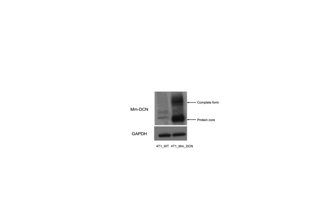
Figure 1: Overexpression of DCN in 4T1 breast cancer cells as compared to WT.
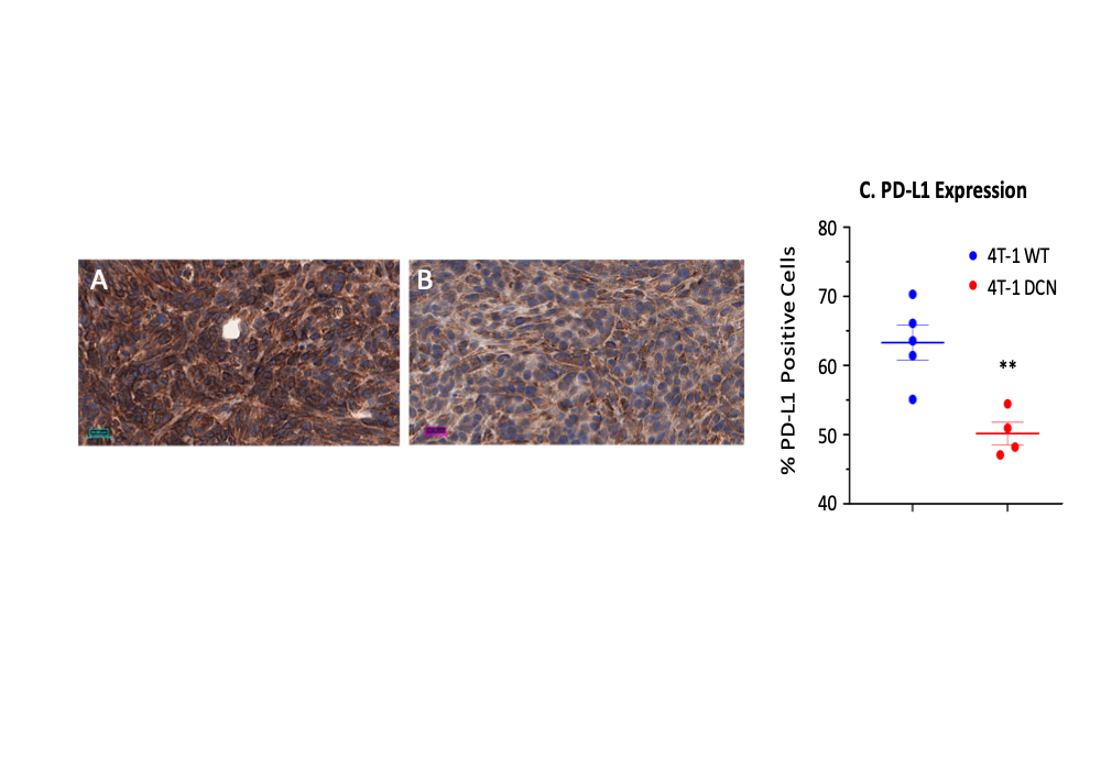
Figure 2: DCN overexpression decreases PD-L1 expression. Representative PD-L1 immunostained tumor section from (A) 4T1 wild type tumor (4T1-WT) and (B) DCN overexpressing (4T1-DCN) tumor. (C) A significant decrease of PD-L1 expression in IHC stained sections was observed with DCN overexpression in 4T1 tumors. **p<0.01
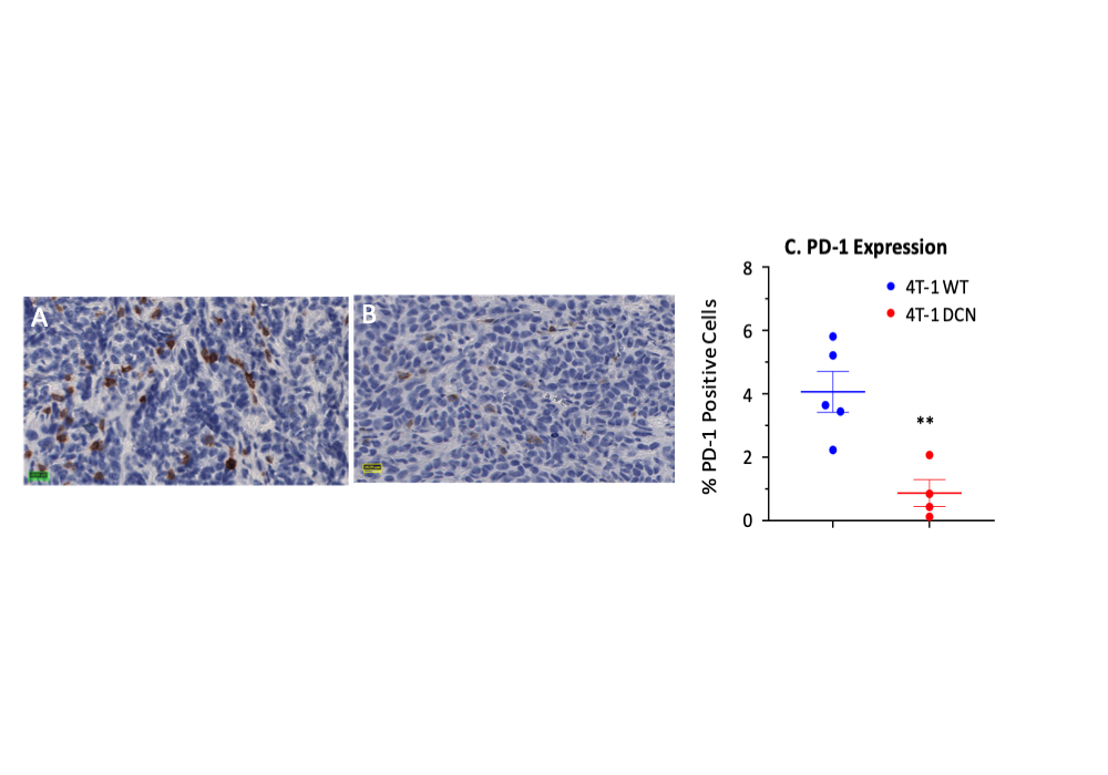
Figure 3: DCN overexpression decreases PD-1 expression. Representative PD-1 immunostained tumor section from (A) 4T1 wild type tumor (4T1-WT) and (B) DCN overexpressing (4T1-DCN) tumor. (C) A significant decrease of PD-1 in IHC stained sections was observed with DCN overexpression in 4T1 tumors. **p<0.01
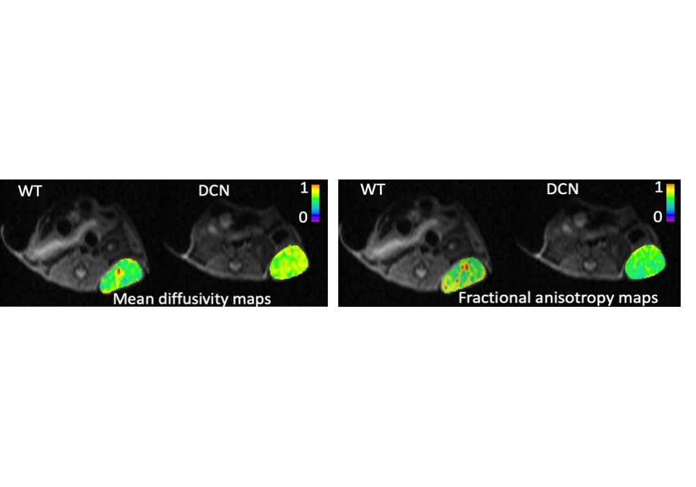
Figure A: Representative MD maps of WT and DCN overexpressing tumors overlaid with the corresponding B0 image. Figure B: Representative FA maps of WT and DCN tumors overlaid with the corresponding B0 image.
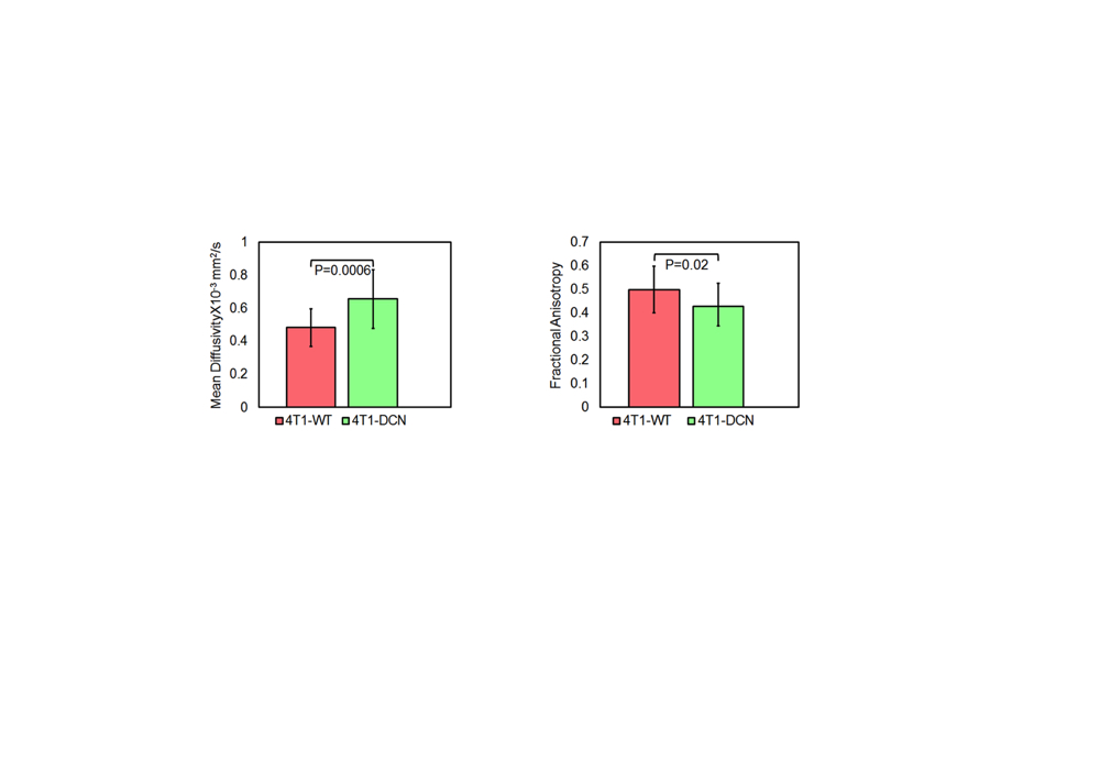
Figure A: MD increases with DCN overexpression. Figure B: FA decreases with DCN overexpression.
DOI: https://doi.org/10.58530/2024/2578