2575
Quantification of non-Gaussian diffusion in the human heart in vivo1Leeds Institute of Cardiovascular and Metabolic Medicine, University of Leeds, Leeds, United Kingdom, 2Cardiff University Brain Research Imaging Centre (CUBRIC), School of Psychology, Cardiff University, Cardiff, United Kingdom, 3Siemens Healthcare Ltd, Camberly, United Kingdom, 4Siemens Healthcare GmbH, Erlangen, Germany, 5Medical Radiation Physics, Clinical Sciences Lund, Lund University, Lund, Sweden
Synopsis
Keywords: DWI/DTI/DKI, Diffusion/other diffusion imaging techniques, Cardiac diffusion MRI, non-Gaussian diffusion, strong gradients, Diffusion Kurtosis imaging
Motivation: Diffusion tensor modeling, which is based on Gaussian diffusion, is commonly used in cardiac diffusion MRI (dMRI). However, the tissue's microstructure (cells, membranes, etc.) restricts the water molecules and deviates the signal from Gaussian behavior.
Goal(s): This effect may be observed for higher b-values, which are presently outside the realm of routine cardiac dMRI due to the limited gradient strength of clinical scanners.
Approach: Cardiac-gated, second-order motion-compensated dMRI were performed with $$$\mathrm{b_{max}=1500\,s/mm^2}$$$ in healthy volunteers on a 3T MRI scanner with $$$\mathrm{G_{max}=300\,mT/m}$$$.
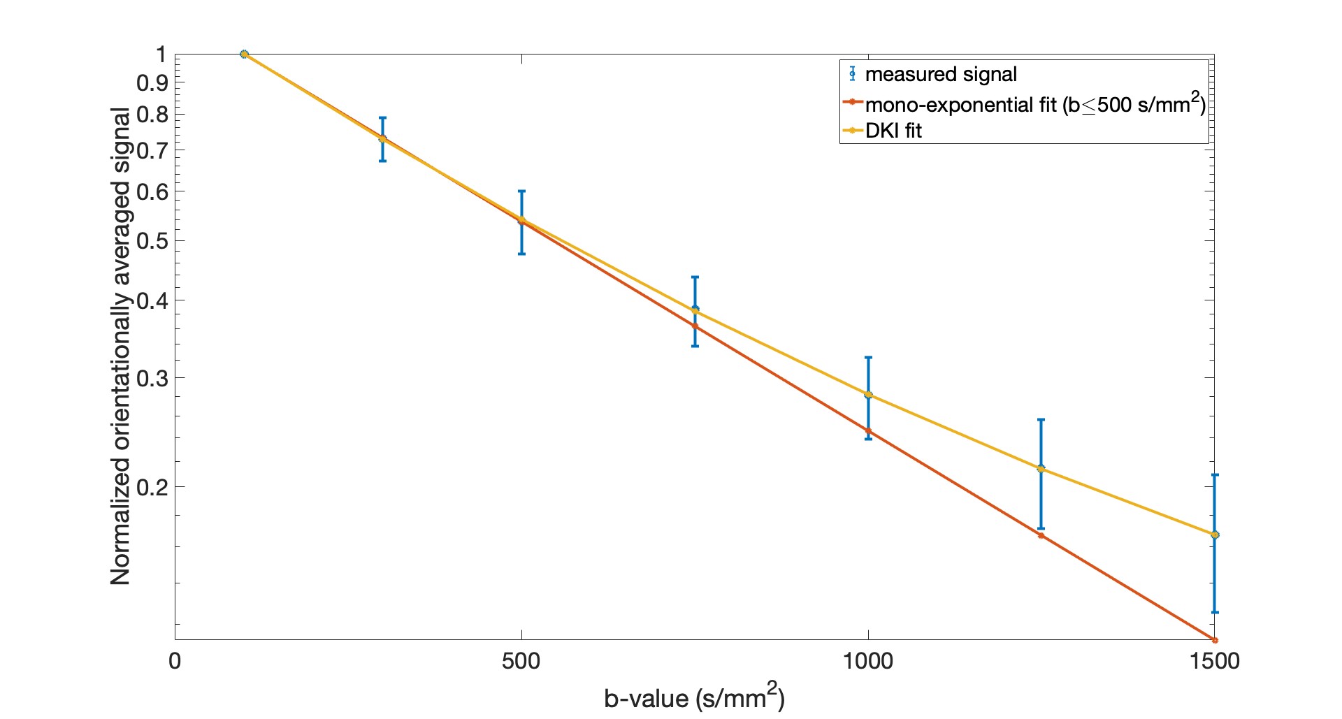
Results: We demonstrate deviation of the signal from Gaussian decay at $$$\mathrm{b>500\,s/mm^2}$$$ confirming the presence of non-Gaussian diffusion at higher b-values.
Impact: This work demonstrates feasibility of quantifying non-Gaussian diffusion in the human heart in vivo $$$\mathrm{\textbf{at realistic echo times}}$$$, using Connectom scanner ($$$\mathrm{G_{max}=300\,mT/m}$$$, 4-8 times stronger than clinical scanners). It may open the field for new biomarkers in cardiac diffusion MRI.
Introduction
Diffusion magnetic resonance imaging is a non-invasive technique to study tissue microstructure1. Cardiac diffusion tensor imaging (cDTI) which is most commonly used in microstructural investigations of the heart is based on Gaussian diffusion of water molecules2. However, due to the cell membranes and other restrictions in the biological tissue, the diffusion of water molecules deviates from the Gaussian model3-5. Diffusion kurtosis imaging (DKI), which has been proposed4 to quantify non-Gaussian diffusion, may offer metrics that are more sensitive to the presence of restrictions such as cell membranes and organelles. However, non-Gaussian analysis of in vivo human heart data has been limited. Teh et al.,6 have recently shown the feasibility of quantification of non-Gaussian diffusion in the human heart in vivo, however, the echo time (TE) of their experiment was very long ($$$>100 \, \mathrm{ms}$$$) resulting in low SNR at high b-values. The aim of this study is to investigate non-Gaussian diffusion in healthy human hearts in vivo using strong gradients (300 mT/m) at a TE = 77 ms (equivalent to the ones commonly used for cDTI) and $$$\mathrm{b_{max} = 1500 \, s/mm^2}$$$ for a second order motion compensated waveform.Methods
Data acquisitionCardiac diffusion-weighted images (cDWI) were acquired on a Connectom 3T MR imaging system with a maximum gradient strength of 300 mT/m and slew rate of 200 T/m/s. Data were acquired from two healthy volunteers who provided written consent. Diffusion gradient waveforms were designed using the NOW toolbox7-9 (https://github.com/jsjol/NOW) to provide optimized, Maxwell- and second-order motion compensated waveforms that can reach a specific b-value in the shortest echo time.cDWI was performed with a prototype pulse sequence that enables diffusion encoding with user-defined gradient waveforms and an EPI readout10. The diffusion-weighted imaging parameters were: TR = 3RR-intervals, TE = 77 ms, field‐of‐view = $$$320 \times 195 \,\mathrm{mm^2}$$$, resolution = $$$2.3 \times 2.3 \, \times 8 \mathrm{mm^3}$$$, slice gap = 8 mm, 3 short axis slices (base, mid, and apical), partial Fourier = 7/8, no parallel imaging, bandwidth = 2012 Hz/pixel. Each data set comprised 7 b-values [b = 100, 300, 500, 750, 1000, 1250, 1500 $$$\mathrm{s/mm^2}$$$] in 30 directions per shell with 4 repetitions, except for the lowest b-value which only had 3 directions. The total acquisition time was around one hour. Both magnitude and phase data were collected and used to generate the complex-valued images.
Data analysis
The phase variation was removed11 and real-valued diffusion-weighted images were then corrected for motion by a 2D rigid image registration and the outliers were removed12,13. The DKI model was fit to the data and the diffusion metrics such as fractional anisotropy (FA), mean diffusivity (MD), mean kurtosis ($$$\mathrm{\bar{K}}$$$), and axial ($$$\mathrm{K_{\mid\mid}}$$$) and radial kurtosis ($$$\mathrm{K_{\perp}}$$$) were calculated for each voxel14,15.
Results
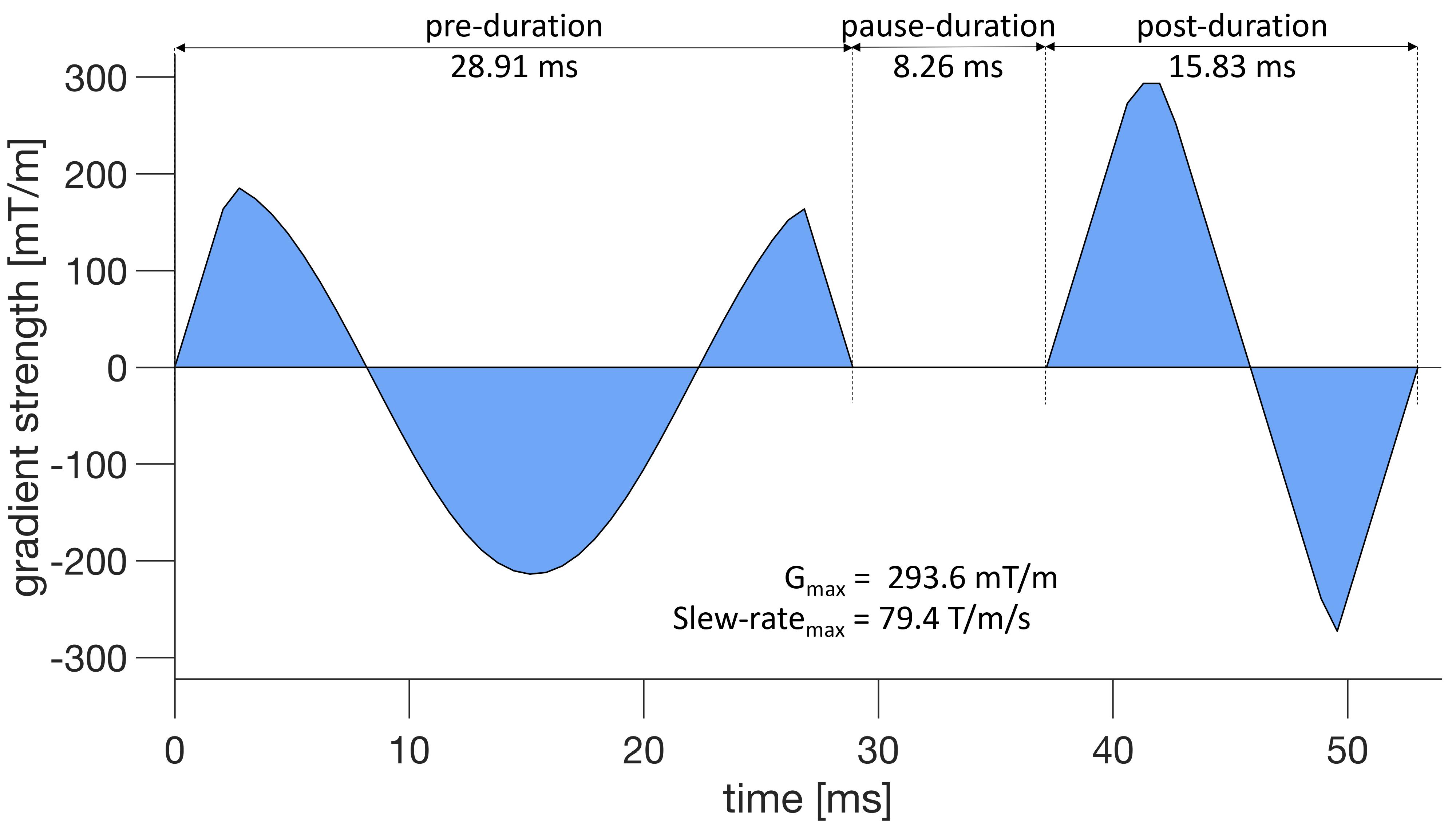
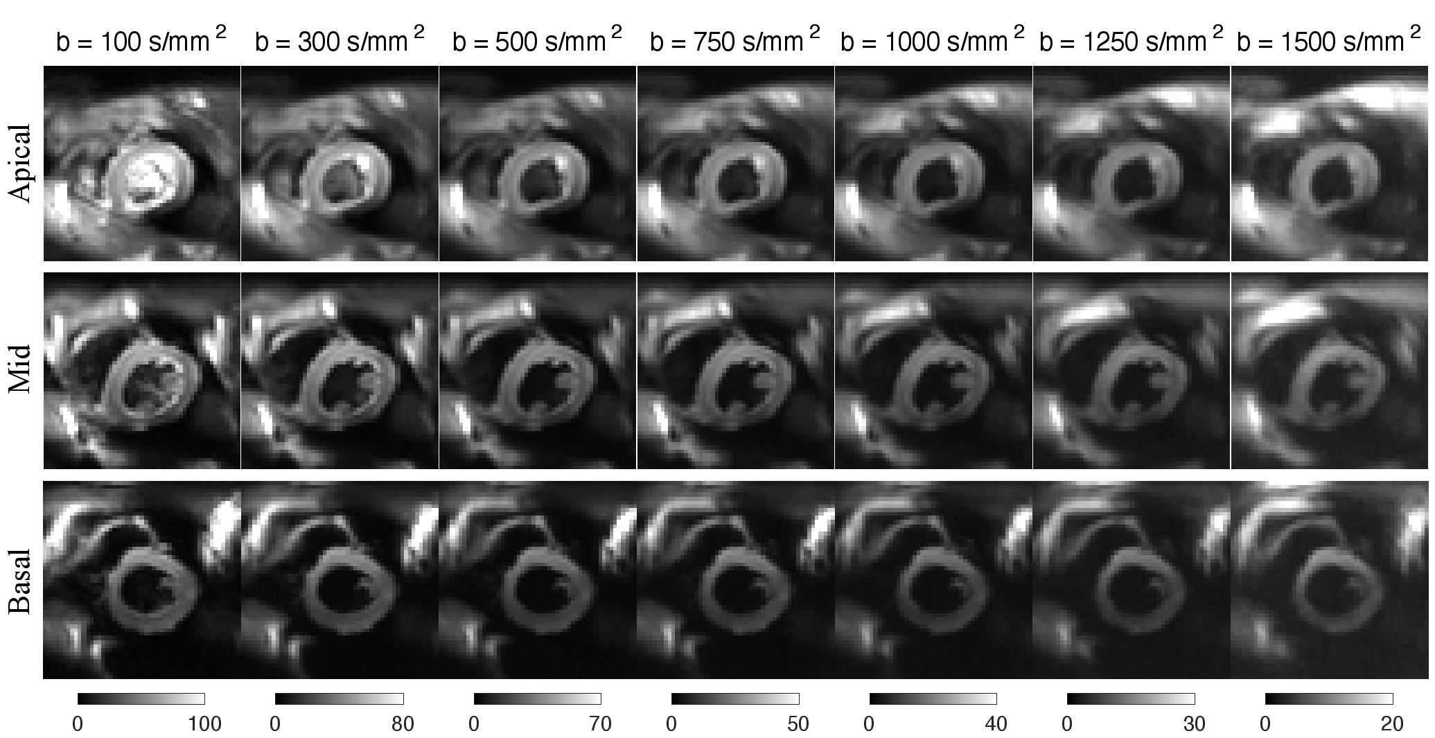
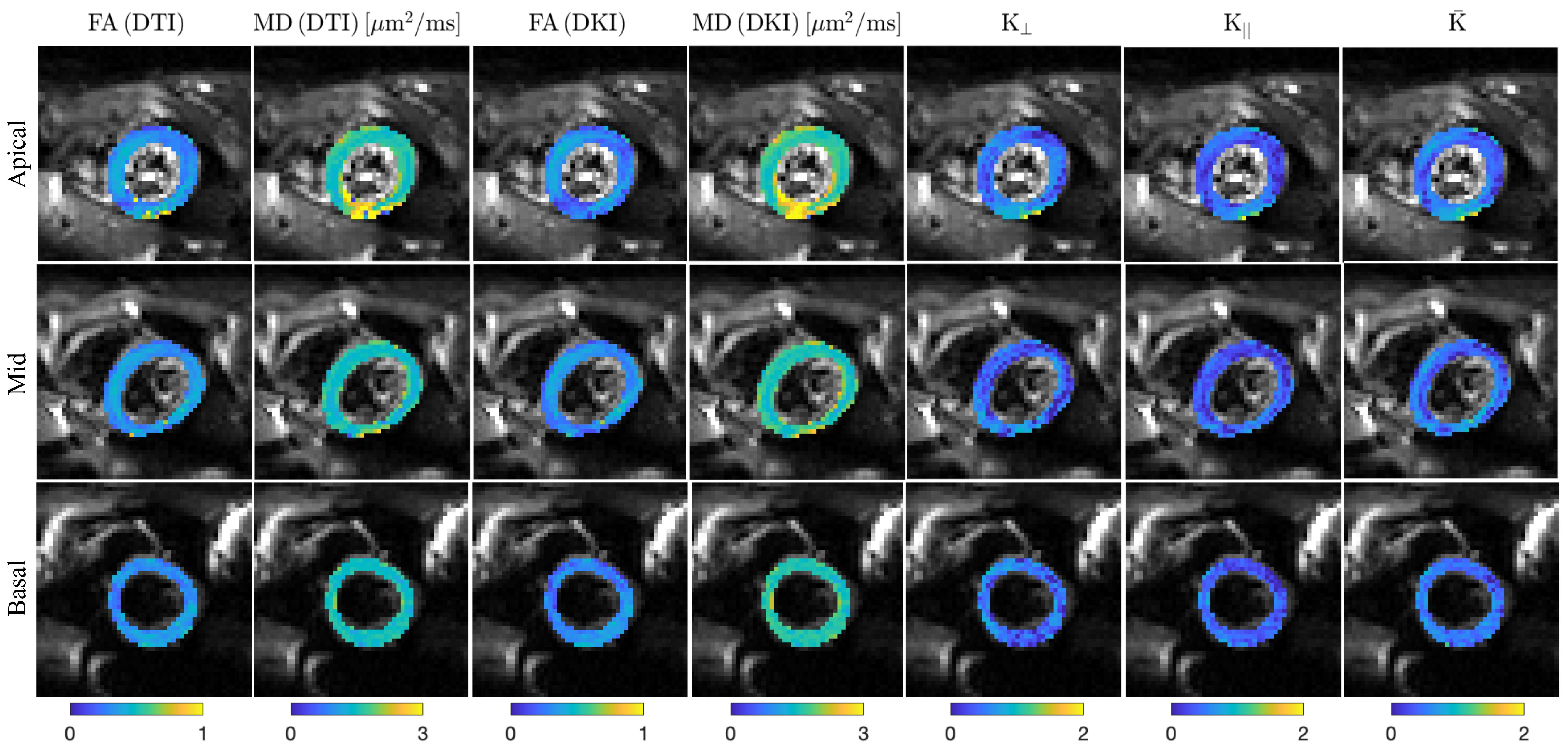
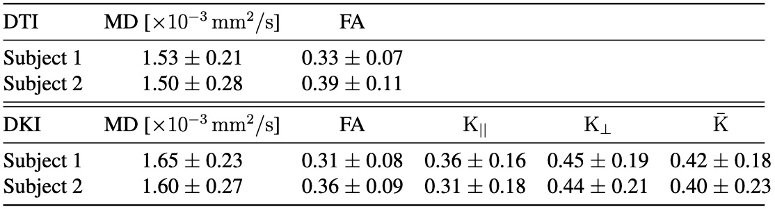
Figure 1 shows the diffusion gradient waveform optimized for second-order motion compensation with $$$\mathrm{b_{max} = 1500 \, s/mm^2}$$$. Figure 2 shows representative diffusion-weighted images obtained in short axis view with different b-values. Figure 3 shows the average signal over the left ventricle. The blue dots and error bars indicate the mean and standard deviation of the measured signal while the mono-exponential fit to the signal from $$$\mathrm{b\leq 500 \, s/mm^2}$$$ is shown in red and the DKI fit in yellow. The measured signal clearly deviates from the mono-exponential decay for $$$\mathrm{b>500 \, s/mm^2}$$$. Figure 4 shows the maps for estimated FA and MD for DTI ($$$\mathrm{b = 100 \, and \, 500 \, s/mm^2}$$$) and MD, FA, $$$\mathrm{K_{\perp}}$$$, $$$\mathrm{K_{\mid\mid}}$$$, and $$$\mathrm{\bar{K}}$$$ for DKI (all b-values). The average value of the DTI and DKI metrics are presented in Table 1.Discussion and Conclusion
In this study, we investigated the existence of non-Gaussian diffusion in human hearts in vivo using strong gradients (300 mT/m). In all hearts, radial kurtosis ($$$\mathrm{K_{\perp}}$$$) was observed larger than axial kurtosis ($$$\mathrm{K_{\mid\mid}}$$$). This may be due to the fact that there are more restrictions perpendicular to the cardiomyocyte orientations than parallel orientations. These results are in agreement with a previous study on in vivo human heart6, rat heart5 and ex vivo pig hearts16. The use of high-performance gradient system in this study, significantly improved the SNR efficiency of in vivo cardiac non-Gaussian diffusion imaging. The achieved sequence timings were comparable to standard cDTI acquisitions at more than three times ($$$> 3 \times$$$) higher b-values.Acknowledgements
We thank Siemens Healthcare for the pulse sequence development environment. This work was supported by Wellcome Trust Investigator Award (219536/Z/19/Z), EPSRC (EP/M029778/1), The Wolfson Foundation, and the British Heart Foundation (PG/19/1/34076).References
1. Moseley ME, Cohen Y, Kucharczyk J, Mintorovitch J, Asgari H, Wendland M, et al. Diffusion-weighted MR imaging of anisotropic water diffusion in cat central nervous system. Radiology. 1990;176(2):439-45.
2. Basser PJ, Mattiello J, LeBihan D. MR diffusion tensor spectroscopy and imaging.Biophysical journal. 1994;66(1):259-67.
3. Alexander D, Barker G, Arridge S. Detection and modeling of non-Gaussian apparent diffusion coefficient profiles in human brain data. Magnetic Resonance in Medicine: An Official Journal of the International Society for Magnetic Resonance in Medicine. 2002;48(2):331-40.
4. Jensen JH, Helpern JA, Ramani A, Lu H, Kaczynski K. Diffusional kurtosis imaging: the quantification of non-Gaussian water diffusion by means of magnetic resonance imaging. Magnetic Resonance in Medicine: An Official Journal of the International Society for Magnetic Resonance in Medicine. 2005;53(6):1432-40.
5. McClymont D, Teh I, Carruth E, Omens J, McCulloch A, Whittington HJ, et al. Evaluation of non-Gaussian diffusion in cardiac MRI. Magnetic resonance in medicine. 2017;78(3):1174-86.
6. Teh I, Shelley D, Boyle JH, Zhou F, Poenar AM, Sharrack N, et al. Cardiac q-space trajectory imaging by motion-compensated tensor-valued diffusion encoding in human heart in vivo. Magnetic Resonance in Medicine. 2023;90(1):150-65.
7. Sjölund J, Szczepankiewicz F, Nilsson M, Topgaard D, Westin CF, Knutsson H. Constrained optimization of gradient waveforms for generalized diffusion encoding. Journal of Magnetic Resonance. 2015;261:157-68.
8. Szczepankiewicz F, Westin CF, Nilsson M. Maxwell-compensated design of asymmetric gradient waveforms for tensor-valued diffusion encoding. Magnetic resonance in medicine. 2019;82(4):1424-37.
9. Szczepankiewicz F, Sjölund J, Dall’Armellina E, Plein S, Schneider JE, Teh I,et al. Motion-compensated gradient waveforms for tensor-valued diffusion en-coding by constrained numerical optimization. Magnetic resonance in medicine. 2021;85(4):2117-26.
10. Szczepankiewicz F, Sjölund J, Ståhlberg F, Lätt J, Nilsson M. Tensor-valued diffusion encoding for diffusional variance decomposition (DIVIDE): Technical feasibility in clinical MRI systems. PLoS One. 2019;14(3):e0214238.
11. Eichner C, Cauley SF, Cohen-Adad J, Möller HE, Turner R, Setsompop K, et al. Real diffusion-weighted MRI enables true signal averaging and increased diffusion contrast. NeuroImage. 2015;122:373-84.
12. Marstal K, Berendsen F, Staring M, Klein S. SimpleElastix: A user-friendly, multi-lingual library for medical image registration. In: Proceedings of the IEEE conference on computer vision and pattern recognition workshops; 2016. p. 134-42.
13. Coveney C, Kelly C, Teh I, Afzali M, Mueller L, Das A, et al. Semi-automated rejection of corrupted images in cardiac diffusion tensor imaging. Proceedings of the Annual Meeting of ISMRM. 2023.
14. Hansen B, Shemesh N, Jespersen SN. Fast imaging of mean, axial, and radial diffusion kurtosis. Neuroimage. 2016;142:381-93.
15. Henriques RN, Jespersen SN, Jones DK, Veraart J. Toward more robust and reproducible diffusion kurtosis imaging. Magnetic resonance in medicine. 2021;86(3):1600-13.
16. Mazzoli V, Froeling M, Nederveen AJ, Nicolay K, Strijkers GJ. Cardiac diffusion MRI beyond DTI. In: Proceedings of the 22nd Annual Meeting of ISMRM, Milan, Italy; 2014.
Figures