2573
Time-dependent diffusion MRI-based microstructural mapping of cervical cancer: correlation with immunohistochemical biomarkers1MRI, The First Affiliated Hospital of Zhengzhou University, Zhengzhou, Zhengzhou, China, 2Advanced Technical Support, Philips Healthcare, Beijing, China, 3the First Affiliated Hospital of Zhengzhou University, Zhengzhou, China
Synopsis
Keywords: Microstructure, Diffusion/other diffusion imaging techniques, cervical cancer, immunohistochemistry, time-dependent diffusion MRI, microstructural mapping, oscillating gradient spin-echo
Motivation: Some immunohistochemical (IHC) markers are gradually accepted as vital prognostic factors guiding therapy in cervical cancer (CC).
Goal(s): This study aimed to evaluate the relationship of time-dependent diffusion MRI (td-dMRI)-based microstructural mapping and IHC status of CC.
Approach: Quantitative information on cell microstructure are obtained by fitting the IMPULSED model to the td-dMRI data.
Results: Intracellular fraction (fin) and cellularity were significantly correlated with the PD-L1 expression, respectively. In addition, fin and cellularity were significantly correlated with Ki-67. The results suggested that microstructural mapping might facilitate the establishment of novel diffusion-derived biomarkers to estimate IHC status and orient treatment of CC.
Impact: The td-dMRI-based microstructural mapping showed the potential to estimate the IHC status of cervical cancer for the first time.
Introduction
Recently, numerous immunohistochemical (IHC) biomarkers associated with hallmarks of cervical cancer and prognosis have been identified. The Ki-67 is biomarker associated with proliferation, the human epidermal growth factor receptor 2 (HER2) is associated with tumourigenesis and apoptosis, and the biomarker p53 is associated with apoptosis. Evasion of immune destructing has been recognised as a new hallmark of cancer, and the IHC biomarker programmed death ligand 1 (PD-L1) is involved in immune escape pathways. Pembrolizumab, a programmed death-1 receptor inhibitor, is widely investigated and has received US Food and Drug Administration approval for patients with persistent, recurrent, or metastatic cervical cancer with a PD-L1 combined positive score (CPS) of ≥1 based on Keynote-158 [1]. This study aimed to investigate whether the tumor microstructural properties obtained from diffusion time-dependent diffusion MRI (td-dMRI) could be used to reflect the expression of prognostic IHC biomarkers in cervical cancer.Methods
Seventy-one patients with pathologically confirmed cervical cancer underwent MRI on a 3T system (Ingenia Elition, Philips Healthcare, Best, the Netherlands). A house-made oscillating gradient spin-echo (OGSE) dMRI sequence with trapezoid-cosine gradients and pulse gradient spin-echo (PGSE) dMRI sequence were implemented with 2D echo-planar imaging acquisition. OGSE data were acquired at 17 Hz (effective td = 15 ms, 1 cycle, b = 0/250/500/750/1000 s/mm2), and 33 Hz (effective td = 7.5 ms, 2 cycles, b = 0/100/200/300 s/mm2). PGSE was acquired with b-value of 0/250/500/750/1000/1400/1800 s/mm2. Quantitative information on cell microstructure are obtained by fitting the IMPULSED (Imaging Microstructural Parameters Using Limited Spectrally Edited Diffusion) model to the td-dMRI data [2]. The microstructural parameters included mean cell diameter (d), intracellular fraction (fin), and extracellular diffusivity (Dex), while intracellular diffusivity (Din) was fixed at 1.58 μm2/ms to ensure fitting stability. Cellularity was represented as fin/d for simplicity. In addition, apparent diffusion coefficient (ADC) was calculated with all b values at each td. The regions-of-interest (ROIs) of tumor tissue were manually delineated on a representative single slice of DW images showing the maximum tumor area. The necrotic area or surrounding tissue was carefully excluded from the segmentation. The averaged values of microstructural parameters within the tumor ROIs (d, fin, Dex, and ADC) were recorded. Spearman correlation was applied to evaluate the association between MR parameters and PD-L1 expression, parameters and Ki-67 LI. Parameter differences were assessed between different HER2 and P53 status.Results
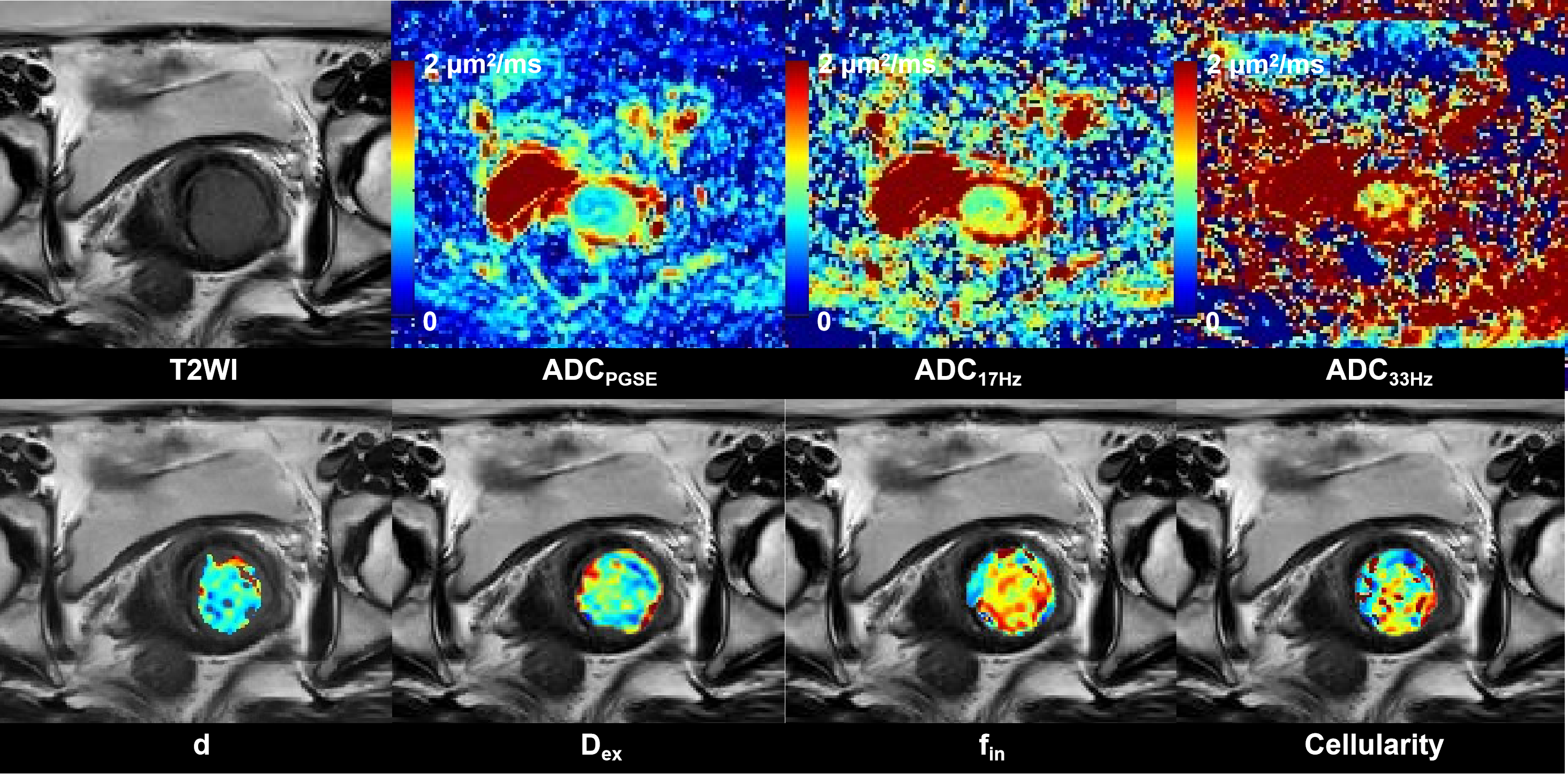
The Spearman correlation analysis showed that fin and cellularity were significantly correlated with the PD-L1 expression (r = 0.332 and 0.358, respectively, both p < 0.05). ADC values for OGSE/17Hz (ADC17Hz), and PGSE (ADCPGSE) were negatively correlated with the PD-L1 expression (r = -0.361 and 0.-369, respectively, both p < 0.05). No statistically significant associations were found between other parameters (d, Dex, ADC33Hz) and PD-L1 expression. In addition, fin and cellularity were significantly correlated with Ki-67 LI (r = 0.566 and 0.442, respectively, both p < 0.05). The d, Dex, ADC17Hz, ADC33Hz and ADCPGSE had no significant correlation with Ki-67 LI. None of the parameters differ significantly between HER2-positive and HER2-negaitive tumor, between P53-positive and P53-negaitive tumor. Representative images from conventional T2W, ADC, and td-dMRI-based microstructural mapping are shown in Figure 1.Discussion
In this study, our results demonstrated that both td-dMRI-based microstructural parameters and conventional ADCPGSE can be used to reflect the expression of PD-L1 in cervical cancer. It is reported that poorly differentiated cervical cancer had higher PD-L1 expression compared with grade 2 tumors [3]. The higher cell density and more significant cellular atypia in poorly differentiated tumor may result in the increased cellularity, intracellular fraction and decreased ADC. In addition, high PD-L1 levels was associated with high CD8 positive lymphocytic infiltrates in cervical cancer [3], which may also increased the cellularity in tumor tissue. Our study found that the microstructural parameters had advantages over the ADCPGSE in the prediction of Ki-67 LI. This result may indicate that td-dMRI-based microstructural mapping may be better than conventional PGSE dMRI in reflecting cell proliferation level in cervical cancer. OGSE dMRI can shorten the diffusion times by using rapidly oscillating gradients and is sensitive to smaller spatial scales in the tissues compared with conventional PGSE dMRI [4]. By fitting a combination of OGSE and PGSE signals to specific biophysical models, we can estimate important microstructural properties such as cell size, cell volume fraction, and cellularity, which are closely related to the pathological changes of tumor.Conclusion
Our study demonstrated the feasibility of reflecting prognostic IHC biomarkers status in cervical cancer using the td-dMRI-based microstructural parameters for the first time.Acknowledgements
NoReferences
- Chung HC, Ros W, Delord JP, et al. Efficacy and Safety of Pembrolizumab in Previously Treated Advanced Cervical Cancer: Results From the Phase II KEYNOTE-158 Study. J Clin Oncol 2019;37:1470-1478.
- Xu J, Jiang X, Li H, et al. Magnetic resonance imaging of mean cell size in human breast tumors. Magn Reson Med 2020;83:2002-2014.
- Saglam O, Zhou J, Wang X, et al. PD-L1 Expression Correlates With Young Age and CD8+ TIL Density in Poorly Differentiated Cervical Squamous Cell Carcinoma. Int J Gynecol Pathol 2020;39:428-435.
- Ejima F, Fukukura Y, Kamimura K, et al. Oscillating Gradient Diffusion-Weighted MRI for Risk Stratification of Uterine Endometrial Cancer. J Magn Reson Imaging 2023; Epub ahead of print. PMID: 37886909.
Figures