2566
Multi-Exponential Diffusion Image Analysis (MEDIA) of the human kidney: A clinical feasibility study1Department of Diagnostic and Interventional Radiology, Düsseldorf University Hospital, Düsseldorf, Germany
Synopsis
Keywords: IVIM, Microstructure
Motivation: Multi-exponential signal analysis is utilised to identify underlying present diffusion components in diffusion-weighted MRI signals. Various techniques emerged in recent years, but a detailed in-vivo comparison of the fitting approaches employed in the kidney is still missing.
Goal(s): Thus, we comparatively applied frequently used fitting methods and novel techniques towards precise in-vivo appliance.
Approach: The study comprised 15 healthy volunteers, intended for comparison with tumour patients. Besides NLLS and NNLS techniques, the pyramidal approach was employed
Results: Results demonstrated improvements for renal in-vivo data, a distinction between cortex and medulla was also accomplished. These encouraging findings conduct further investigations and comparison with pathologies
Impact: Identifying the most reliable and stable MEDIA approaches will pave the way for novel techniques. These advancements will enhance in-vivo applications, potentially allowing to distinguish between healthy and diseased tissue, recognise pathologies and long-term replace the need for biopsies.
Introduction
The multi-exponential diffusion signal is composed of various components originating from diffusion processes in the underlying tissue1. Various techniques have emerged in recent years for multi-exponential signal analysis in diffusion-weighted MRI signals2,3. However, a comprehensive in-vivo comparison of the fitting approaches employed in the kidney is still lacking. In this study, we applied frequently used fitting methods and novel techniques comparatively to achieve precise in-vivo application on DWI data. Applied approaches particularly include common least-square approaches NLLS (non-linear) and NNLS (non-negative least-squares), as well as the pyramidal approach and a novel pre-fitting technique for multi-level fitting. . In this study we compared the performance of NLLS and NNLS approach for the quantification of diffusion signal quantification in healthy kidney and renal tumour tissue. The findings of this study will enhance in-vivo applications, potentially enabling the distinction between healthy and diseased tissue and identification of various pathologies.Methods
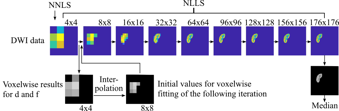
Based on literature4,5 and considering the optimal parameter sets for MEDIA determined in an earlier study by our own study group6, a number of 16 b-values were selected. For comparability, we incorporated two b-value ranges, including the common range of up to 750 mm²/s and an expanded range up to a maximum b-value of 950 mm²/s. The NNLS utilised 300 logarithmically spaced diffusion components and the fitting range was selected between Dmin = 0.7*10-3 mm²/s and Dmax = 300*10-3 mm²/s. The study involved 15 healthy volunteers, intended for future comparison with tumour patients. Four distinct fitting approaches were compared. The commonly used NLLS requires a priori knowledge regarding the number of diffusion compartments, restricting its use due to the influence of pathophysiological conditions on the number of diffusion compartments within the tissue5. Conversely, NNLS resolves these issues as it does not necessitate a specification of the diffusion coefficients a priori7. An advanced NNLS algorithm developed by our own study group in earlier work6 was employed and compared both pixel-wise and ROI-wise application. Another approach analysed is the pyramidal technique8, promising more stable results due to iterative NLLS fitting of downsampled versions of the original image (Figure 1). Finally, we tested a novel fitting approach utilising pre-fitting. The NNLS algorithm employs initial starting parameters obtained from a ROI-wide NLLS evaluation. This combination of both techniques results in a multi-level fitting procedure. To enable comparison, all outcomes were evaluated regarding their coefficient of variation (CoV).Results
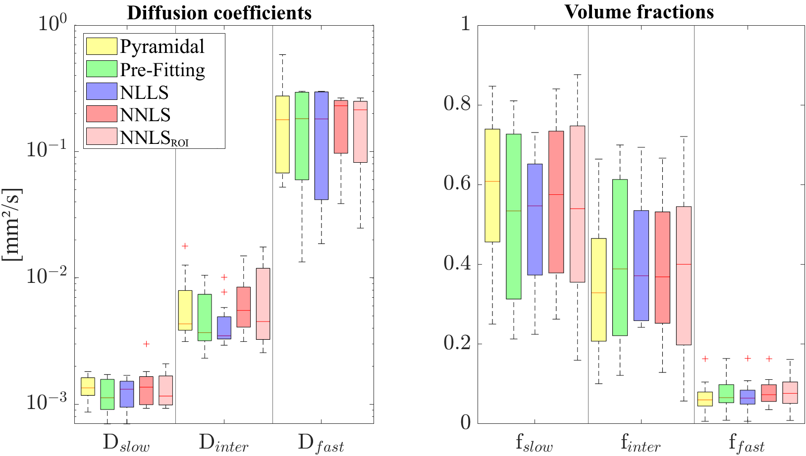
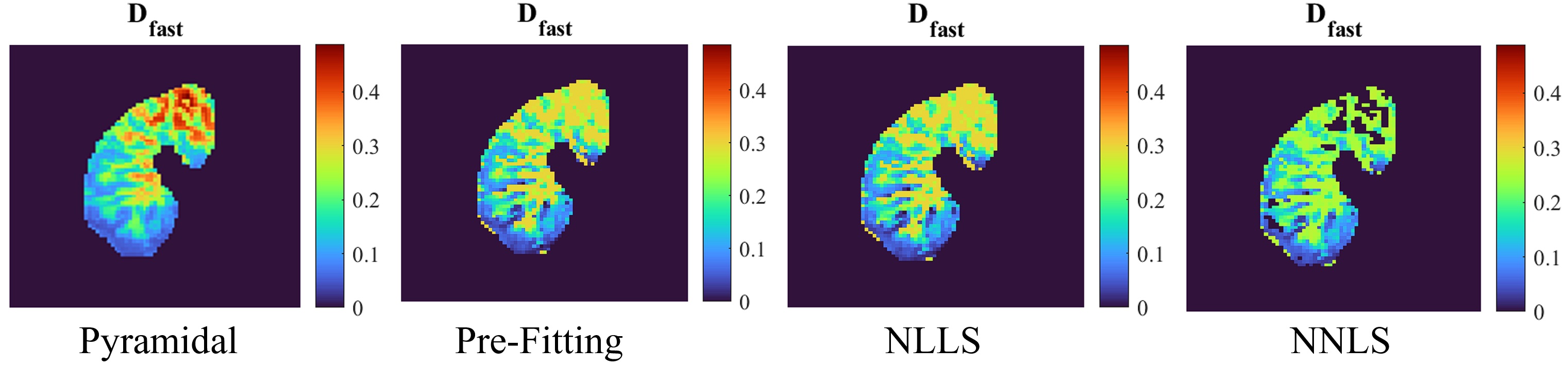
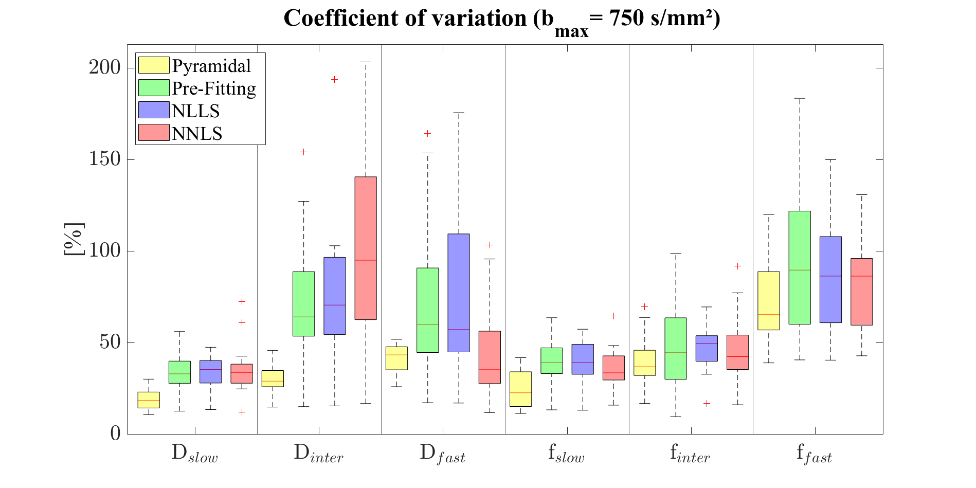
Although all methods produced similar results for D and f (Figure 2), only the NLLS, NNLSROI and the pre-fitting approach at bmax = 750 mm²/s were capable of distinguishing between cortex and medulla, considering Dinter in this cohort of 15 participants. These structures were also demonstrated in the heatmaps (Figure 3). The NLLS algorithm exhibited considerable variations overall (Figure 4). NNLS showed reliable results, but had difficulty identifying the intermediate component, resulting in high CoV values for Dinter. The pyramidal approach yielded the smallest variations for all parameters. Using the pre-fitting approach did not lead to an improvement in results. Both b-value ranges produced similar results, with less variation for the slow and intermediate components at a bmax of 950 mm²/s.Discussion
All methods yielded results consistent with previous research. The three volume fractions were distinguishable for every method employed at bmax of 750 mm²/s. With Dinter it became feasible to differentiate between cortex and medulla using NNLSROI, NLLS and the pre-fitting approach. Reliability was assessed using the CoV for all methods, indicating the pyramidal approach as the most suitable for achieving optimum and consistent overall outcomes. It is, however, important not to underestimate the potential of NNLS’s free fitting approach suitable for pathologies causing an alteration of diffusion components. The pre-fitting approach did not yield relevant benefits. Comparing the two b-value ranges demonstrates a maximum value of 750 mm²/s allowing for differentiation between the cortex and medulla and is a reasonable choice for renal imaging purposes.Conclusion
In conclusion, the pyramidal approach showcased superior reliability and consistency for multi-exponential diffusion image analysis in the human kidney. A maximum b-value of 750 mm²/s is sufficient for in-vivo applications and outperformed the higher threshold of bmax = 950 mm²/s. In the further course of this work, pathologies will be analysed through MEDIA and our results will be compared with those of tumour patients.Acknowledgements
The author of this work, Jonas Jasse, received a doctoral grant from the Jürgen-Manchot-Stiftung.References
1. Whittall KP, MacKay AL. Quantitative interpretation of NMR relaxation data. Journal of Magnetic Resonance (1969). 1989/08/01/ 1989;84(1):134-152. doi:10.1016/0022-2364(89)90011-5
2. MacKay A, Laule C, Vavasour I, Bjarnason T, Kolind S, Madler B. Insights into brain microstructure from the T2 distribution. Magn Reson Imaging. May 2006;24(4):515-25. doi:10.1016/j.mri.2005.12.037
3. van Baalen S, Leemans A, Dik P, Lilien MR, ten Haken B, Froeling M. Intravoxel incoherent motion modeling in the kidneys: Comparison of mono-, bi-, and triexponential fit. Journal of Magnetic Resonance Imaging. 2017;46(1):228-239. doi:10.1002/jmri.25519
4. Chevallier O, Wang YXJ, Guillen K, Pellegrinelli J, Cercueil JP, Loffroy R. Evidence of Tri-Exponential Decay for Liver Intravoxel Incoherent Motion MRI: A Review of Published Results and Limitations. Diagnostics (Basel). Feb 23 2021;11(2)doi:10.3390/diagnostics11020379
5. Periquito J, Gladytz T, Millward J, et al. Continuous diffusion spectrum computation for diffusion-weighted magnetic resonance imaging of the kidney tubule system. Quantitative Imaging in Medicine and Surgery. 07/01 2021;11:3098-3119. doi:10.21037/qims-20-1360
6. Jasse J, Wittsack H-J, Thiel T, et al. Toward optimal fitting parameters for multi-exponential DWI image analysis of the human kidney: A simulation study comparing different fitting algorithms. Magn Reson Med. 2023;(Manuscript submitted for publication)
7. Bjarnason TA, Mitchell JR. AnalyzeNNLS: magnetic resonance multiexponential decay image analysis. J Magn Reson. Oct 2010;206(2):200-4. doi:10.1016/j.jmr.2010.07.008
8. Stabinska J, Zollner HJ, Thiel TA, Wittsack HJ, Ljimani A. Image downsampling expedited adaptive least-squares (IDEAL) fitting improves intravoxel incoherent motion (IVIM) analysis in the human kidney. Magn Reson Med. Mar 2023;89(3):1055-1067. doi:10.1002/mrm.29517
Figures