2565
Spatial habitats features derived from IVIM for the HER2 status prediction of breast cancer: a preliminary study1Department of Radiology, The Fourth Hospital of Hebei Medical University, Shijiazhuang, China, 2MR Research Collaboration, Siemens Healthineers Ltd., Beijing, China, 3East China Normal University, Shanghai, China, 4MR Application Predevelopment, Siemens Healthineers AG, Erlangen, Germany
Synopsis
Keywords: IVIM, Diffusion/other diffusion imaging techniques, IVIM,MRI, breast tumor, the HER2 factor, habitats
Motivation: Besides immunohistochemistry, a non-invasive method based on tumor habitats is worth exploring. It predicted the expression of HER2 factor in malignant breast cancer.
Goal(s): We determined whether a prediction model, which was based on tumors habitats, could be used to predict the expression of HER2 factor.
Approach: A predictive model based on tumors habitats was developed and used to diagnose HER2 status. The IVIM-based imaging technique could extract features.
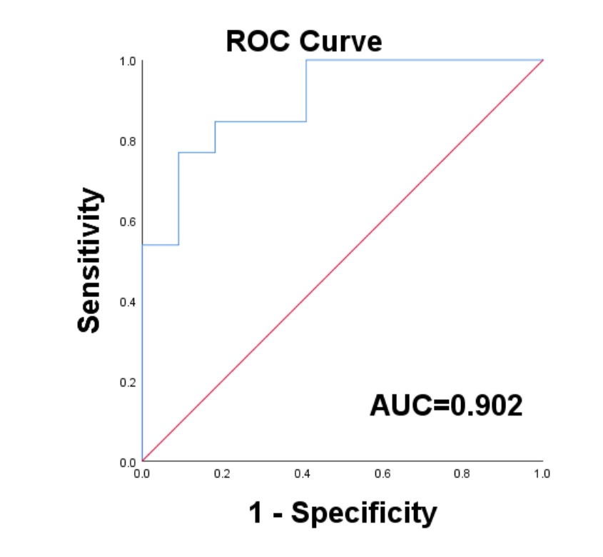
Results: The model of tumor habitats predicted the expression of HER2 factor in breast cancer by using the following parameters: AUC of 0.902, sensitivity of 0.709, and specificity of 0.909.
Impact: In this experiment, a predictive model based on features of habitats could determine the expression of HER2 factor in patients with malignant breast cancer. Thus, this non-invasive approach is a better treatment option in clinical practice.
Introduction
Previous studies have reported that about the heterogeneity of tumors in HER2-positive breast cancer patients1. The prognosis of these patients is better when they are accurately identified at an early stage, and targeted therapy is provided. This implies that HER2 factor is an important reference indicator 2. Meanwhile, heterogeneous tumor perfusion involves adverse events, such as an insufficient supply of microcirculation within the lesion. This leads to the development of hypoxic condition3 . To predict the prognosis of cancer, researchers should analyze the microenvironment of tumors. It is usually characterized by local hypoxia, low pH, and nutrient deficiency. Tumor habitat analysis is a novel data driven method, which is used to classify voxels with similar features in a tumor. Tumor subregions are classified with clustering methods, and tumor segmentation is achieved. Then, the characteristics of the subregions are extracted to evaluate tumor heterogeneity. Intravoxel incoherent motion (IVIM) is an enhanced functional imaging method, which evaluates the diffusion of water molecules in tissues (D) and the microvascular perfusion fraction of tumors (FP). The results reflect the consumption and supply of oxygen. In this study, our aim was to explore the application value of first-order radiomics features, which were extracted from tumor subregions, by these imaging-defined IVIM technique. Thus, the expression of HER2 factor was predicted in malignant breast cancer tissues.Methods
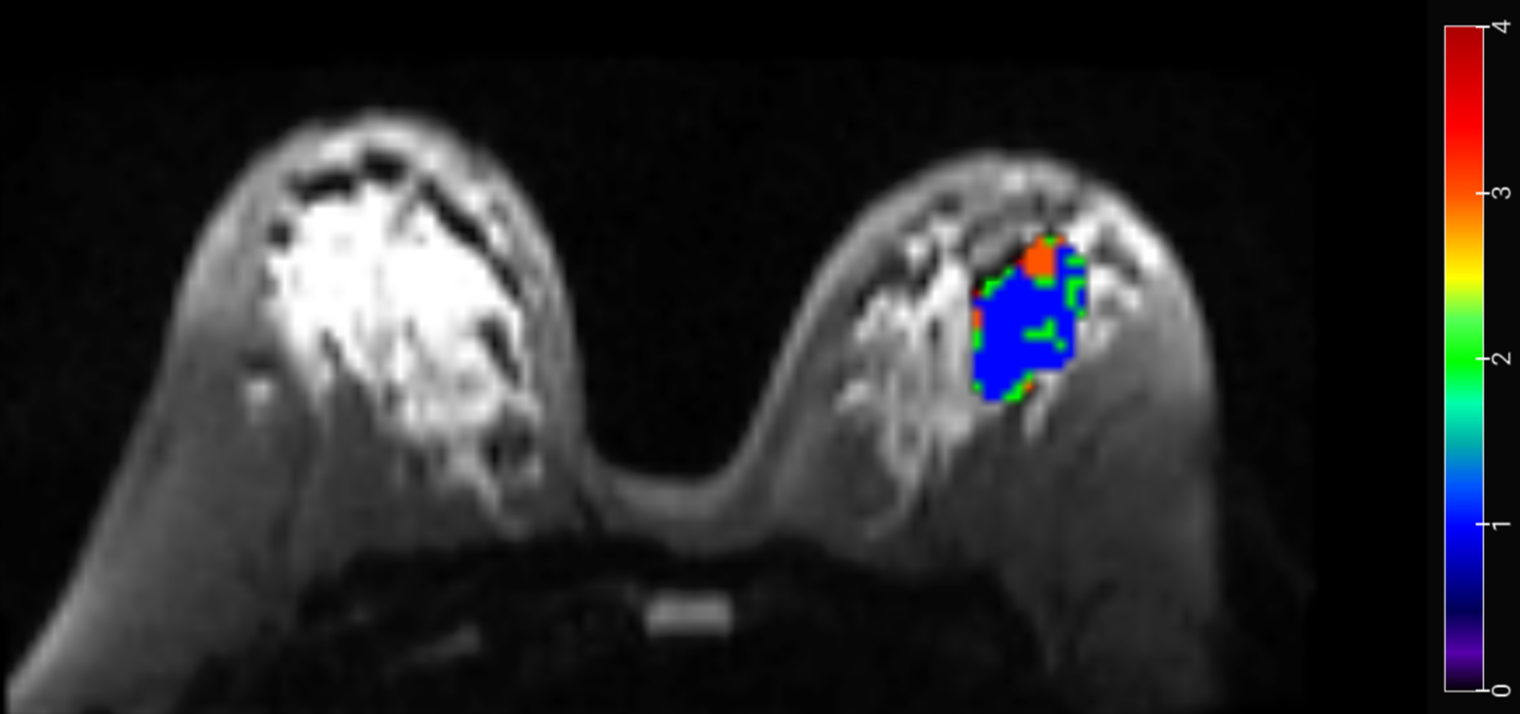
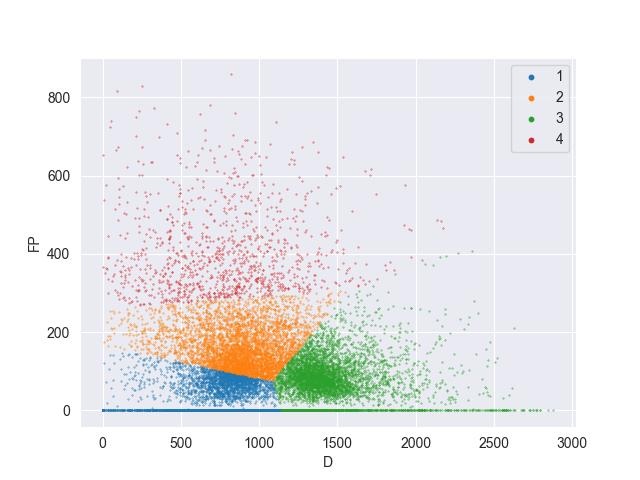
We recruited 35 cases of malignant breast cancer. Invasive ductal carcinoma was confirmed by surgery and pathology. Twenty two patients were HER2-negative, and thirteen patients were HER2-positive. A 3T MR scanner (MAGNETOM Vida, Siemens Healthineers, Erlangen, Germany) was used to perform an MRI scan on the breasts of all the patients. The IVIM data was acquired by a single-shot spin-echo echo planar imaging (SE EPI) sequence, with 9 b values of 0, 20, 40, 80, 120, 200, 400, 800, 1200 s/mm2 (TR = 6000 ms, TE = 61 ms, slice thickness = 4.5 mm, slice gap = 0.9 mm, FOV = 340 × 180 mm, matrix = 128 × 128). The matrices of IVIM (D, D* and FP) were calculated with a research software (MR Body Diffusion Toolbox v1.4.0, Siemens Healthineers, Erlangen, Germany). Then, ROI that included whole tumors on b0 images was manually drawn with the 3D Slicer software (version 5.2.2). For each voxel, the D and FP of IVIM were used to segment the tumor into subregions. A K-means clustering method was performed with an in-house software named nnFAE. We identified subregion1 with low D and low FP (DlowFPlow), subregion2 with moderate D and moderate FP (DmoderateFPmoderate), subregion3 with high D and low FP (DhighFPlow), and subregion4 with low D and high FP (DlowFPhigh) (Figure 1, 2). Then, we extracted the first-order radiomics features from all four subregions. All the features were expressed as mean ± standard deviation. To evaluate the inter-group differences, we performed the nonparametric Mann-Whitney U Test analysis. Logistic regression analysis was conducted to develop a predictive model based on indicators with significant differences. Then, the performance of this model was determined by estimating the area under the receiver operating characteristic curve (AUC).Results
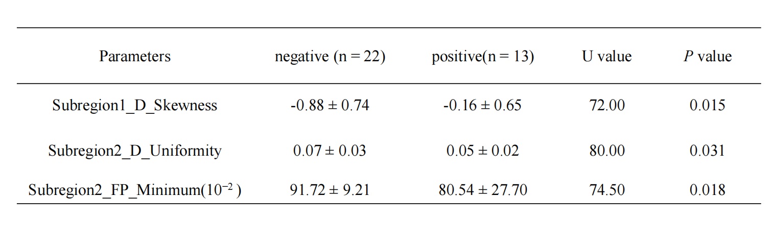
For the HER2-negative group and HER2-positive group, significant differences exist between the features of subregions with DlowFPlow and DmoderateFPmoderate. (P<0.05) (Table 1). The logistic regression model performed excellently in predicting the expression of HER2. The parameters were as follows: an AUC of 0.902, sensitivity of 0.709, and specificity of 0.909 (Table 2, Figure 3).Discussion and conclusion
We evaluated whether tumor microenvironment (habitats) could predict the expression of HER2 in breast cancer tissues. Using the clustering method and IVIM metrics (D and FP), we segmented the tumor into four subregions. The features of DlowFPlow and DmoderateFPmoderate subregions represented the hypoperfusion (low oxygen supply) and hypercellularity (high oxygen consumption) regions, respectively. The skewness of D of DlowFPlow subregion was higher in the HER2-negative group than in the HER2-positive group. Therefore, the intra-tumor spatial heterogeneity was higher in the HER2-negative group. Compared to the HER2-positive group, the uniformity of D in the DmoderateFPmoderate subregion was higher for the HER2-negative group. Therefore, the intra-tumor heterogeneity was higher in the HER2-positive group. Compared to the HER2-positive group, the minimum of FP of DmoderateFPmoderate subregion was higher in the HER2-negative group. Therefore, the HER2-positive tumor was more aggressive; its microcirculation perfusion had declined; and it lacked the supply of oxygen and nutrients. Such a tumor was more likely to degrade 4,5.Therefore, the logistic regression model, which was based on the features of habitats, can predict the HER2 status and the prognosis of malignant breast cancer tissues.Acknowledgements
These authors are grateful to thank these patients of this entire study. And they thank the guarantors who have assisted in data interpretation.References
1.Takada M, Toi M. Neoadjuvant treatment for HER2-positive breast cancer. Chin Clin Oncol. 2020;9(3):32. doi: 10.21037/cco-20-123. Epub 2020 Jun 3.
2.Mann RM, Cho N, Moy L. Breast MRI: State of the Art. Radiology. 2019;292(3):520-536. doi: 10.1148/radiol.2019182947. Epub 2019 Jul 30.
3.Mo T, Brandal SHB, Köhn-Luque A, et al . Quantification of Tumor Hypoxia through Unsupervised Modelling of Consumption and Supply Hypoxia MR Imaging in Breast Cancer. Cancers (Basel). 2022 4;14(5):1326. doi: 10.3390/cancers14051326.
4.Mo T, Brandal SHB, Köhn-Luque A, et al . Quantification of Tumor Hypoxia through Unsupervised Modelling of Consumption and Supply Hypoxia MR Imaging in Breast Cancer. Cancers (Basel). 2022;14(5):1326. doi: 10.3390/cancers14051326.
5.Jalnefjord O, Montelius M, Arvidsson J, et al. Data-driven identification of tumor subregions based on intravoxel incoherent motion reveals association with proliferative activity. Magn Reson Med. 2019 ;82(4):1480-1490. doi: 10.1002/mrm.27820. Epub 2019 May 13.
Figures