2564
Time-Dependent Diffusion MRI for Characterizing Microstructural Mapping of Prostate cancer Located in the Transition Zone1Department of Radiology, The First Affiliated Hospital, Sun Yat-Sen University, Guangzhou, China
Synopsis
Keywords: Microstructure, Prostate, Time-dependent diffusion magnetic resonance imaging; microstructure;transition zone
Motivation: Time-dependent diffusion MRI (td-dMRI) has the potential to characterize the microstructure mapping of tissue in vivo.
Goal(s): Whether it can benefit accurate and accessible risk stratification for prostate cancer especially in trasition zone has not been established.
Approach: In this study, patients who underwent prostate MRI including td-dMRI were enrolled. Correlation between the td-dMRI-based microstructural parameters and International Society of Urological Pathology grade groups (ISUP GG) were investigated and their performance in discriminating clinically significant prostate cancer (csPCa) from indolent disease was also evaluated.
Results: The results showed that Td-dMRI measurements has good differential diagnostic power, and cellularity achieved the highest diagnostic performance.
Impact: Time-dependent diffusion MRI–derived microstructural parameters showed high discrimination between clinically significant prostate cancer from indolent disease in the transition zone, which might thereby add an extra dimension in the development of prostate cancer biomarkers.
Introduction
There are 25%–40% of prostate cancer (PCa) arise from the transition and central zones1. At magnetic resonance imaging (MRI), the appearance of the transition zone can be heterogeneous due to the co-existence of stromal hyperplasia, prostatitis, and varying degrees of glandular, which increases difficulty for the detection,and risk strafification of transition zone cancer2. Histopathologically, PCa is characterized by the aggregation of physical deformation of glandular architecture on the micrometer scale. And numerous studies have demonstrated that the tissue composition and cellularity changed with the increase of International Society of Urological Pathology grade groups (ISUP GG)3. Thus identification of the cellular microstructure in lesions might be helpful for differentiating the transition zone cancers from noncancerous tissue. The recently developed time-dependent diffusion MRI (td-dMRI) can capture the non-Gaussian behavior of water molecules at varying diffusion time (td) by using a combination of specialized diffusion encoding schemes, thus offers the deeper-level microstructural information about tumor pathology, such as cell size, cell volume fraction and transcytolemmal water exchange4,5. Technical feasibility of td-dMRI has been confirmed in various tumors including head and neck tumors6, breast cancer7, and PCa4,8. Preliminary work shows that td-dMRI-based microstructural parameters could be used for noninvasively characterizing cellular properties of PCa and for discriminating between clinically significant PCa and clinically insignificant disease4. However, the utility of td-dMRI for evaluation of those lesions located in transition zone lesion is now established. Therefore, the purpose of this study was to evaluate the utility of td-dMRI-based microstructural parameters in the characterization of transition zone lesions.Materials and Methods
Men with a clinical suspicion of PCa were enrolled prospectively between March 2023 and August 2023. Td-dMRI data were acquired with pulsed and oscillating gradient diffusion MRI sequences at an equivalent diffusion time of 7.5-30 msec on a 3.0-T scanner(Premier, GE). td-dMR-based microstructural parameters, including cell diameter, intracellular volume fraction (fin), extracellular diffusivity (Dex), and cellularity, were estimated with a two-compartment model. In addition, parameters of diffusion tensor imaging (DTI) at different td (33 Hz, 17 Hz, and 0 Hz) were also acquired. Correlation between the td-dMRI-based microstructural parameters and ISUP GG were investigated and their performance in discriminating clinically significant PCa (csPCa) (GG ≥2) from indolent disease (benign and GG 1) was also evaluated.Results
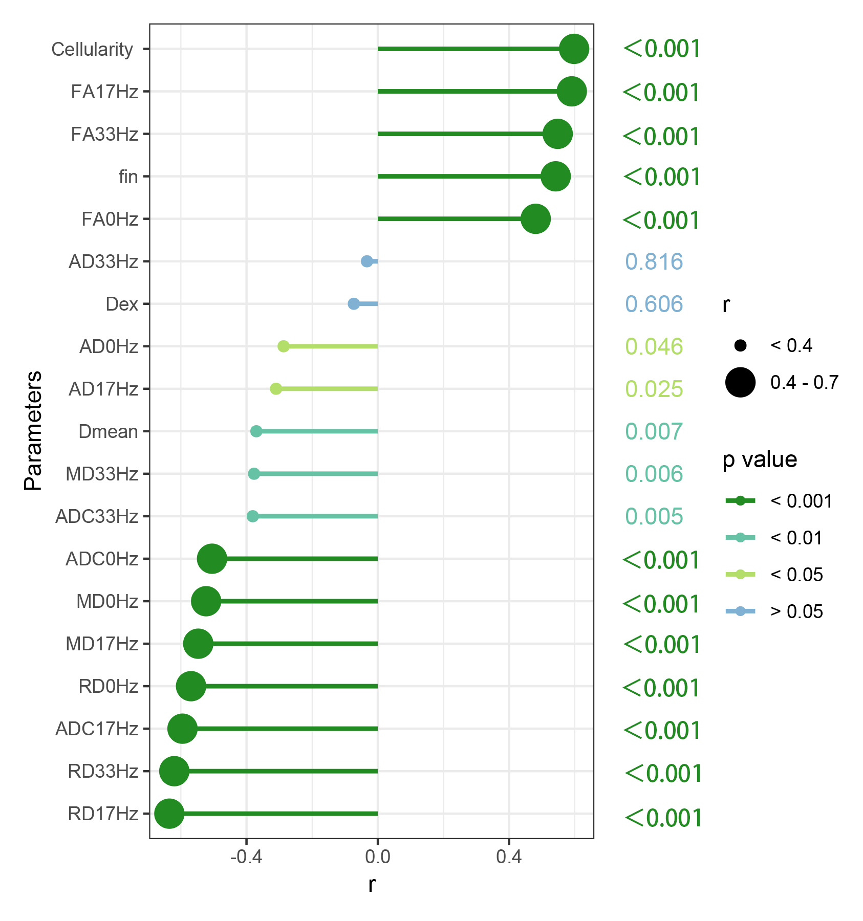
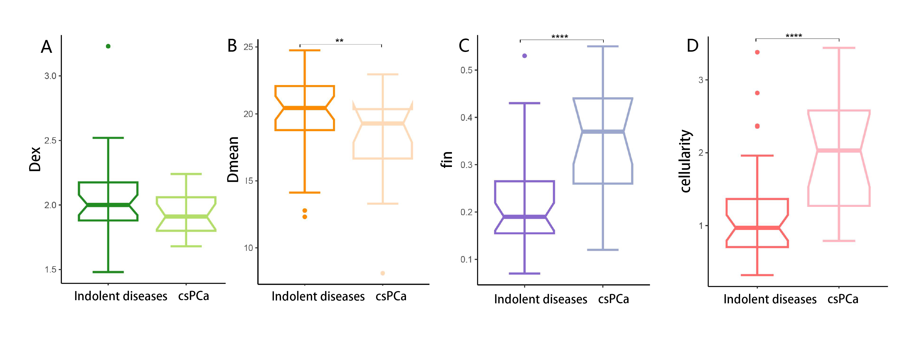
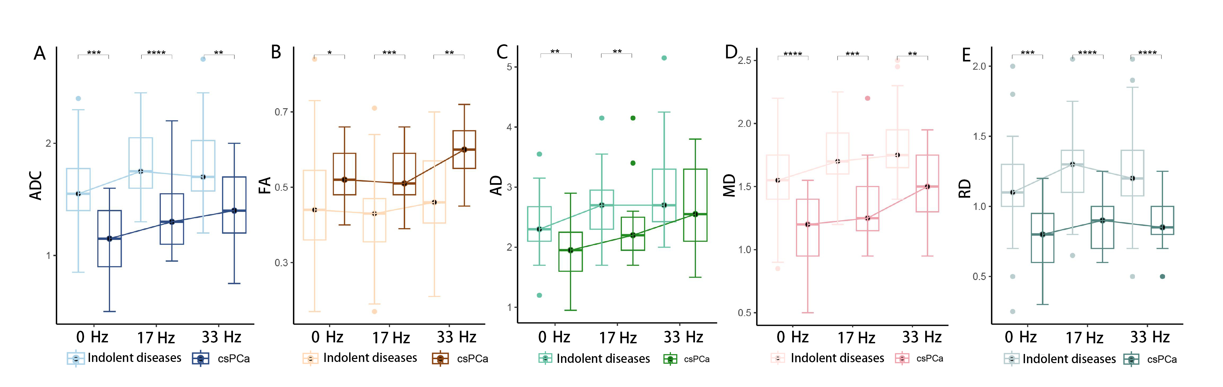
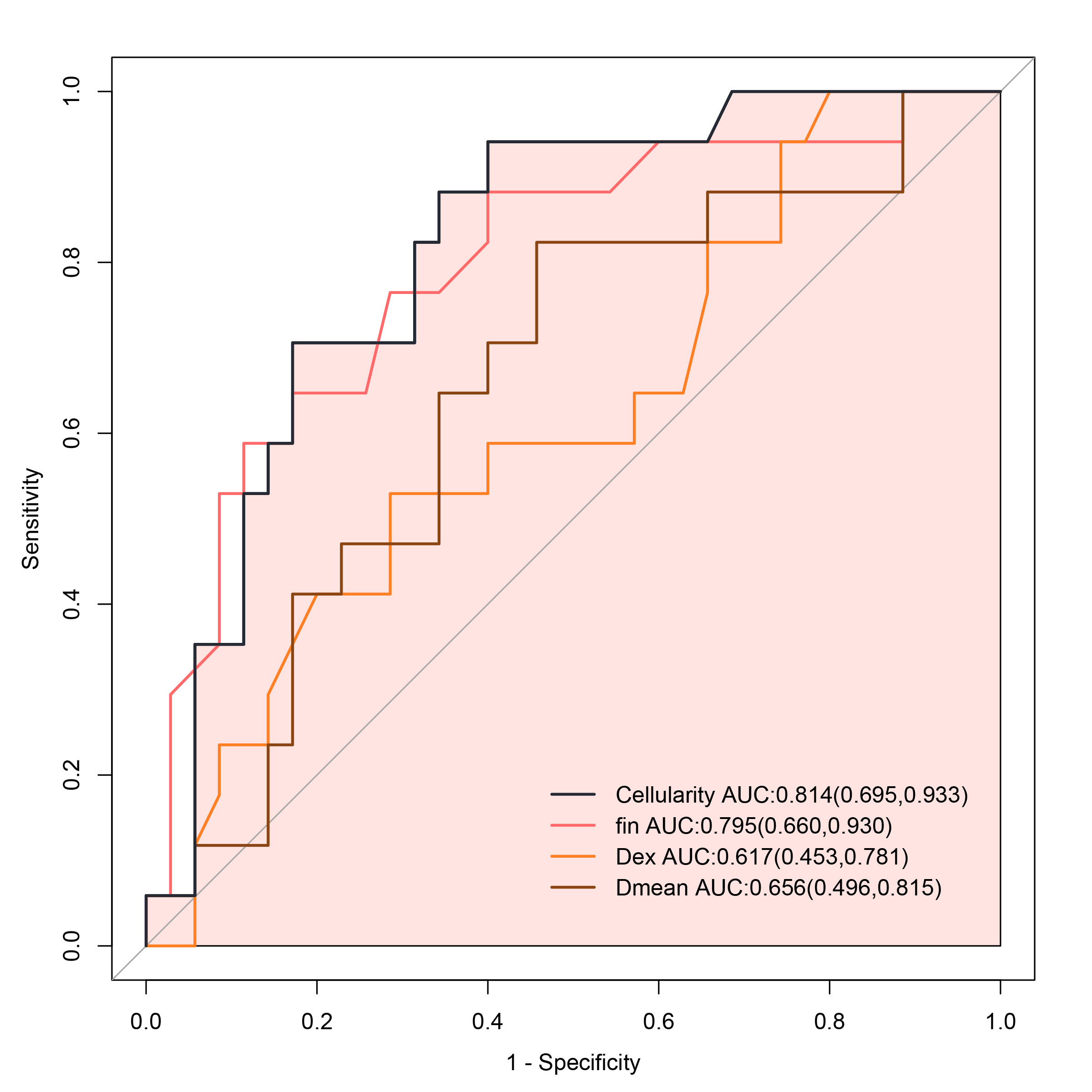
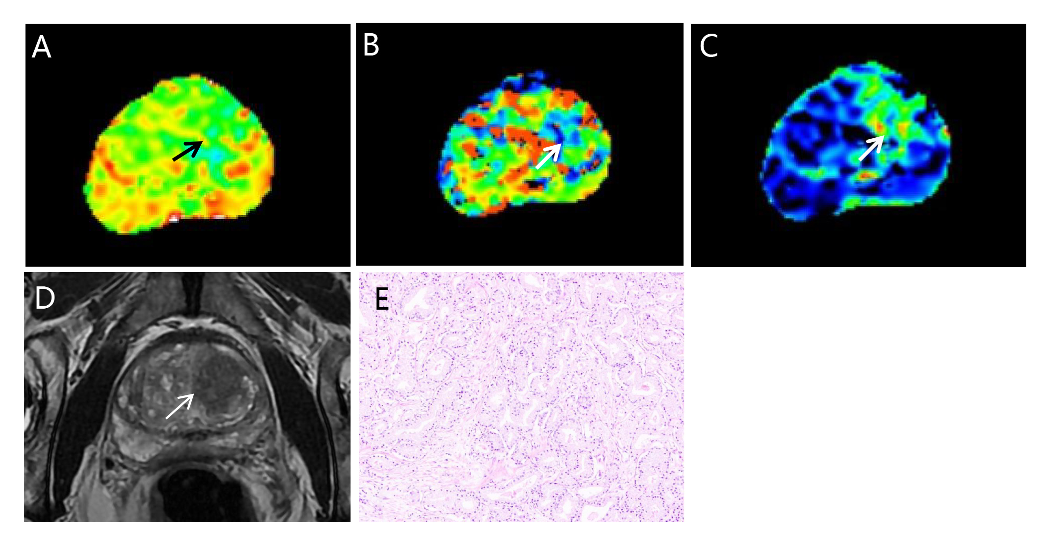
A total of 44 patients with 52 lesions were enrolled in this prospective study. The td-dMRI measurements showed that higher GG was correlated with higher cellularity and fin (r = 0.598, and 0.542, p<0.001), while cell diameter was negatively correlated with the ISUP GGs (r = -0.370, p = 0.007). No significant relationship was found between Dex and ISUP GGs. Approximate upward trends were observed in the DTI parameters at the increased frequency, which reflects the diffusion time dependency of prostate tissues. For the differentiation between csPCa from indolent disease, among all measurements derived from td-dMRI, cellularity achieved the highest diagnostic performance, with the area under the receiver operating characteristic curve of 0.814 (95% CI: 0.695, 0.933).Discussion
Non-Gaussian diffusion is closer to the movement and distribution of water molecules within biologic tissues. Its diffusion signal depends both on the diffusion weighting wave vector q and diffusion time t8. Diffusion time is an essential parameter of a diffusion-weighted imaging (DWI) sequence that determines the diffusion length of water molecules. The microstructural features of tissue, such as restriction size, permeability, and cellularity, can influenced the diffusion distance of water molecules at a given t. Td-dMRI can capture the microstructural restriction at different diffusion length scale by varying t, which is sensitive to the physical deformation due to cancer and might provide complimentary information to q-space imaging. A high b-value is needed to probe restricted diffusion in the conventional DWI that based on pulsed gradient spin-echo (PGSE), leading to a long diffusion time due to the limited maximum achievable gradient strength in clinical MRI systems. In contrast, the recently developed oscillating gradient spin-echo (OGSE) method can shorten the diffusion time by substituting the long-lasting diffusion-sensitizing gradients used in PGSE methods with rapid oscillation gradients, thus allows the exploration of the diffusion time dependencies at the short diffusion time regime9,10. At the study of Wu et al.4, the diagnostic value of td-dMRI for characterizing the microstructural features of PCa has been confirmed in clinical settings, which showed highly correlation with the pathologic findings. Our study further corroborated the feasibility and utility of td-dMRI for characterizing prostatic lesions located in transition zone .Conclusion
Td-dMRI based microstructural mapping may be useful for characterizing prostatic lesions in transition zone, which can further benifit the accurate detection and risk stratification.for prostate cancer.Acknowledgements
Not applicable.References
1. Panda A, Obmann VC, Lo WC, et al. MR Fingerprinting and ADC Mapping for Characterization of Lesions in the Transition Zone of the Prostate Gland. Radiology. 2019;292(3):685-694.
2. Akin O, Sala E, Moskowitz CS, et al. Transition zone prostate cancers: features, detection, localization, and staging at endorectal MR imaging. Radiology. 2006;239(3):784-792.
3. Chatterjee A, Watson G, Myint E, Sved P, McEntee M, Bourne R. Changes in Epithelium, Stroma, and Lumen Space Correlate More Strongly with Gleason Pattern and Are Stronger Predictors of Prostate ADC Changes than Cellularity Metrics. Radiology. 2015;277(3):751-762.
4. Wu D, Jiang K, Li H, et al. Time-Dependent Diffusion MRI for Quantitative Microstructural Mapping of Prostate Cancer. Radiology. 2022;303(3):578-587.
5. Zhang H, Liu K, Ba R, et al. Histological and molecular classifications of pediatric glioma with time-dependent diffusion MRI-based microstructural mapping. Neuro-oncology. 2023;25(6):1146-1156.
6. Iima M, Yamamoto A, Kataoka M, et al. Time-dependent diffusion MRI to distinguish malignant from benign head and neck tumors. Journal of magnetic resonance imaging : JMRI. 2019;50(1):88-95.
7. Ba R, Wang X, Zhang Z, et al. Diffusion-time dependent diffusion MRI: effect of diffusion-time on microstructural mapping and prediction of prognostic features in breast cancer. European radiology. 2023;33(9):6226-6237.
8. Lemberskiy G, Rosenkrantz AB, Veraart J, Taneja SS, Novikov DS, Fieremans E. Time-Dependent Diffusion in Prostate Cancer. Investigative radiology. 2017;52(7):405-411.
9. Wu D, Martin LJ, Northington FJ, Zhang J. Oscillating gradient diffusion MRI reveals unique microstructural information in normal and hypoxia-ischemia injured mouse brains. Magnetic resonance in medicine. 2014;72(5):1366-1374.
10. Baron CA, Beaulieu C. Oscillating gradient spin-echo (OGSE) diffusion tensor imaging of the human brain. Magnetic resonance in medicine. 2014;72(3):726-736.
Figures