2562
Comparative study of time-dependent diffusion MRI and conventional DWI for microstructural characterization of breast lesions1Department of Radiology, Beijing Hospital, National Center of Gerontology, Institute of Geriatric Medicine, Chinese Academy of Medical Sciences, Beijing, China, 2Key Laboratory for Biomedical Engineering of Ministry of Education, Department of Biomedical Engineering, College of Biomedical Engineering & Instrument Science, Zhejiang University, Zhejiang, China, 3MR Research Collaboration, Siemens Healthineers, Beijing, China
Synopsis
Keywords: Microstructure, Diffusion/other diffusion imaging techniques, apparent diffusion coefficient; MRI; breast lesions
Motivation: Time-dependent diffusion MRI (dMRI), thanks to its distinct advantages in characterizing tissue microstructure, has gained increasing popularity in clinical research in recent years.
Goal(s): However, its potential for distinguishing breast lesions remains uncertain.
Approach: The present study was conducted to compare the diagnostic performance of time-dependent dMRI parameters and ADC metrics derived from conventional DWI for breast lesions.
Results: The study results suggest that microstructural parameters based on time-dependent dMRI are superior to conventional DWI measurements in diagnosing breast lesions, and that the addition of time-dependent dMRI parameters improves the performance of conventional DWI in differentiating breast lesions.
Impact: Herein, the microstructural characteristics of breast lesions were investigated using time-dependent dMRI technique and compared with conventional DWI. It was found that that adding time-dependent dMRI parameters could improve the performance of conventional DWI in the diagnosis of breast cancer.
Background and Purpose
Diffusion magnetic resonance imaging (dMRI) is currently one of the most promising methods for the microstructural imaging of biological tissues1. Traditional dMRI such as conventional diffusion-weighted imaging (DWI) has been reported to be of great value in the diagnosis of breast cancer as a noninvasive and unenhanced imaging modality1. However, pulsed gradient spin echo (PGSE) sequence is extensively adopted in conventional DWI, and MRI systems have limited maximum gradient strength in clinical practice, which results in longer diffusion time for PGSE, thereby limiting the minimum microstructural size that can be measured2,3. Oscillating gradient spin echo (OGSE) sequence is a new technique developed on the basis of traditional dMRI, which reduces the effective diffusion time of measurable water molecules by applying an oscillating gradient, and obtains indicators of tissue microstructural parameters, such as extracellular water molecule diffusion coefficient (Dex), cell diameter, intracellular water molecule volume fraction (fin), cellularity and other microstructural characteristics, through mathematical modeling3,4. In this case, time-dependent dMRI may provide more detailed information about breast tissue microstructure, but its value in the diagnosis of breast lesions has been rarely reported5,6. First, the present study was carried out to evaluate the diagnostic performance of time-dependent dMRI parameters and quantitative apparent diffusion coefficient (ADC) metrics extracted from conventional DWI for the characterization of breast lesions, and secondly, the complementarity of time-dependent diffusion MRI with conventional DWI in distinguishing benign from malignant breast lesions would be investigated.Materials and Methods
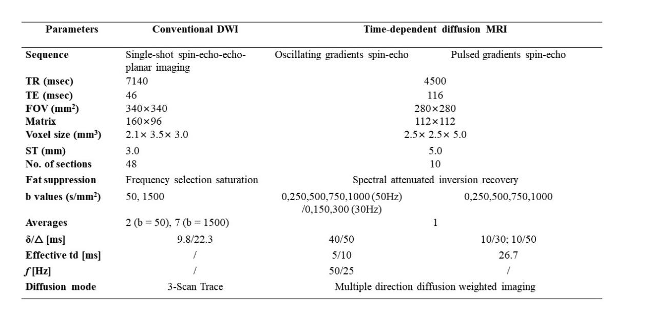
The study was approved by the Institutional Ethics Committee. Fifty-three women (mean age, 51.17 ± 11.61 [SD] years; range, 31-76 years) with suspicious breast lesions (BI-RADS 3-5) were hereby prospectively recruited between February and September 2023. Conventional DWI and time-dependent diffusion MRI were performed on a 3.0-T MR scanner (MAGNETOM Prisma, Siemens Healthcare, Erlangen, Germany) with a high-performance gradient system (maximum gradient = 80 mT/m per axis, maximum slew rate = 200 mT/m) using the parameters shown in Figure 1. The time-dependent dMRI data were fitted with a two-compartment model to calculate microstructural parameters (i.e., fin, Dex, diameter, cellularity, ADC50 ms, and tauin). Minimum, maximum, and mean ADC values (ADCmin, ADCmax, ADCmean) were obtained by measuring the ADC map of DWI (b = 0 and 1500 s/mm). ADCheterogeneity was then obtained by the following formula: ADCheterogeneity = (ADCmax - ADCmin) / ADCmean. Histology was taken as the reference standard. Besides, diagnostic performance of OGSE and conventional DWI for breast lesions was assessed using the area under the receiver operating characteristic curve (AUC) and compared using the Delong test.Results
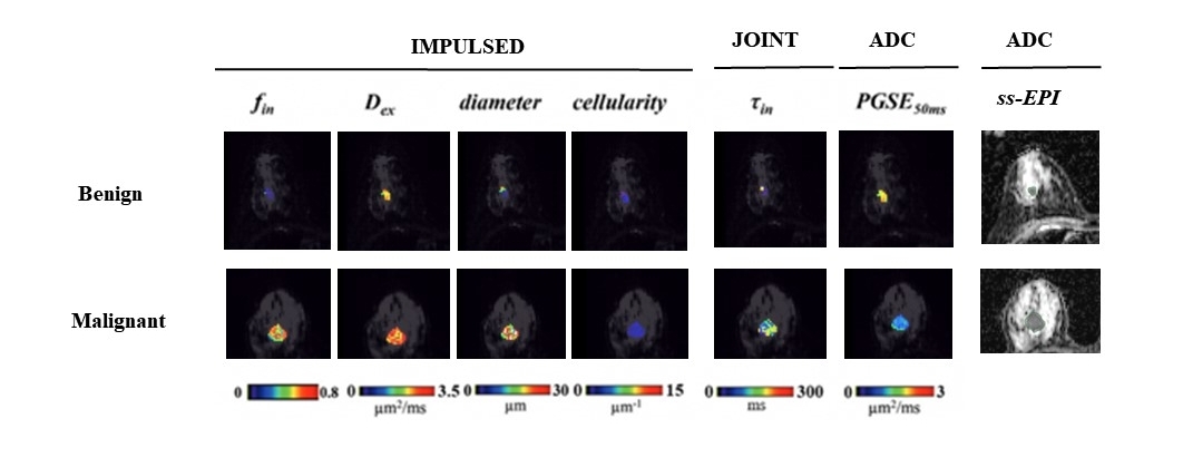
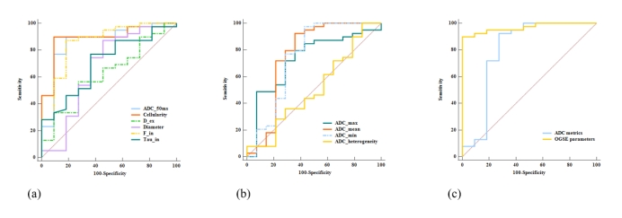
Compared with benign breast lesions (n = 14), malignant lesions (n = 39) showed significantly higher fin, cellularity, and tauin, and significantly lower ADC50 ms, ADCmin, ADCmax, and ADCmean. The cellularity achieved the highest AUC of 0.888 (95% Confidence Interval [CI]: 0.767-0.960) among all parameters, while ADCmin, ADCmax and ADCmean presented AUCs of 0.744 (95% CI: 0.605-0.854), 0.729 (95% CI: 0.589-0.842), and 0.760 (95% CI: 0.623-0.867), respectively. The combination of quantitative DWI parameters yielded the AUC, sensitivity, and specificity of 0.766, 87.18%, and 71.43%, respectively. With the addition of time-dependent dMRI parameters (fin, cellularity, ADC50 ms, and Tauin), the AUC increased to 0.972 (95% CI: 0.881-0.998), with sensitivity and specificity of 94.87% and 90.91%, respectively.Discussion and Conclusions
Conventional DWI is effective in detecting pathological changes in breast cancer but has limitations when it comes to describing the cellular microstructure of tumors1. Time-dependent dMRI, on the other hand, allows for a more detailed characterization of breast tissue microstructure and paves the way for new avenues in breast cancer research5,6. Although previous studies have reported that ADC differences7 can reflect the inherent biological heterogeneity of breast cancer, it remains unclear which of the two modalities, time-dependent dMRI or conventional DWI metrics, offers superior performance in characterizing breast cancer microstructure. In the present study, a time-dependent dMRI technique was employed to evaluate the microstructural features of breast lesions, which were also compared with parameters derived from conventional ADC maps. These microstructural parameters, especially cellularity index, achieved superior performance in differentiating benign from malignant breast lesions, with an AUC of 0.88, superior to conventional ADC measurements. This indicated the unique advantage of the time-dependent dMRI technique in depicting cellular microstructures. In addition, it was found that combining time-dependent dMRI parameters could improve diagnostic performance (AUC increased from 0.766 to 0.972) compared to conventional ADC parameters alone. In conclusion, time-dependent dMRI-based microstructural properties are superior to conventional DWI measurements in diagnosing breast lesions. Moreover, the addition of time-dependent dMRI parameters improves the performance of conventional DWI in differentiating breast lesions.Acknowledgements
Not applicableReferences
1. Iima M, Honda M, Sigmund EE, Ohno Kishimoto A, Kataoka M, Togashi K. Diffusion MRI of the breast: Current status and future directions. J Magn Reson Imaging. 2020;52(1):70-90. doi:10.1002/jmri.26908
2. Zhu A, Shih R, Huang RY, et al. Revealing tumor microstructure with oscillating diffusion encoding MRI in pre-surgical and post-treatment glioma patients. Magnetic Resonance In Medicine. 2023;90(5):1789-1801. doi:10.1002/mrm.29758
3. Ejima F, Fukukura Y, Kamimura K, et al. Oscillating Gradient Diffusion-Weighted MRI for Risk Stratification of Uterine Endometrial Cancer. J Magn Reson Imaging. 2023;doi:10.1002/jmri.29106
4. Kamimura K, Kamimura Y, Nakano T, et al. Differentiating brain metastasis from glioblastoma by time-dependent diffusion MRI. Cancer Imaging : the Official Publication of the International Cancer Imaging Society. 2023;23(1):75. doi:10.1186/s40644-023-00595-2
5. Xu J, Jiang X, Li H, et al. Magnetic resonance imaging of mean cell size in human breast tumors. Magnetic Resonance In Medicine. 2020;83(6):2002-2014. doi:10.1002/mrm.28056
6. Iima M, Kataoka M, Honda M, et al. The Rate of Apparent Diffusion Coefficient Change With Diffusion Time on Breast Diffusion-Weighted Imaging Depends on Breast Tumor Types and Molecular Prognostic Biomarker Expression. Investigative Radiology. 2021;56(8):501-508. doi:10.1097/RLI.0000000000000766
7. Kim JJ, Kim JY, Suh HB, et al. Characterization of breast cancer subtypes based on quantitative assessment of intratumoral heterogeneity using dynamic contrast-enhanced and diffusion-weighted magnetic resonance imaging. Eur Radiol. 2022;32(2):822-833. doi:10.1007/s00330-021-08166-4
Figures