2561
Comparison of single-shot, FOCUS single-shot, MUSE, and FOCUS MUSE diffusion weighted imaging for pulmonary lesions: a pilot study1Department of Radiology, Second Affiliated Hospital of Naval Medical University, Shanghai, China, 2College of Health Sciences and Engineering, University of Shanghai for Science and Technology, Shanghai, China, 3MR Research,GE Healthcare, Beijing, China, 4Department of Thoracic Surgery, Second Affiliated Hospital of Naval Medical University, Shanghai, China
Synopsis
Keywords: DWI/DTI/DKI, Diffusion/other diffusion imaging techniques, Magnetic resonance imaging; Diffusion weighted imaging; Lung; Pulmonary lesions
Motivation: DWI has been used for pulmonary lesion assessment. But clinical single-shot (SS) EPI DWI for lung is prone to susceptibility distortions. FOCUS SS, MUSE, and FOCUS MUSE are improved EPI-based techniques which are less sensitive to susceptibility artifacts. However, their performance for lung DWI remains unknown.
Goal(s): Performance comparison among the four sequences for lung DWI.
Approach: 44 patients were recruited, and each was imaged with the four sequences. Image quality and diagnostic performance of benign and malignant lesion discrimination were compared.
Results: FOCUS MUSE had least distortions and best diagnostic performance.
Impact: Application of FOCUS MUSE would be beneficial for lung lesion assessment.
Introduction
Lung cancer remained the leading cause of cancer-related death worldwide1. Thus, it would have clinical significance to detect pulmonary lesions, distinguish malignant from benign lesions for guiding treatment decisions selection, and predict prognosis. DWI has become part of state-of-the-art multiparametric MRI protocols for assessment of the chest2. Single-shot (SS) EPI-DWI is routinely used in clinic because of its fast imaging and decreased motion artifact properties. However, SS-EPI-DWI is prone to susceptibility induced distortions which makes it particularly challenge for lung with complex air-tissue interfaces. FOCUS allows decreasing the required readout duration for SS-EPI resulting with better DWI images3. MUSE is a technique inherently estimating both linear and nonlinear phase variations from multi-shot segmented EPI-DWI which can achieve high resolution and SNR, and alleviating distortions3. FOCUS MUSE is a novel technique which integrated FOCUS with MUSE 3. Despite DWI has emerging applications in the lung, it remains unknown if the improved EPI-based DWI techniques could be beneficial for lung lesion evaluation. Thus, the aim of this study was to compare the performance of FOCUS SS, MUSE, and FOCUS MUSE with routinely used SS-EPI DWI for lung lesions.Materials and Methods
Patients44 patients (35 malignant and 9 benign) were used in this study.
MRI Acquisition
All MR experiments were performed on a 3.0T MRI scanner (SIGNA Premier; GE Healthcare, CA, USA) with dedicated 21-channel AIRTM flexible coil (GE Healthcare). Scanning parameters: (1) SS: FOV=40cm×40cm, matrix size=168×168, shots=1; (2) FOCUS SS: FOV=32cm×16cm, matrix size=132×68, shots=1; (3) MUSE: FOV=36cm×36cm, matrix size=148×148, shots=2; (4) FOCUS MUSE: FOV=32cm×16cm, matrix size=132×68, shots=2. The b value and number of average for all DWIs were 0/50/800 s/mm2 and 1/2/6, respectively.
Data Analysis
All assessments were performed on the workstation (AW4.7; GE Healthcare, Milwaukee, USA). The SNR and distortion ratio (DR) of pulmonary lesion were used to objectively assess the image quality of DWIs4. DR was defined as the ratio of the maximum lesion displacement in the phase-encoded direction between DWI and T2WI to the lesion diameter in the phase-encoded direction on T2WI. The ADC map was fitted using the workstation. The mean ADC value of lesion was extracted from the ADC map using the same lesion ROI for SNR.
Statistical analysis was performed using SPSS (version 27.0; IBM, NY, USA) and MedCalc (version 22.0; Mariakerke, Belgium). P <0.05 was considered statistically significant. The Friedman test was used to assess the differences of SNR, DR, and ADC value among the four DWI sequences, and the Dunn-Bonferroni post hoc was used to adjust all paired comparisons with significantly difference. The Mann-Whitney was used to compare the ADC difference between benign and malignant pulmonary lesions. The receiver operating characteristic curve (ROC) was carried out and area under curve (AUC) was used to assess the performance of ADC value for discriminating malignant from benign lesions.
Results
Compared with SS, FOCUS SS and MUSE, FOCUS MUSE demonstrated significantly smaller DR (P<0.001) (Figure 1-3). The SNR was comparable among SS, FOCUS SS, and FOCUS MUSE (P>0.05) while MUSE presented with significantly higher SNR over them (P<0.01) (Figure 1). FOCUS MUSE was better than the other three sequences in delineating and detecting of lesions (Figure 2-3). No significant difference of lesion ADC was shown among the four sequences (P>0.05). The ADC of malignant was significantly smaller than that of benign for all the four sequences (P<0.05). ROC analysis showed FOCUS MUSE (AUC=0.820) was relatively superior to SS (AUC=0.748), FOCUS SS (AUC=0.778), and MUSE (AUC=0.729) for discriminating malignant from benign lesions (Figure 5).Discussion and Conclusion
In this study, we compared the performance of four EPI-based DWIs for lung. The results demonstrated FOCUS MUSE had sufficient SNR and better image quality over SS, FOCUS SS, and MUSE, in terms of distortions, pulmonary lesion delineation and detection, and malignant and benign discrimination. Therefore, the application of FOCUS MUSE would be beneficial for lung DWI.Previous study had reported the detection rate was 86.4% and dropped to 43.8% for pulmonary lesions with size of 6-9 mm and 3-5 mm, respectively 5. Our results showed FOCUS MUSE can detect small lesions which were omitted on the other three sequences indicating its role in increasing the detection rate of small pulmonary lesions. Consistent with previous studies 6,7, our results showed the ADC was significantly lower for malignant than benign lesions. The FOCUS MUSE was significantly least distorted which may contribute to its best performance for malignant and benign discrimination.
In conclusion, FOCUS MUSE possessed sufficient SNR and was better over SS, FOUCS SS, and MUSE for characterizing pulmonary lesions.
Acknowledgements
References
1. Sung H, Ferlay J, Siegel RL, et al. Global Cancer Statistics 2020: GLOBOCAN Estimates of Incidence and Mortality Worldwide for 36 Cancers in 185 Countries. CA Cancer J Clin. 2021;71(3):209-249.
2. Broncano J, Steinbrecher K, Marquis KM, et al. Diffusion-weighted Imaging of the Chest: A Primer for Radiologists. Radiographics. 2023;43(7):e220138.
3. Bai Y, Pei Y, Liu WV, et al. MRI: Evaluating the Application of FOCUS-MUSE Diffusion-Weighted Imaging in the Pancreas in Comparison With FOCUS, MUSE, and Single-Shot DWIs. J Magn Reson Imaging. 2023;57(4):1156-1171.
4. Wan Q, Lei Q, Wang P, et al. Intravoxel Incoherent Motion Diffusion-Weighted Imaging of Lung Cancer: Comparison Between Turbo Spin-Echo and Echo-Planar Imaging. J Comput Assist Tomogr. 2020;44(3):334-340.
5. Regier M, Schwarz D, Henes FO, et al. Diffusion-weighted MR-imaging for the detection of pulmonary nodules at 1.5 Tesla: intraindividual comparison with multidetector computed tomography. J Med Imaging Radiat Oncol. 2011;55(3):266-274.
6. Zhu Q, Ren C, Xu JJ, Li MJ, Yuan HS, Wang XH. Whole-lesion histogram analysis of mono-exponential and bi-exponential diffusion-weighted imaging in differentiating lung cancer from benign pulmonary lesions using 3 T MRI. Clin Radiol. 2021;76(11):846-853.
7. Mahdavi Rashed M, Nekooei S, Nouri M, Borji N, Khadembashi A. Evaluation of DWI and ADC Sequences' Diagnostic Values in Benign and Malignant Pulmonary Lesions. Turk Thorac J. 2020;21(6):390-396.
Figures
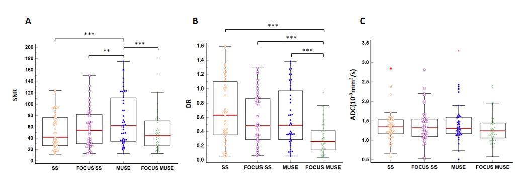
Figure 1: SNR, DR, and ADC values of the four DWI sequences. *, P<0.05; **, P<0.01; ***, P<0.001.
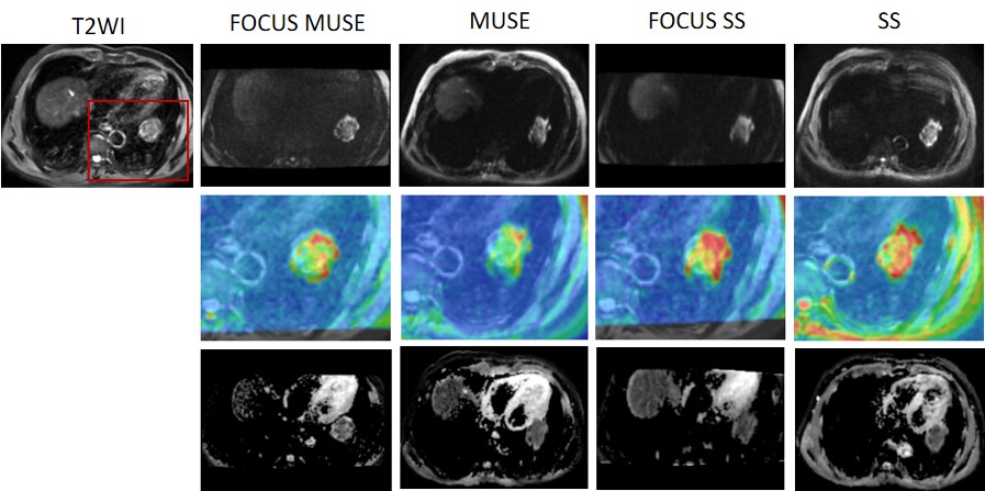
Figure 2: Seventy-five years old man with nonidentified NSCLC in the left lower lobe. The diameter of tumor is about 4 cm. The first row was T2WI and location matched FOCUS MUSE, MUSE, FOCUS SS, and SS DWI images sequentially. The second row was corresponded and enlarged images with DWI overlayed on T2WI. The location of the enlarged region was shown in red rectangle on the T2WI image. The third row was the ADC map accordingly.
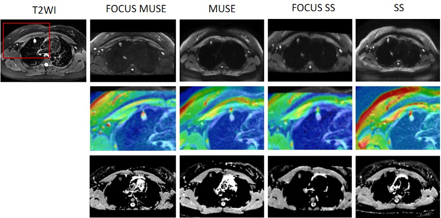
Figure 3: Thirty-one years old woman with adenocarcinomas adjacent to chest wall in right lower lobe. The diameter of tumor is about 1.5 cm. The first row was T2WI and location matched FOCUS MUSE, MUSE, FOCUS SS, and SS DWI images sequentially. The second row was corresponded and enlarged images with DWI overlayed on T2WI. The location of the enlarged region was shown in red rectangle on the T2WI image. The third row was the ADC map accordingly.
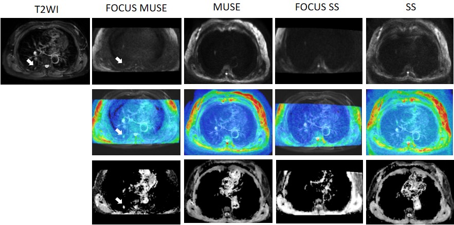
Figure 4: Seventy-two years old woman with adenocarcinomas in the right upper lobe. The diameter of tumor is about 0.8 cm. The first row was T2WI and location matched FOCUS MUSE, MUSE, FOCUS SS, and SS DWI images sequentially. The second row was corresponded images with DWI overlayed on T2WI. The third row was the ADC map accordingly. The white arrows pointed the location of lesion. The lesion can be detected convincedly on FOCUS MUSE but not on SS, FOCUS SS, and MUSE.
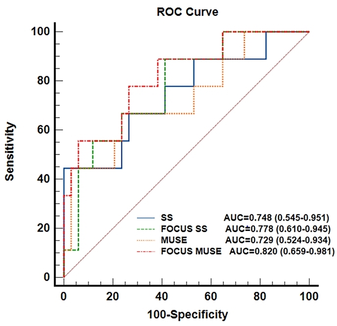
Figure 5: The ROC curves of the four DWI sequences for distinguishing malignant from benign lesions.