2560
Time-dependent diffusion MRI for microstructural mapping to distinguish high-grade serous ovarian cancer and serous borderline ovarian tumor1The First Affiliated Hospital of Nanjing Medical University, Nanjing, China, 2Philips Healthcare, Shanghai, China
Synopsis
Keywords: Microstructure, Diffusion/other diffusion imaging techniques, microstructure, IMPULSED
Motivation: The intraoperative frozen sections have shortcomings in distinguishing between high-grade serous ovarian cancer (HGSOC) and serous borderline ovarian tumor (SBOT). Time-dependent diffusion MRI (td-dMRI) can depict the microstructural parameters of tumors and might play a role in preoperative differentiation of HGSOC from SBOT.
Goal(s): To investigate the value of td-dMRI in discriminating HGSOC from SBOT.
Approach: Td-dMRI uses the combination of oscillating gradient spin-echo (OGSE) and pulsed gradient spin-echo (PGSE) sequences. Td-dMRI signals were fitted by the IMPULSED model, to extract microstructural parameters. The diagnostic performance was evaluated.
Results: Microstructural parameters derived from td-dMRI had good diagnostic performance in differentiating HGSOC from SBOT.
Impact: Time-dependent diffusion MRI can depict the microstructural parameters of tumors. It may further assist in preoperative diagnosis and treatment decision-making in clinical practice in the future.
Background
High-grade serous ovarian cancer (HGSOC) patients often undergo more aggressive treatments such as radical surgery and cytoreductive surgery than serous borderline ovarian tumor (SBOT) patients1,2. Therefore, it is important to distinguish HGSOC from SBOT preoperatively to ensure appropriate treatment. However, the differential diagnosis of HGSOC and SBOT still depends on the morphological evaluation through histopathology. Although the analysis of intraoperative frozen sections is widely used for the assessment of tumor cell morphology, it does not provide a completely accurate diagnosis in SBOT, often resulting in discrepant final pathological findings3. Thus, there is a need for developing noninvasive tools for pre-operative differential diagnosis of HGSOC and SBOT. Time-dependent diffusion MRI can depict the microstructural parameters of tumors and might play a role in preoperative differentiation of HGSOC from SBOT.Material and Methods
Totally 34 HGSOC and 12 SBOT cases who received preoperative pelvic MRI were consecutively included in this study. Two radiologists delineated the tumors to obtain the regions of interest (ROIs). All scans were performed on 3.0 T scanner (Ingenia CX, Phillips, Best, the Netherlands). Time-dependent diffusion MRI uses the combination of oscillating gradient spin-echo (OGSE) and pulsed gradient spin-echo (PGSE) sequences. OGSE data were acquired at 25 Hz (effective diffusion time, 10 ms; 1 cycle; b = 0, 100, 200, 400, 500 or 1000 s/mm2) or 50 Hz (effective diffusion time, 5 ms; 2 cycles; b = 0, 50, 100, 150, 200 or 300 s/mm2). PGSE data were acquired with diffusion duration and separation of 42.7 and 59.3 ms (b = 0, 200, 300, 500, 800 or 1000 s/mm2). The following parameters were used for both sequences: three diffusion directions; repetition time/echo time, 4500/120 ms; field of view, 365 × 365 mm; sampling resolution, 2 × 2 mm2; thickness, 3 mm. Time-dependent diffusion MRI signals were fitted by the IMPULSED (imaging microstructural parameters using limited spectrally edited diffusion) model, encompassing two compartments: S = fin ∙ Sin + (1 - fin) ∙ Sex, to extract microstructural parameters, including fraction of the intracellular component (fin), cell diameter (d), cellularity and extracellular diffusivity (Dex)(Fig 1). Apparent diffusion coefficient (ADC) values were also obtained from standard diffusion-weighted imaging (DWI). The parameters of HGSOCs and SBOTs were compared, and the diagnostic performance was evaluated. The associations of microstructural indexes with immunopathological parameters were assessed, including Ki-67, P53, Pax-8, ER and PR.Results
In this study, fin, cellularity, Dex and ADC had good diagnostic performance levels in differentiating HGSOC from SBOT, with AUCs of 0.936, 0.909, 0.902 and 0.914(Fig 2-3,Table 1-2), respectively. There were no significant differences in diagnostic performance among these parameters. Spearman analysis revealed in the HGSOC group, cellularity had a significant positive correlation with P53 expression (P = 0.028, r = 0.389) and Dex had a significant positive correlation with Pax-8 expression (P = 0.018, r = 0.415). ICC showed excellent agreement for all parameters.Discussion
Many efforts have been deployed to depict the microstructure of cells using time-dependent diffusion MRI. For example, the vascular, extracellular, and restricted diffusion for cytometry (VERDICT) method can reflect vascular and intra- and extracellular volume fractions4. However, the range of diffusion time detected by VERDICT is narrower than that of IMPULSED because it uses higher b-values and could not describe indicators such as cell size and cell density5. The IMPULSED method used in this study was based on a two-compartment model. It simplifies mathematical calculations by considering cancer cells spheres. Wu et al. 6applied the IMPULSED method based on time-dependent diffusion MRI for clinically distinguishing between significant prostate cancer and clinically insignificant disease. Meanwhile, microstructural parameters were shown to correlate with pathological findings. Zhang et al. 7also applied the IMPULSED method in classifying low- and high-grade pediatric gliomas. fin and d were also consistent with histological results. In this study, as shown above, fin, cellularity and Dex both differed between HGSOCs and SBOTs. HGSOC exhibits higher cell atypia and more significant cell proliferation compared with SBOT19,26, so cell density and the nuclear-cytoplasmic ratio are higher, corroborating the current findings.fin, cellularity and Dex both showed high diagnostic performance in differentiating HGSOC from SBOT. However, no significant differences were found in diagnostic performance among these parameters. However, these results reveal a potential value for time-dependent diffusion MRI as a non-invasive technique for differentiating HGSOC from SBOT.
Conclusion
Time-dependent diffusion MRI had a value in evaluating the microstructures of HGSOC and SBOT and could discriminate between these tumors. Time-dependent diffusion MRI may be a noninvasive method with potential value for the early assessment of tumor proliferation and patient prognosis.Acknowledgements
No AcknowledgementsReferences
1. Tropé CG, Kaern J, Davidson B. Borderline ovarian tumours. Best Pract Res Clin Obstet Gynaecol 2012;26:325-336.
2. Hacker NF, Rao A. Surgery for advanced epithelial ovarian cancer. Best Pract Res Clin Obstet Gynaecol 2017;41:71-87.
3. Song T, Choi CH, Kim HJ, et al. Accuracy of frozen section diagnosis of borderline ovarian tumors. Gynecol Oncol 2011;122:127-131.
4. Panagiotaki E, Chan RW, Dikaios N, et al. Microstructural characterization of normal and malignant human prostate tissue with vascular, extracellular, and restricted diffusion for cytometry in tumours magnetic resonance imaging. Invest Radiol 2015;50:218-227
5. Xu J, Jiang X, Li H, et al. Magnetic resonance imaging of mean cell size in human breast tumors. Magn Reson Med 2020;83:2002-2014.
6. Wu D, Jiang K, Li H, et al. Time-Dependent Diffusion MRI for Quantitative Microstructural Mapping of Prostate Cancer. Radiology 2022;303:578-587.
7. Zhang H, Liu K, Ba R, et al. Histological and molecular classifications of pediatric glioma with time-dependent diffusion MRI-based microstructural mapping. Neuro Oncol 2023;25:1146-1156.
Figures
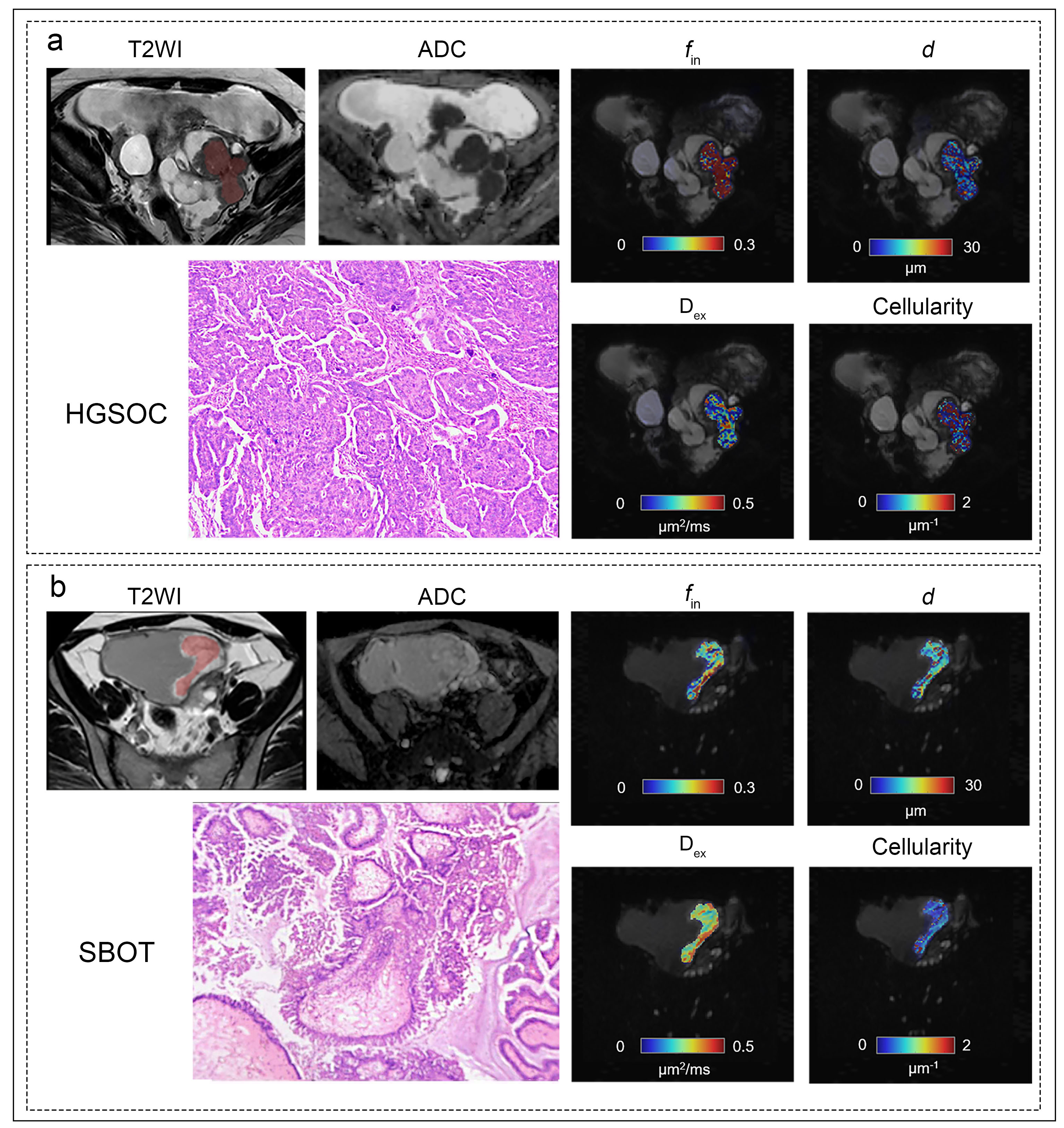
Fig 1. T2WI scan, ADC map and microstructural features derived from IMPULSED model (including fin, d, cellularity and Dex), and corresponding microstructural maps for (a) a HGSOC patient (59-year-old) and (b) a SBOT patient (25-year-old).
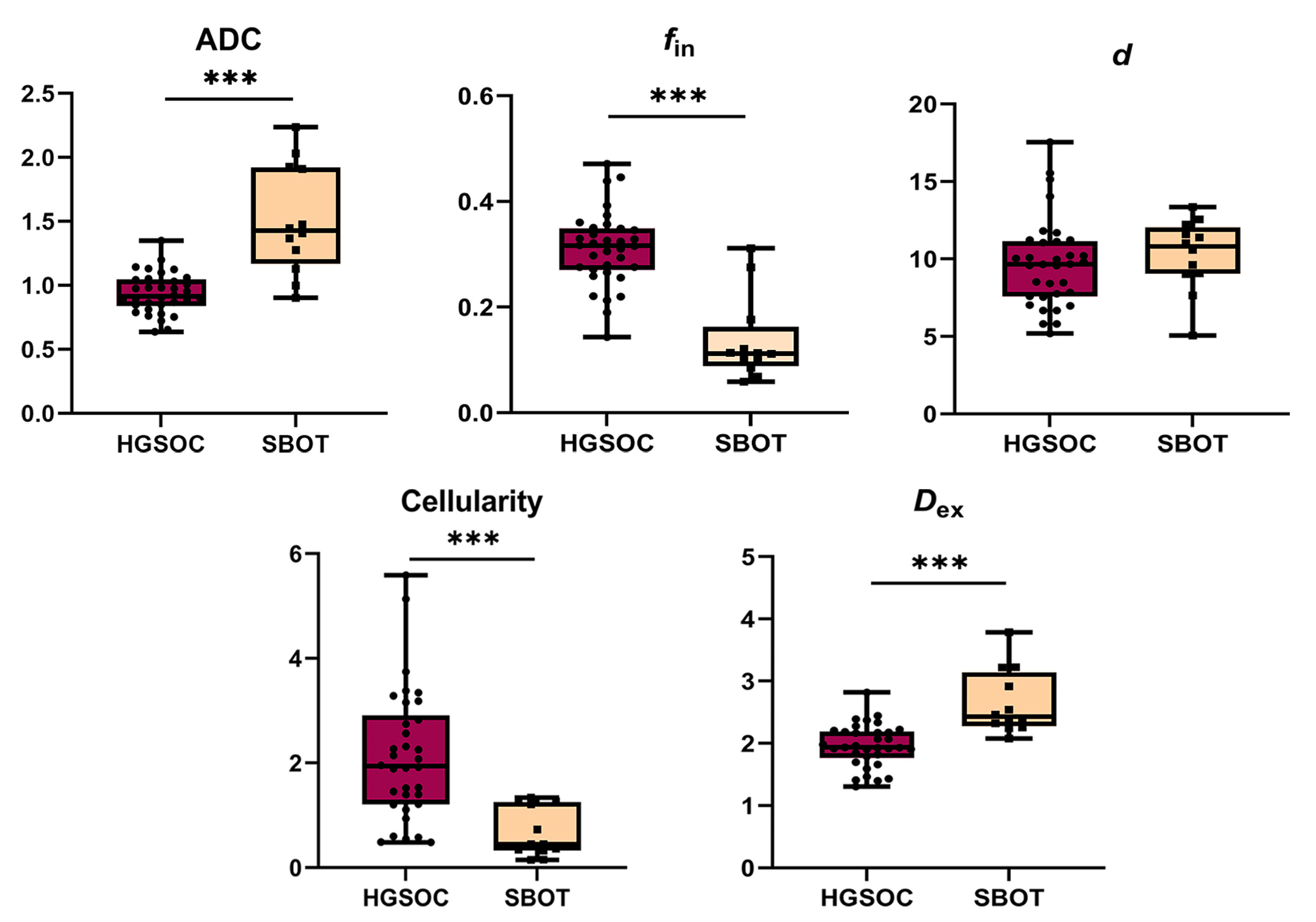
Fig 2. Box plots showing group differences between high-grade serous ovarian cancer (HGSOC) and serous borderline ovarian tumor (SBOT). *** P < 0.001.
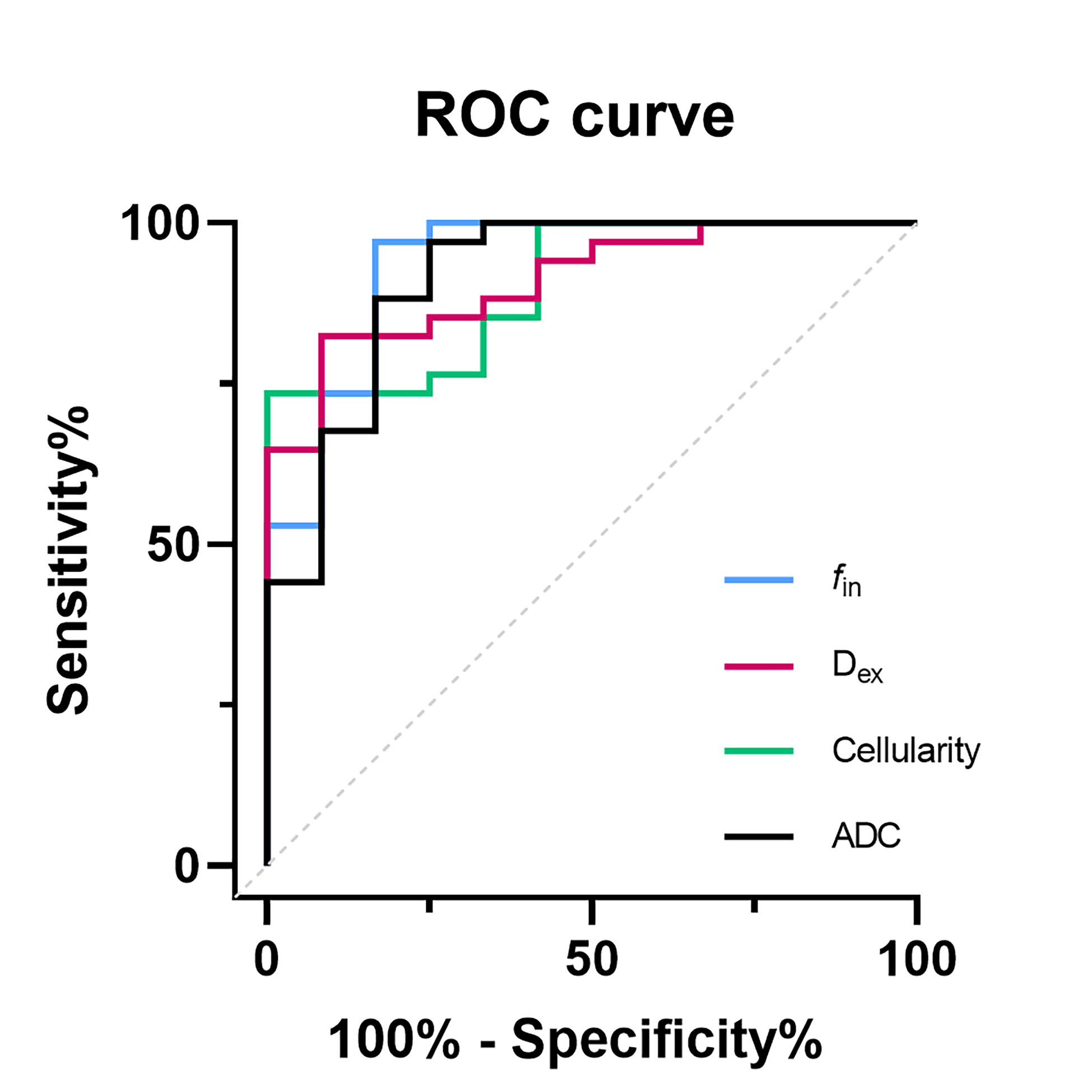
Fig 3. ROC curve analysis of factors differentiating high-grade serous ovarian cancer (HGSOC) from serous borderline ovarian tumor (SBOT).
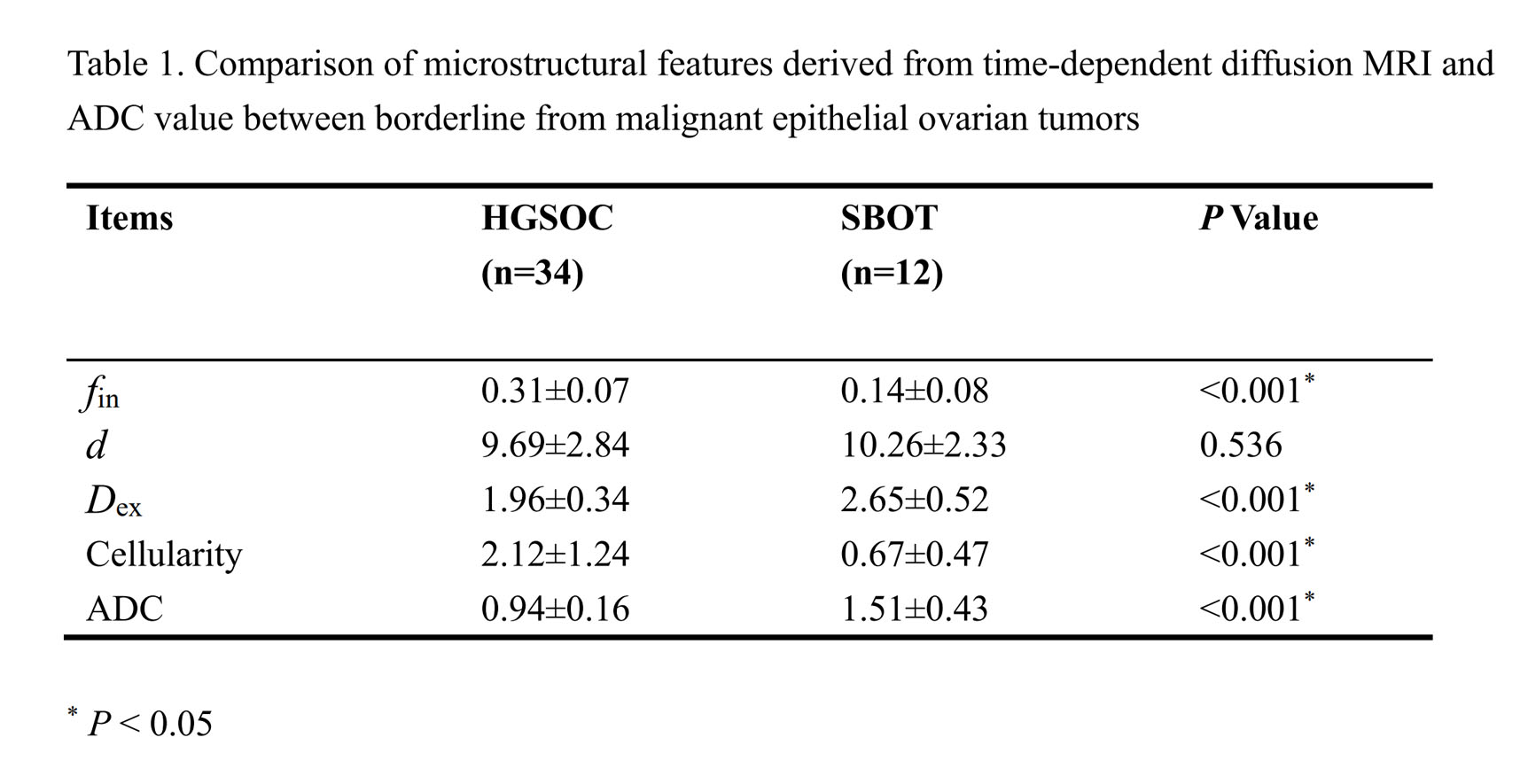
Table 1. Comparison of microstructural features derived from time-dependent diffusion MRI and ADC value between borderline from malignant epithelial ovarian tumors
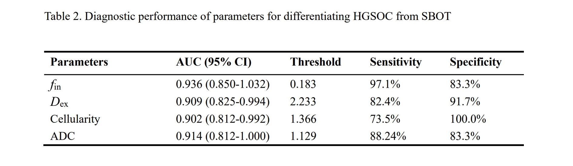
Table 2. Diagnostic performance of parameters for differentiating HGSOC from SBOT