2555
Association with Maternal Diet Quality during Pregnancy and Neonatal Brain White Matter Development1Radiology, University of Arkansas for Medical Sciences, Little Rock, AR, United States, 2Pediatrics, University of Arkansas for Medical Sciences, Little Rock, AR, United States, 3Arkansas Children’s Nutrition Center, Little Rock, AR, United States, 4Arkansas Children’s Research Institute, Little Rock, AR, United States
Synopsis
Keywords: Neonatal, Neuroscience
Motivation: Maternal nutrient intake is important for fetal growth and development.
Goal(s): This study examined associations between maternal diet quality during pregnancy and neonatal brain white matter development.
Approach: Forty-four healthy pregnant woman/newborn dyads were included in the study. The mothers had nutrition intake assessed by Healthy Eating Index-2015 (HEI-2015) throughout pregnancy. Correlations between neonatal MRI diffusion tensor imaging (DTI)-measured fractional anisotropy (FA) and HEI-2015 scores were evaluated.
Results: Significant correlations between maternal sodium intake at 1st trimester of pregnancy and neonatal white matter FA values were found, indicating potential influence of maternal sodium intake during early pregnancy on neonatal brain white matter development.
Impact: We found significant correlations between maternal sodium intake at first trimester of pregnancy and neonatal DTI-measured FA values, indicating sodium intake better aligned with the Dietary Guidelines of Americans during early pregnancy is associated with better neonatal white matter development.
Introduction
Healthy nutrition helps pregnant women with extra demanding on their own bodies and the growing fetuses. A balanced and nutrient-rich diet during pregnancy is essential for optimal fetal health. In this study, we aimed to investigate whether there is an association between maternal dietary quality at different time points assessed by Healthy Eating Index-2015 (HEI-2015) during pregnancy and neonatal brain white matter (WM) development.Method
We recruited healthy pregnant women without medical complications and their newborns. Inclusion criteria: second parity, singleton pregnancy; ≥ 21 years of age; conceived without assisted fertility treatments. Exclusion criteria: pre-existing medical conditions such as diabetes mellitus, seizure disorder, and serious psychiatric disorders; drug abuse issues or alcohol use or smoking during pregnancy; sexually transmitted diseases; medical complications developed during pregnancy such as gestational diabetes and pre-eclampsia. The women had their dietary intake estimated using 3-day food records at 1st, 2nd and 3rd trimester during pregnancy respectively. The food records were obtained and reviewed by trained research assistants, and analyzed using the Nutrient Data System for Research (NDSR) from the University of Minnesota [1]. Estimation of dietary intake was calculated and the HEI-2015 was derived from published formulas [2, 3]. HEI-2015 is a measurement for assessing dietary quality, specifically the degree to which the dietary intake aligns with the recommendations in the 2015-2020 Dietary Guidelines for Americans (DGA 2015-2020). At ~2 weeks of postnatal age, the newborns underwent a brain MRI examination. The MRI was performed on a Philips Acheiva 1.5T scanner during newborn’s natural sleep without sedation, using protocol designed specifically for neonatal brain. A 3D T1-weighted scan covering the whole brain was implemented for incidental screening. In addition, a single-shot spin echo EPI sequence with TR/TE 4200ms/66ms and 180 mm x 180 mm field of view, 90 x 90 acquisition matrix and 3 mm slice thickness (30-36 continuous axial slices for each brain scan) and diffusion-weighting gradients in 15 uniformly distributed directions with a b-value of 700 s/mm2 was used to acquire DTI data in a very short scan time. Overall, 44 pregnant women had their HEI-2015 scores recoded, and their newborns (23 boys/21 girls) successfully completed the MRI scan. DTI data were exported for pre-processing including brain extraction, eddy currents and movements corrections, and eigenvalues for the diffusion tensors were then computed, followed by the generation of DTI parameter maps using FSL. Tract-based spatial statistics (TBSS) methods were used for DTI data analysis [4]. In brief, the fractional anisotropy (FA, a main DTI parameter sensitive to white matter microstructural integrity with great contrast between white and gray matters) maps were processed and aligned to a neonatal template. All FA maps were then skeletonized to illustrate their major white matter tracts (defined as FA ≥ 0.1, which was adjusted from adult threshold to reflect lower FA values in newborns). Randomization tests with the threshold-free cluster enhancement (TFCE) option and 5000 permutations for both positive and negative correlations were used to investigate voxel-level associations between neonatal FA values and all components of HEI-2015 scores at each trimester of pregnancy respectively. Newborn’s sex, postmenstrual age at MRI and maternal BMI were included as covariates due to their effects on offspring brain development [5-8]. Family-wise error rate (FWE) was used for multiple comparisons. Clusters identified with FWE-corrected two-tailed P ≤ 0.05 in the voxel-level analyses and with size ≥ 40 voxels were tested for post-hoc region-of-interest (ROI) analyses. Mean FA value of each ROI was extracted for each cluster per subject. Partial Spearman’s Rank correlation test using Matlab software (The Mathworks Inc., Natick, MA) with the same covariates controlled was performed to confirm their significance (P ≤ 0.05).Results
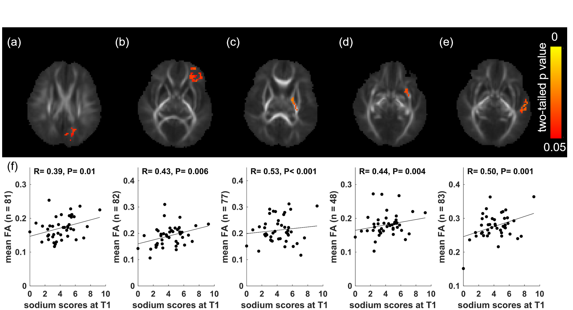
For voxel-level analyses, positive correlations (two-tailed P ≤ 0.05, FWE corrected) of neonatal FA values with sodium scores at first trimester were identified in multiple clusters (with size ≥ 40 voxels) in the brain WM using the TFCE. After post-hoc analyses, correlations (P ≤ 0.05) between maternal sodium scores at first trimester and neonatal mean FA values remained significant in left parietal white matter (R = 0.39, P = 0.01), anterior corona radiata (R = 0.43, P = 0.006), posterior limb of internal capsule (R = 0.53, P < 0.001), external capsule (R = 0.44, P = 0.004) and temporal white matter (R = 0.50, P = 0.001) (Figure 1).Conclusions
Our results show that less sodium intake during uncomplicated early pregnancy is associated with better neonatal brain white matter microstructural development, and suggest that maternal sodium intake during early pregnancy may impact offspring brain development.Acknowledgements
This project was supported by NIH 1R01HD099099 and USDA-ARS 6026-51000-012-06S.References
1. Diaz, E.C., et al., Breastfeeding duration modifies the association between maternal weight status and offspring dietary palmitate oxidation. American Journal of Clinical Nutrition, 2022. 116(2): p. 404-414.
2. Krebs-Smith, S.M., et al., Update of the Healthy Eating Index: HEI-2015. Journal of the Academy of Nutrition and Dietetics, 2018. 118(9): p. 1591-1602.
3. Reedy, J., et al., Evaluation of the Healthy Eating Index-2015. Journal of the Academy of Nutrition and Dietetics, 2018. 118(9): p. 1622-1633.
4. Smith, S.M., et al., Tract-based spatial statistics: voxelwise analysis of multi-subject diffusion data. Neuroimage, 2006. 31(4): p. 1487-505.
5. Ou, X., et al., Gestational Age at Birth and Brain White Matter Development in Term-Born Infants and Children. American Journal of Neuroradiology, 2017. 38(12): p. 2373-2379.
6. Ou, X., et al., Maternal adiposity negatively influences infant brain white matter development. Obesity (Silver Spring), 2015. 23(5): p. 1047-54.
7. Na, X., et al., Maternal Obesity during Pregnancy is Associated with Lower Cortical Thickness in the Neonate Brain. American Journal of Neuroradiology, 2021. 42(12): p. 2238-2244.
8. Salzwedel, A.P., et al., Maternal Adiposity Influences Neonatal Brain Functional Connectivity. Front Hum Neurosci, 2018. 12: p. 514.
Figures