2552
Altered Functional Connectivity in the Brain Induced by Early-life Mild Necrotizing Enterocolitis1Department of Biomedical Imaging and Radiological Sciences, National Yang Ming Chiao Tung University, Taipei, Taiwan, 2Department of Pharmacology, Tapei Medical University, Taipei, Taiwan, 3Department of Medical Imaging and Radiological Sciences, College of Medicine, Chang Gung University, Taoyuan, Taiwan, 4Department of Medical Imaging and Intervention, Chang Gung Memorial Hospital at Linkou, Taoyuan, Taiwan, 5Department of Pediatrics, National Cheng Kung University Hospital, College of Medicine, National Cheng Kung University, Tainan, Taiwan
Synopsis
Keywords: Neuro, Preclinical
Motivation: According to the clinical findings, the survivors of necrotizing enterocolitis (NEC) may suffer from long-term neurodevelopmental impairment. However, how the local intestine inflammation impacts brain function needs further investigation.
Goal(s): To probe the long-term alteration in the brain network induced by NEC in early life.
Approach: After employing a neonatal rat model of NEC with lipopolysaccharide at postnatal 7 days, we measured the functional connectivity in the brain in its adolescent age (about P40).
Results: Neurodevelopmental impairment induced by mild early-life NEC was reflected by rsfMRI in adolescent rats.
Impact: We provide evidence showing that preterm NEC may induce the subsequent alteration in the functional brain network. Our results indicate the association between the early-life local intestine inflammation and long-term neurodevelopmental impairment, suggesting the interaction of the gut-brain axis.
Introduction
Necrotizing enterocolitis (NEC) is a common illness with a high mortality rate in preterm infants. Severe local inflammation observed after NEC is presumed to be initiated by the altered microbiota in the immature intestine in preterms.10 Despite improving health care, the mortality has been reported to be approximately 30-50%. In addition, 45% of survivors suffer from neurodevelopmental impairment including cerebral palsy, deafness, blindness, lower mental developmental index, and lower psychomotor developmental index.8,11-15 To date, how NEC in the local intestine impacts the functional impairment of the brain is still uncharacterized. In the current study, we would like to assess the altered brain network induced by early-life NEC. We employed a neonatal rat model of NEC with Lipopolysaccharide (LPS), a commonly used endotoxin, and hypoxia1-2,4-5, and measured the functional connectivity in the brain in the corresponding adolescent age. Our results demonstrated that the mild early-life local inflammation in the intestine would lead to long-term neurodevelopmental impairment.Methods
Twenty-two Sprague-Dawley rats were used in the current study. To induce mild NEC, neonatal rats were orally fed with LPS (E.coli O55:B5, 2mg/kg, n=12) twice daily from P1 to P7. On the same days, after the LPS feeding, the rats were treated with hypoxia (100% nitrogen) twice daily.1-2,4-5 Rats in the control group (n=10) were treated with normal saline (NS) from P1 to P7 and without experience in hypoxia. Resting-state functional MRI (rsfMRI) was performed at postnatal day 40 (P40) on a Bruker 7T PharmaScan. After the bolus injection of dexmedetomidine (0.015mg/kg), continuous infusion of dexmedetomidine (0.03mg/kg/h) was performed with 0.25%-0.5% isoflurane during the whole MR protocol.9 rsfMRI was acquired using the single-shot echo-planar imaging (EPI) with TR/TE=1000/12 ms, FOV= 2.0×2.0 cm, matrix size= 96×96, 16 slices, slice thickness of 1 mm and number of repetitions= 300 for total scan time= 5 min. T2-weighted images with the same geometry were also acquired. rsfMRI data (n=5 in the NS group, n=11 in the LPS group) were analyzed using gICA with 25-50 components by GIFT. Dual regression was further used to obtain the z values based on subject differences and calculate the representative sICA maps. The functional connectivity matrices were also calculated through Pearson’s correlation coefficients. Open field test for 10 min was performed in the animals after MRI acquisition. Statistical analysis was performed using an independent t-test with Bonferroni correction to determine the difference between the two groups.Results & Discussion
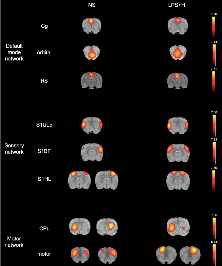
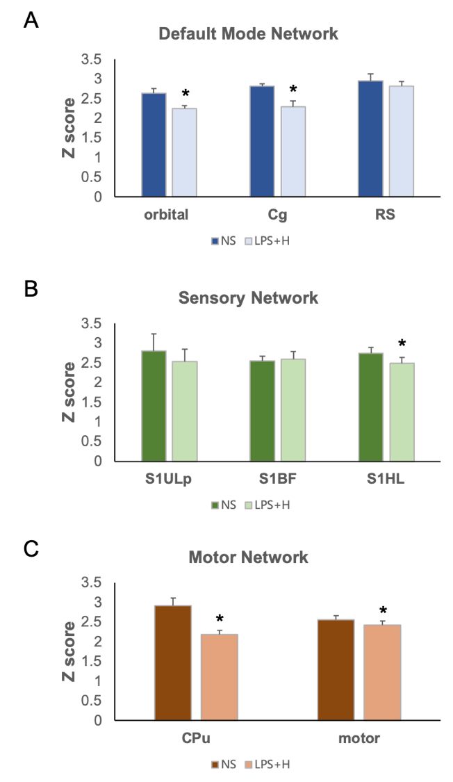
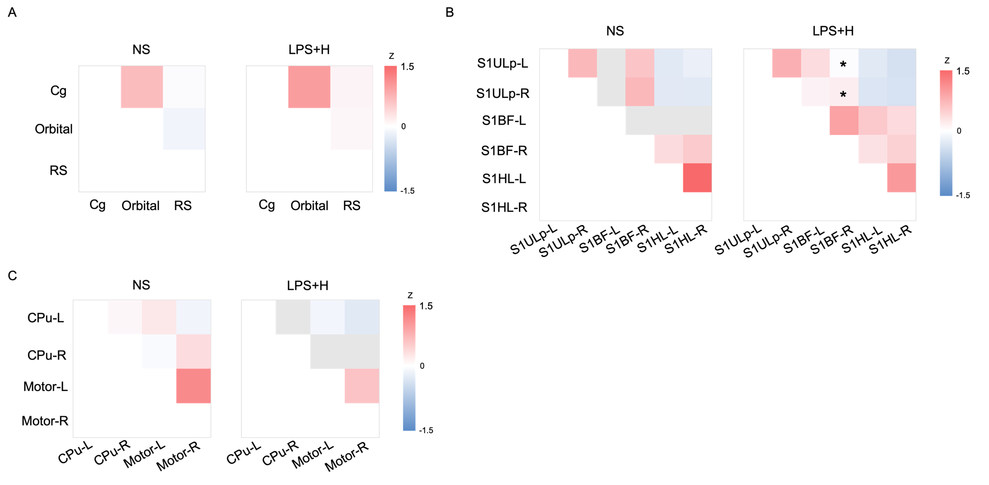
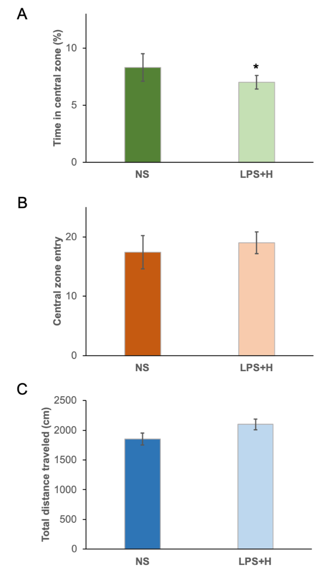
In the adolescent age, the identified major networks are similar to that in the adult rats. The major components of our interest including (1) the default mode network (DMN) with the cingulate cortex (Cg), the orbital cortex, and the retrosplenial cortex (RSC), (2) the sensory network with the upper lip region (S1ULp), the barrel field region (S1BF), and the hindlimb region (S1HL) of the primary somatosensory cortex, and (3) the motor network with the caudate putamen (CPu) and the motor cortex (Figure 1) were observed in both groups of animals. In the DMN, a significant decrease in functional connectivity in the NEC group was observed in the orbital cortex and Cg (P<0.006) (Figure 2A), suggesting that cognitive impairment may occur after the NEC.6,16 A significant decrease in functional connectivity after NEC induction was observed in the S1HL (P<0.006) while no significant difference was found in the S1ULp and S1BF (Figure 2B). In the motor network, a significant decrease could be observed in both the CPu and the motor cortex in the NEC group (P<0.006) (Figure 2C). In general, the decrease in functional connectivity in the DMN, sensory, and motor network may lead to behavioral abnormality due to inflammation.3,7 The interregional connectivity between all regions in DMN increased slightly in the NEC group (Figure 3A) perhaps due to the functional compensation for the significantly lower connectivity strength within the regions. In the sensory network, the interregional connectivity was significantly lower between the S1BF right region and both sides of S1ULp (P<0.005) while no significant difference was found among other regions after NEC induction (Figure 3B). In addition, all the interregional connectivity in the motor network after NEC induction was slightly lower (Figure 3C). In the open field test, animals in the NEC group spent less time in the central zone (P<0.05) (Figure 4A), indicating more anxiety-like behavior after NEC induction. Taken together, our results suggested that preterm NEC may result in decreased DMN functional connectivity and prominent anxiety in the adolescent age in rats.6,16 Our future studies will further analyze DTI images to validate the microstructure changes in the brain after NEC-induced neuroinflammation.Acknowledgements
This study was funded in part by the Ministry of Science and Technology ( NSTC 112-2314-B-A49-061), Taipei,Taiwan.References
1. Biouss, G., Antounians, L., Li, B., O’Connell, J. S., Seo, S., Catania, V. D., Guadagno, J., Rahman, A., Zani-Ruttenstock, E., Svergun, N., Pierro, A., & Zani, A. (2019). Experimental necrotizing enterocolitis induces neuroinflammation in the neonatal brain. Journal of Neuroinflammation, 16(1), 97. https://doi.org/10.1186/s12974-019-1481-9
2. Chen, C. L., Yu, X., James, I. O., Zhang, H. Y., Yang, J., Radulescu, A., Zhou, Y., & Besner, G. E. (2012). Heparin-binding EGF-like growth factor protects intestinal stem cells from injury in a rat model of necrotizing enterocolitis. Lab Invest, 92(3), 331-344. https://doi.org/10.1038/labinvest.2011.167
3. Felger, J. C., Li, Z., Haroon, E., Woolwine, B. J., Jung, M. Y., Hu, X., & Miller, A. H. (2016). Inflammation is associated with decreased functional connectivity within corticostriatal reward circuitry in depression. Mol Psychiatry, 21(10), 1358-1365. https://doi.org/10.1038/mp.2015.168
4. Feng, J., El-Assal, O. N., & Besner, G. E. (2006). Heparin-binding epidermal growth factor-like growth factor decreases the incidence of necrotizing enterocolitis in neonatal rats. J Pediatr Surg, 41(1), 144-149; discussion 144-149. https://doi.org/10.1016/j.jpedsurg.2005.10.018
5. Giannone, P. J., Nankervis, C. A., Richter, J. M., Schanbacher, B. L., & Reber, K. M. (2009). Prenatal lipopolysaccharide increases postnatal intestinal injury in a rat model of necrotizing enterocolitis. J Pediatr Gastroenterol Nutr, 48(3), 276-282. https://doi.org/10.1097/mpg.0b013e31818936b8
6. Guardia-Olmos, J., Soriano-Mas, C., Tormo-Rodriguez, L., Canete-Masse, C., Cerro, I. D., Urretavizcaya, M., Menchon, J. M., Soria, V., & Pero-Cebollero, M. (2022). Abnormalities in the default mode network in late-life depression: A study of resting-state fMRI. Int J Clin Health Psychol, 22(3), 100317. https://doi.org/10.1016/j.ijchp.2022.100317
7. Guevara, E., Pierre, W. C., Tessier, C., Akakpo, L., Londono, I., Lesage, F., & Lodygensky, G. A. (2017). Altered Functional Connectivity Following an Inflammatory White Matter Injury in the Newborn Rat: A High Spatial and Temporal Resolution Intrinsic Optical Imaging Study. Front Neurosci, 11, 358. https://doi.org/10.3389/fnins.2017.00358
8. Hintz, S. R., Kendrick, D. E., Stoll, B. J., Vohr, B. R., Fanaroff, A. A., Donovan, E. F., Poole, W. K., Blakely, M. L., Wright, L., & Higgins, R. (2005). Neurodevelopmental and growth outcomes of extremely low birth weight infants after necrotizing enterocolitis. Pediatrics, 115(3), 696-703. https://doi.org/10.1542/peds.2004-0569
9. Lu, H., Zou, Q., Gu, H., Raichle, M. E., Stein, E. A., & Yang, Y. (2012). Rat brains also have a default mode network. Proceedings of the National Academy of Sciences, 109(10), 3979-3984. https://doi.org/doi:10.1073/pnas.1200506109
10. Neu, J., & Walker, W. A. (2011). Necrotizing enterocolitis. N Engl J Med, 364(3), 255-264. https://doi.org/10.1056/NEJMra1005408
11. Rees, C. M., Pierro, A., & Eaton, S. (2007). Neurodevelopmental outcomes of neonates with medically and surgically treated necrotizing enterocolitis. Archives of Disease in Childhood - Fetal and Neonatal Edition, 92(3), F193-F198. https://doi.org/10.1136/adc.2006.099929
12. Shah, D. K., Doyle, L. W., Anderson, P. J., Bear, M., Daley, A. J., Hunt, R. W., & Inder, T. E. (2008). Adverse neurodevelopment in preterm infants with postnatal sepsis or necrotizing enterocolitis is mediated by white matter abnormalities on magnetic resonance imaging at term. J Pediatr, 153(2), 170-175, 175 e171. https://doi.org/10.1016/j.jpeds.2008.02.033
13. Sonntag, J., Grimmer, I., Scholz, T., Metze, B., Wit, J., & Obladen, M. (2000). Growth and neurodevelopmental outcome of very low birthweight infants with necrotizing enterocolitis. Acta Paediatr, 89(5), 528-532. https://doi.org/10.1080/080352500750027790
14. Tobiansky, R., Lui, K., Roberts, S., & Veddovi, M. (1995). Neurodevelopmental outcome in very low birthweight infants with necrotizing enterocolitis requiring surgery. J Paediatr Child Health, 31(3), 233-236. https://doi.org/10.1111/j.1440-1754.1995.tb00792.x
15. Wadhawan, R., Oh, W., Hintz, S. R., Blakely, M. L., Das, A., Bell, E. F., Saha, S., Laptook, A. R., Shankaran, S., Stoll, B. J., Walsh, M. C., Higgins, R. D., & Network, N. N. R. (2014). Neurodevelopmental outcomes of extremely low birth weight infants with spontaneous intestinal perforation or surgical necrotizing enterocolitis. J Perinatol, 34(1), 64-70. https://doi.org/10.1038/jp.2013.128
16. Zhao, X. H., Wang, P. J., Li, C. B., Hu, Z. H., Xi, Q., Wu, W. Y., & Tang, X. W. (2007). Altered default mode network activity in patient with anxiety disorders: an fMRI study. Eur J Radiol, 63(3), 373-378. https://doi.org/10.1016/j.ejrad.2007.02.006
Figures