2551
DKI reveals abnormal gray matter and white matter development in some brain regions of children with ADHD1Children's Hospital of Chongqing Medical University, chongqing, China, 2GE Healthcare, MR Research China, Beijing, BeiJing, China
Synopsis
Keywords: Neuro, Brain
Motivation: Currently, many studies have applied the DKI technique in adults, while fewer studies have applied this technique in children, especially in children with ADHD
Goal(s): Explore the feasibility of applying DKI technology to the brain of children with ADHD
Approach: 72 children with ADHD and 79 age- and sex-matched healthy controls were included in the study. All children were examined by means of 3D-T1weighted image , DKI and conventional sequence scanning
Results: DKI showed abnormal gray matter and white matter development in some brain regions of children with ADHD.
Impact: DKI imaging showed abnormal gray matter and white matter in frontal lobe, temporal lobe, Caudate nucleus and other brain regions of ADHD children. The brain volume of ADHD is lower than that of healthy children.
Abstract
BackgroundCurrently, there are many studies on the neurodevelopment of ADHD patients, most focus on brain function and whether brain gray and white matter are abnormal.However, some of the factors remain unclear , for example, it is unknown that if white matter abnormalities in ADHD children are accompanied by gray matter abnormalities,or gray matter abnormalities in ADHD children are accompanied by white matter abnormalities.
DKI technology can detect not only changes in white matter microstructure but also changes in gray matter microstructure.Many studies have applied the DKI technique in adults, while fewer studies have applied this technique in children, especially in children with ADHD.
In the present study, the gray matter and white matter parameters of each brain region in children with ADHD and healthy children were obtained by DKI, and the brain region parameters of the two groups were compared, to explore the feasibility of applying DKI technology to the brain of children with ADHD.
Patients
72 children with ADHD and 79 age- and sex-matched healthy controls were included in the study.
MRI Acquisition
A GE discovery MR750 3.0T MRI scanner was used. All children were scanned with cerebral 3D-T1W MRI, DKI and routine sequences, including T1 FLAIR, T2 FLAIR and T2 WI sequences on the horizontal axis. The DKI parameters were as follows: FOV, 24 cm; matrix of acquired images, 128×128; slice thickness, 3 mm; TR, 4500 ms; 43 layers; total diffusion direction, 30; B values of 0, 1000, and 2000; and scanning time, 5 minutes and 20 seconds.
Data Analysis
A GE ADW 4.6 workstation was used to import the original DKI sequence data into Functool software to obtain the DKI parameter values. To calculate the values of volumes and DKI parameters in different brain regions, we used the voxel-based morphometry (VBM) method. On the MATLAB 2018a platform, we used SPM12 software to register the 3D-T1W sequence structure diagram with DKI parameter maps and the CAT12 toolkit to segment the registered DKI structure quantitative maps in SPM12 software.
Results
The values of Kr, Ka and FA in the frontal lobe, temporal lobe and caudate nucleus of children with ADHD were lower than those in the corresponding brain regions of healthy children (p<0.05). The values of MD and FAK in the frontal lobe, temporal lobe and caudate nucleus of healthy children were lower than those in the corresponding brain regions of children with ADHD (p<0.05) ; SEX does not exert a significant influence on values such as Kr, Ka, FA, FAK, MD, MK, etc., in children (Q > 0.05)(Table 2, Fig. 2).The total brain volume was lower in children with ADHD than in healthy children (p < 0.05). (Table 3).ROC analysis The values of MK, FA, Kr, and Ka in the frontal lobe, caudate nucleus, temporal lobe and other brain regions could be used to distinguish children with ADHD (AUC > 0.05, P < 0.05). The MK and FA parameters had higher AUC values in children with ADHD;SEX does not have a substantial impact on brain volume among children (Q > 0.05) (Fig. 3, Table 4).
Discussion and Conclusion
The results showed that the values of various parameters in brain regions such as the frontal lobe, caudate nucleus and temporal lobe all differed in children with ADHD. Therefore, we performed ROC analysis of the values of DKI parameters in the above three brain regions. The ROC results showed that the values of MK, FA, Kr and Ka in brain regions such as the frontal lobe, caudate nucleus and temporal lobe could be used to distinguish children with ADHD (AUC > 0.5, P < 0.05). The MK and FA parameters had higher AUC values. Hence, the frontal lobe, caudate nucleus, temporal lobe could be one of the brain area markers in children with ADHD.
The present study also has some limitations. It was not a multicenter study; thus, the findings may not be generalizable. The large age range (5-13 years) of children included in the study and the absence of other age groups (2-4 years,14-18 years) of children with ADHD may have led to some bias in the findings. The above weaknesses will be addressed in future studies.
In conclusion, DKI showed that children with ADHD had abnormal gray matter and white matter development; DKI parameters could be one of the brain area markers in children with ADHD.
Acknowledgements
NONEReferences
[1] Ulrich M, Heckel K, Kölle M, Grön G. Methylphenidate Differentially Affects Intrinsic Functional Connectivity of the Salience Network in Adult ADHD Treatment Responders and Non-Responders. Biology (Basel). 2022 ,11(9):1320-1341.
[2]Montagna A, Karolis V, Batalle D, Counsell S, Rutherford M, Arulkumaran S, Happe F, Edwards D, Nosarti C(2020). ADHD symptoms and their neurodevelopmental correlates in children born very preterm. PLoS One. 15,e0224343. http://10.1371/journal.pone.0224343IF: 3.752 Q2 .
[3]Becker SP(2020). ADHD and sleep: recent advances and future directions. Curr Opin Psychol. 34,50-56. http:// 10.1016/j.copsyc.2019.09.006IF: 6.813 Q1 .
[4] Bruchhage MMK, Bucci MP, Becker EBE. Cerebellar involvement in autism and ADHD. Handb Clin Neurol. 2018;155:61-72. doi: 10.1016/B978-0-444-64189-2.00004-4. PMID: 29891077.
[5] Dupont G, van Rooij D, Buitelaar JK, Reif A, Grimm O. Sex-related differences in adult attention-deficit hyperactivity disorder patients - An analysis of external globus pallidus functional connectivity in resting-state functional MRI. Front Psychiatry. 2022 , 2(13):962911.
[6]Long Y, Pan N, Ji S, Qin K, Chen Y, Zhang X, He M, Suo X, Yu Y, Wang S, Gong Q. Distinct brain structural abnormalities in attention-deficit/hyperactivity disorder and substance use disorders: A comparative meta-analysis. Transl Psychiatry. 2022, 12(1):368-379.
[7]Sonuga-Barke EJS, Becker SP, Bölte S, Castellanos FX, Franke B, Newcorn JH, Nigg JT, Rohde LA, Simonoff E. Annual Research Review: Perspectives on progress in ADHD science - from characterization to cause. J Child Psychol Psychiatry. 2023 ,64(4):506-532.
[8]Mooney MA, Ryabinin P, Morton H, Selah K, Gonoud R, Kozlowski M, Nousen E, Tipsord J, Antovich D, Schwartz J, Herting MM, Faraone SV, Nigg JT. Joint polygenic and environmental risks for childhood attention-deficit/hyperactivity disorder (ADHD) and ADHD symptom dimensions. JCPP Adv. 2023,3(2):e12152.
[9]Nigg JT, Karalunas SL, Mooney MA, Wilmot B, Nikolas MA, Martel MM, Tipsord J, Nousen EK, Schmitt C, Ryabinin P, Musser ED, Nagel BJ, Fair DA. The Oregon ADHD-1000: A new longitudinal data resource enriched for clinical cases and multiple levels of analysis. Dev Cogn Neurosci. 2023 ,60:101222.
[10]Nigg JT, Karalunas SL, Gustafsson HC, Bhatt P, Ryabinin P, Mooney MA, Faraone SV, Fair DA, Wilmot B. Evaluating chronic emotional dysregulation and irritability in relation to ADHD and depression genetic risk in children with ADHD. J Child Psychol Psychiatry. 2020 ,61(2):205-214.
[11]Albajara Sáenz A, Villemonteix T, Massat I. Structural and functional neuroimaging in attention-deficit/hyperactivity disorder. Dev Med Child Neurol. 2019,61(4):399-405.
[12]Samea F, Soluki S, Nejati V, Zarei M, Cortese S, Eickhoff SB, Tahmasian M, Eickhoff CR. Brain alterations in children/adolescents with ADHD revisited: A neuroimaging meta-analysis of 96 structural and functional studies. Neurosci Biobehav Rev. 2019,100:1-8.
[13]Pereira-Sanchez V, Castellanos FX. Neuroimaging in attention-deficit/hyperactivity disorder. Curr Opin Psychiatry. 2021,34(2):105-111.
[14] Lee D, Quattrocki Knight E, Song H, Lee S, Pae C, Yoo S, Park HJ. Differential structure-function network coupling in the inattentive and combined types of attention deficit hyperactivity disorder. PLoS One. 2021,16(12):e0260295.
[15] Chen Y, Lei D, Cao H, Niu R, Chen F, Chen L, Zhou J, Hu X, Huang X, Guo L, Sweeney JA, Gong Q. Altered single-subject gray matter structural networks in drug-naïve attention deficit hyperactivity disorder children. Hum Brain Mapp. 2022 ,43(4):1256-1264.
[16] Shi L, Liu X, Wu K, Sun K, Lin C, Li Z, Zhao S, Fan X. Surface values, volumetric measurements and radiomics of structural MRI for the diagnosis and subtyping of attention-deficit/hyperactivity disorder. Eur J Neurosci. 2021,54(10):7654-7667.
Figures
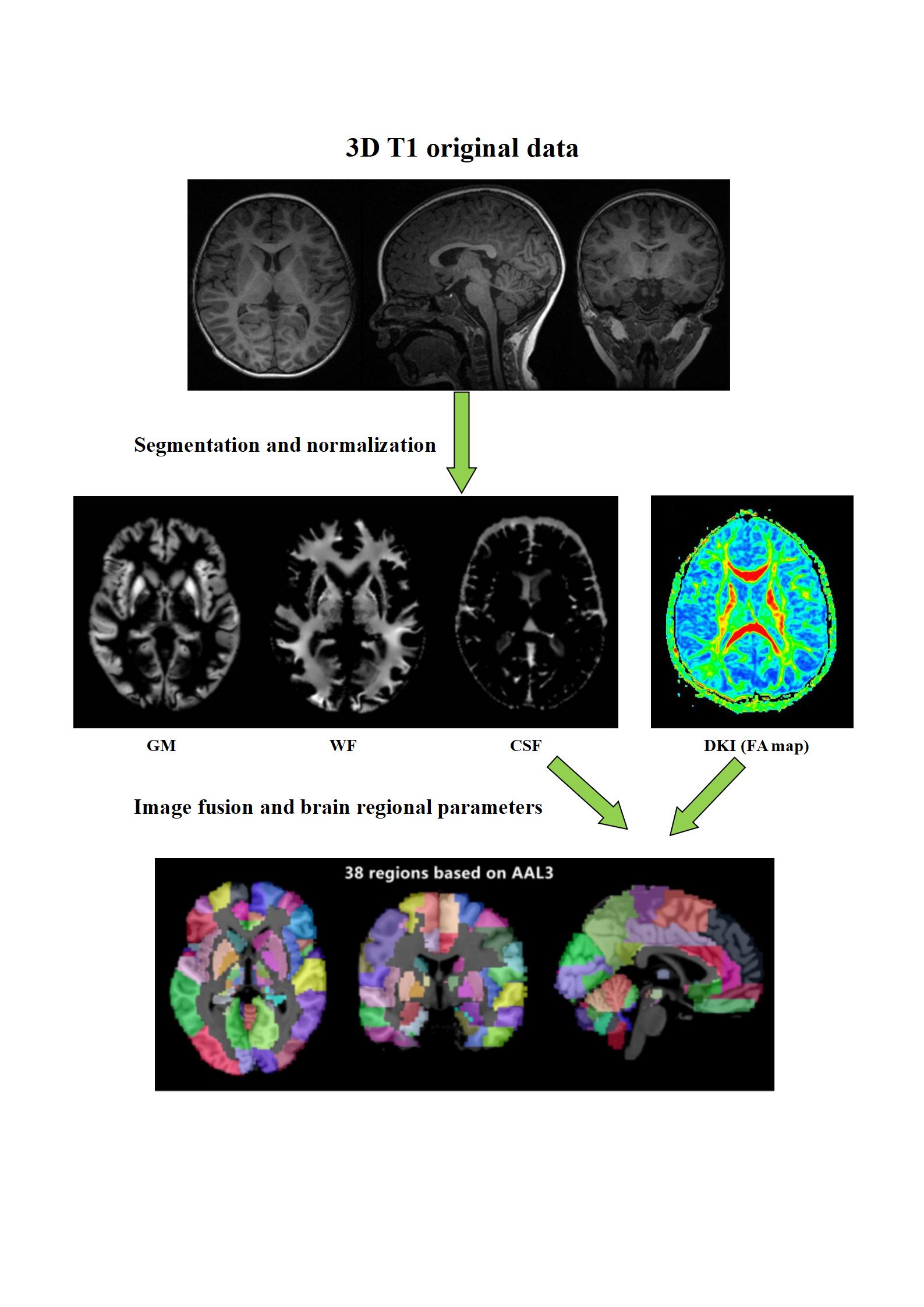
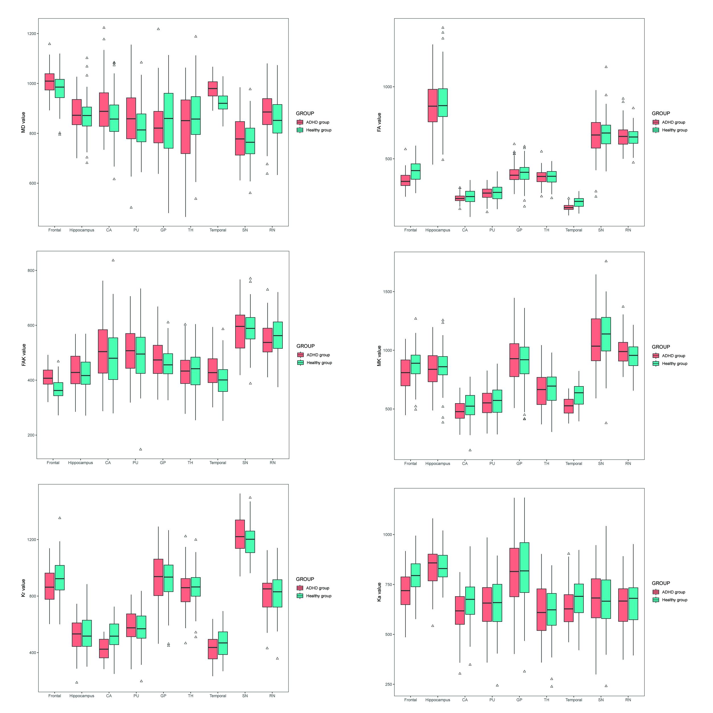
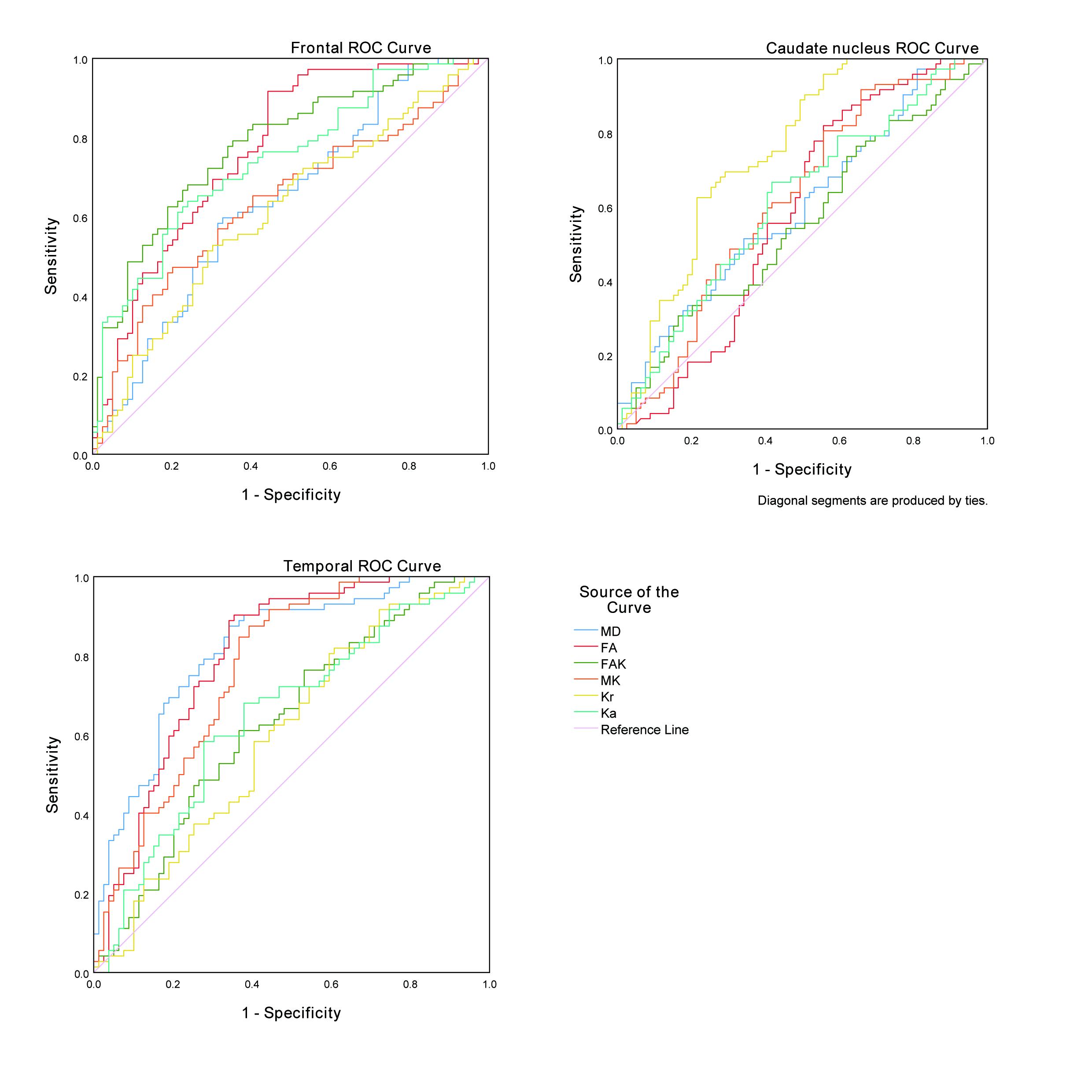
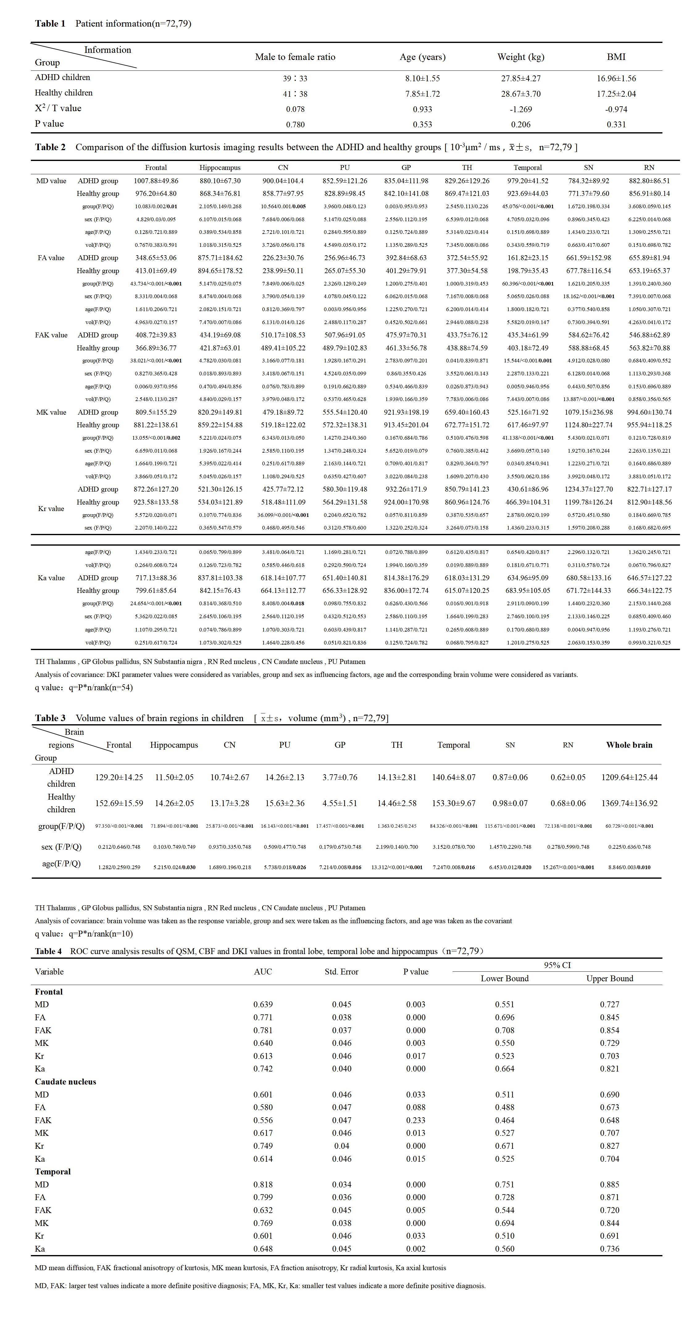
Table 1 Patient information(n=72,79)
Table 2 Comparison of the diffusion kurtosis imaging results between the ADHD and healthy groups [ 10-3μm2 / ms±s, n=72,79 ]
Table 3 Volume values of brain regions in children [`x±s,volume (mm3) , n=72,79]
Table 4 ROC curve analysis results of QSM, CBF and DKI values in frontal lobe, temporal lobe and hippocampus(n=72,79)