2549
Common and distinct cortical thickness alterations in youth with autism spectrum disorder or attention-deficit/hyperactivity disorder1Department of Radiology and Huaxi MR Research Center (HMRRC), West China Hospital, Sichuan University, chengdu, China, 2Department of Radiology, Sir Run Run Shaw Hospital, Zhejiang University School of Medicine, Hangzhou, China, 3Department of Psychiatry, West China Hospital, Sichuan University, chengdu, China
Synopsis
Keywords: Neuro, Brain, autism spectrum disorder, attention-deficit/hyperactivity disorder, cortical thickness, meta-analysis, surface-based morphometry
Motivation: Autism spectrum disorder (ASD) and attention-deficit/hyperactivity disorder (ADHD) are neurodevelopmental disorders with overlapping behavioral features and genetic etiology. Exploring brain cortical thickness (CTh) could help understand the neurobiological basis which builds the bridge between clinical manifestations and genetic liability of the two disorders.
Goal(s): To demonstrate the common and distinct of CTh changes in ASD and ADHD.
Approach: Previous brain structural MRI studies analyzing CTh of ASD and ADHD were included and compared by vertex-based meta-analysis.
Results: The ASD showed disorder-specific increased thickness in parietal lobule, and ADHD-specific decreased CTh was in motor area. Both disorders shared thinner thickness in temporo-parietal junction.
Impact: The shared and different patterns of CTh alterations in ASD and ADHD provide objective evidence for transdiagnosis. The subtle differences in areas with distinct functions could partly explain the divergent behavioral features in the two disorders and elucidate disorder-specific etiologies.
Introduction
Autism spectrum disorder (ASD) and attention-deficit/hyperactivity disorder (ADHD) are neurodevelopmental disorders in children and adolescents. It has been widely recognized that ASD and ADHD have overlapping behavioral features1,2 and genetic liability3. Specifically, social impairment and attention deficits are implicated in both disorders1. While the overlapping and distinct brain mechanisms contributing to these two disorders remain to be clarified.Cortical thickness (CTh) is a sensitive metric for evaluating cortical maturation abnormalities by reflecting the maturation in the columnar organization of the neocortical mantle4,5. Brain CTh alterations have been reported in ASD and ADHD separately and have inconsistent findings across studies6-9. A quantitative meta-analysis is well-suited to identify the most replicable overlapping and specific CTh alterations in these disorders.
Methods
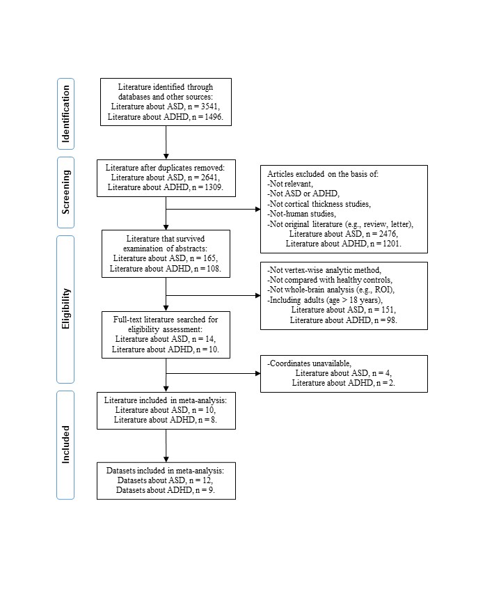
We searched PubMed, Web of Science, Embase, and Science Direct until 18th April 2023, and included MRI studies of CTh comparing youth (age less than 18) with ASD or ADHD with typically developing controls (TDC) in whole brain (Figure 1). There is no difference in demographic information between the two disorder groups. Meta-analysis was performed using seed-based d mapping (SDM) software (version 5.15)10,11. A recently developed mask for surface-based meta-analysis was used, which has been used previously in other neuropsychiatric disorders 12,13. The random-effects analysis was performed in each disorder group. A quantitative comparison of CTh was then performed between the two disorders. The conjunction and disjunction analyses were performed to identify overlapping and divergent abnormalities across disorders relative to TDC. All meta-analyses were conducted with the default threshold (P < 0.005, Z > 1.0), which provides an approximate equivalent to corrected P = 0.05 in SDM14. A more stringent probability threshold was employed for conjunction and disjunction analyses (P < 0.0025)14.Results
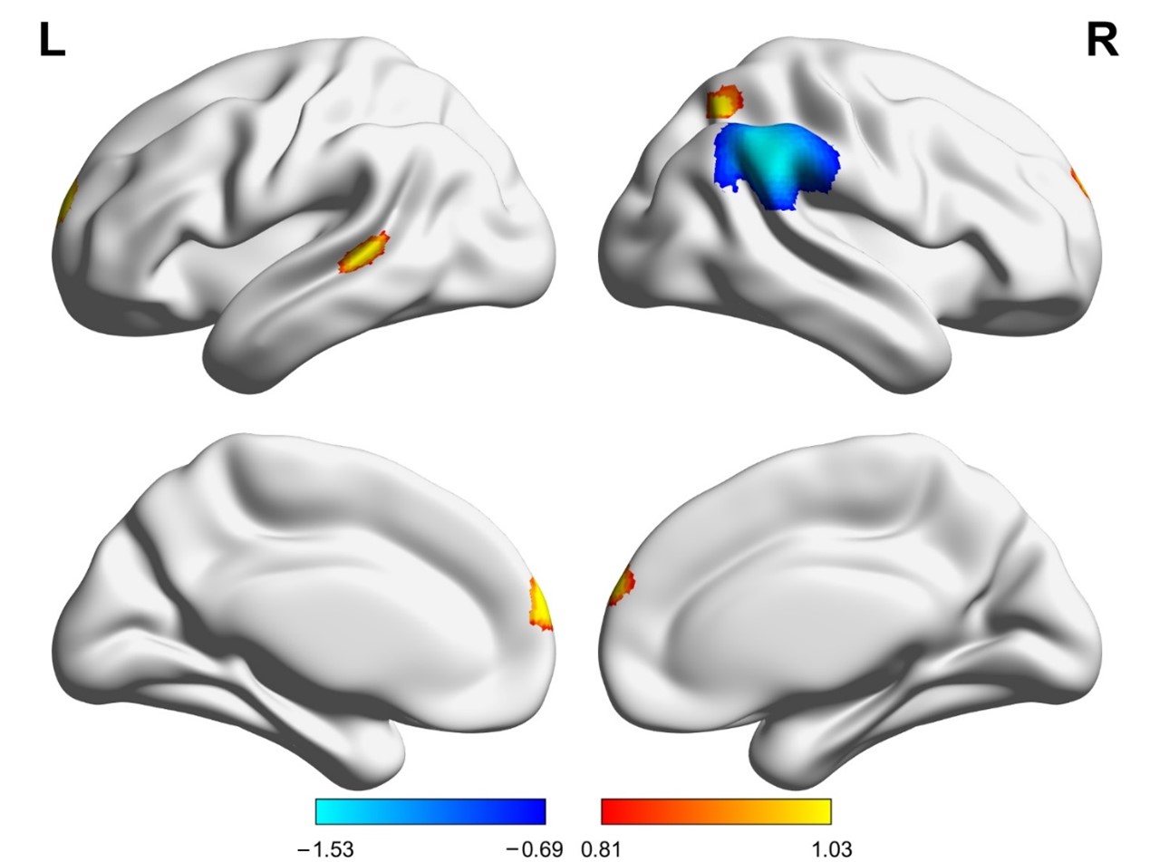
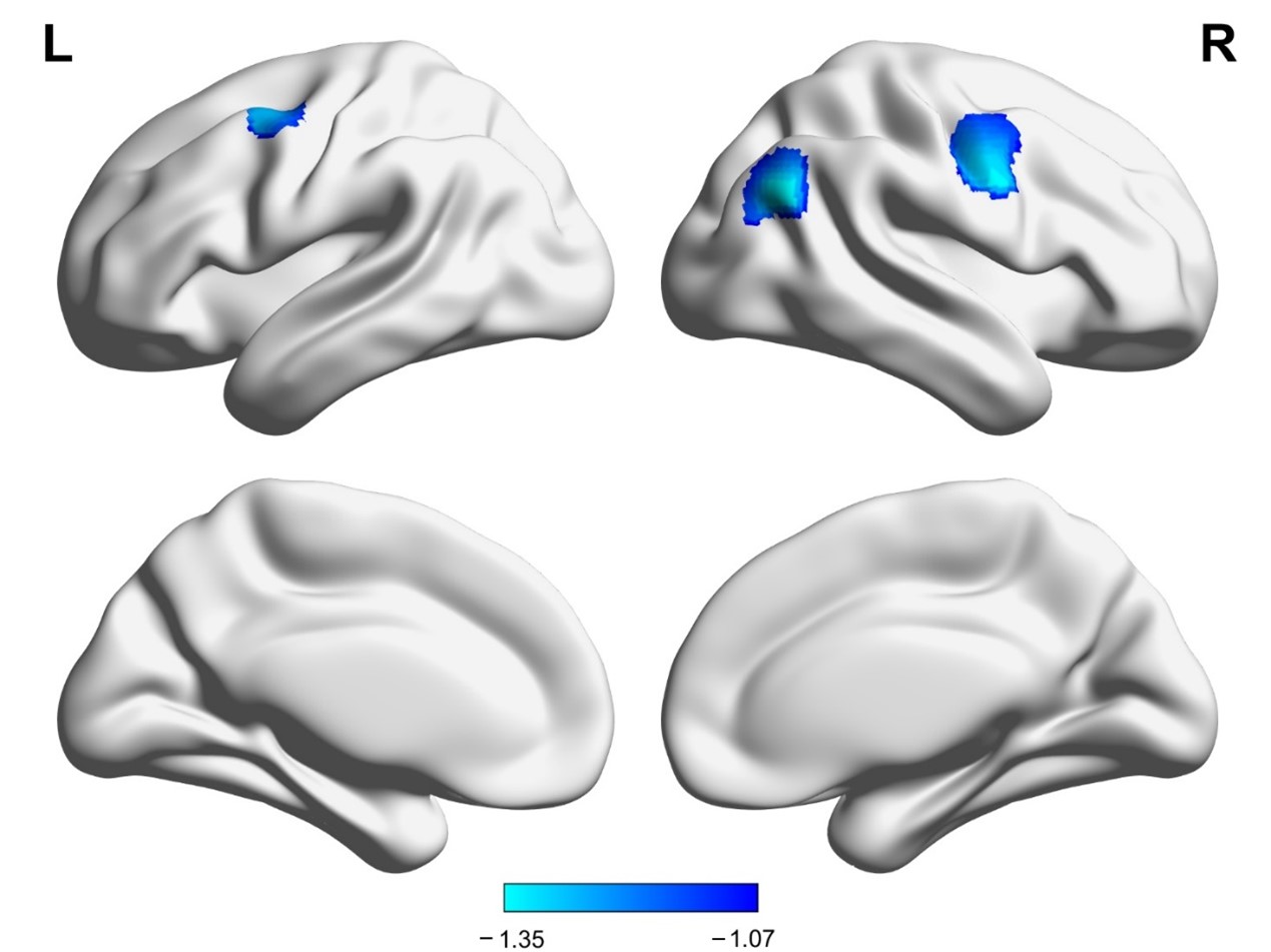
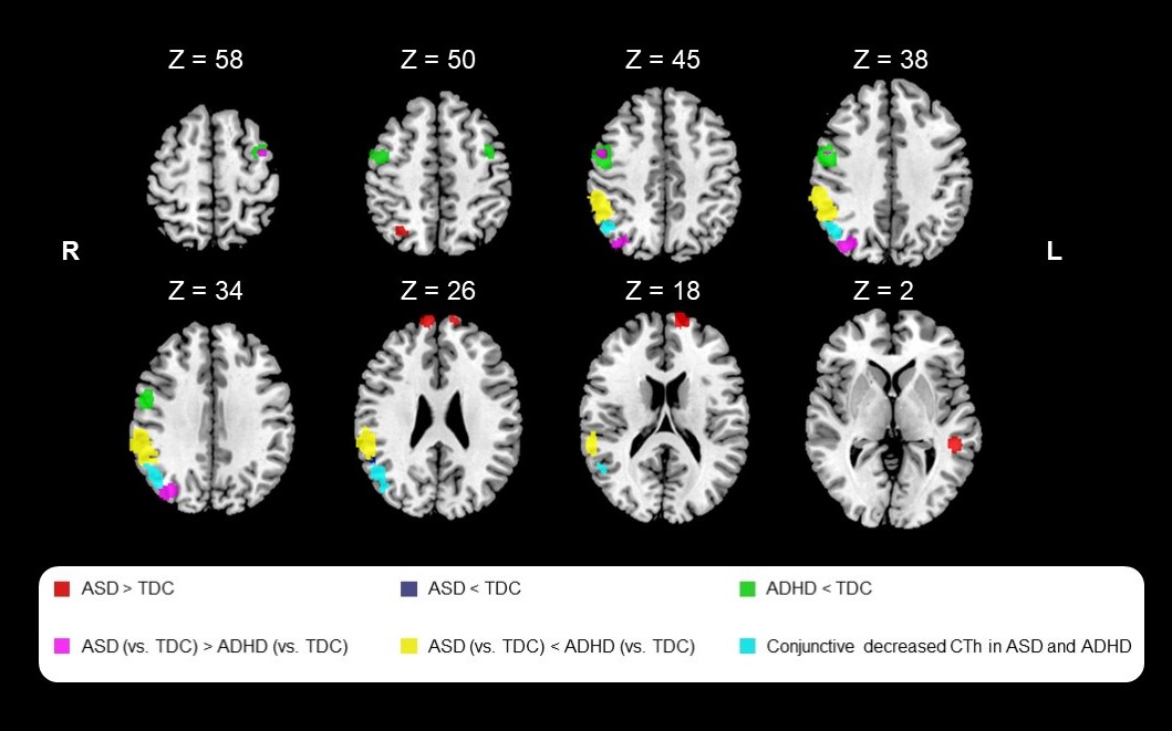
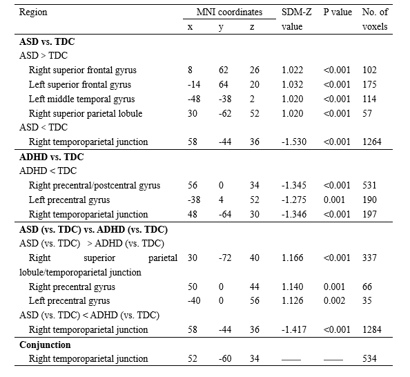
Twelve ASD datasets involving 458 individuals with ASD and nine ADHD datasets involving 322 individuals with ADHD were included in the analysis. Compared to TDC, ASD showed increased CTh in bilateral superior frontal gyrus, left middle temporal gyrus, and right superior parietal lobule, and decreased CTh in right temporoparietal junction (TPJ) (Table 1, Figures 2). ADHD showed decreased CTh in bilateral precentral gyri, right postcentral gyrus, and right TPJ relative to TDC (Table 1, Figures 3). Conjunction analysis showed both disorders shared reduced TPJ CTh located in default mode network (DMN). Comparative analyses indicated ASD had greater CTh in bilateral precentral gyri, right superior parietal lobule, and right TPJ located in dorsal attention network (DAN), and thinner CTh in right TPJ located in ventral attention network (VAN) than ADHD (Table 1, Figures 4).Discussion
Our study found that youth with ASD showed increased CTh in association cortex compared with TDC. This suggests a pattern of brain overgrowth or reduced age-related neuronal pruning, in widespread areas of association cortex 15,16. Participants with ADHD exhibited reduced CTh in bilateral motor cortices compared with TDC, an effect not observed in ASD. Longitudinal studies have shown that the ordered sequence of regional brain development in ADHD is similar to that seen in TDC, but the development was delayed 9. This is consistent with the clinical observation that many individuals have a reduction in ADHD symptoms by early adulthood, by which time delayed maturation of brain systems may be complete 17.The right TPJ is a higher-order area of association cortex including the unimodal visual area V5 responsible for motion processing 18. The dysmaturation of the TPJ region may be a robust transdiagnostic neuroimaging phenotypic biomarker relevant to the behavioral manifestation of both disorders, albeit in somewhat different ways given the subregions affected. Specifically, in ASD, a separate TPJ region with decreased CTh linked to the VAN was observed, while the additional TPJ reduction in ADHD was located in DAN. The VAN mediates the bottom-up attentional processing of novel external stimuli and is involved in detecting and reorienting attention to unexpected stimuli 19. In contrast, DAN mediates top-down attentional processing involving internal guidance of attention based on prior knowledge, willful plans, and current goals 20. These anatomic alterations are consistent with psychological studies demonstrating impaired attentional orienting to external stimulation in ASD 21, and difficulties in guiding voluntary allocation of attention in ADHD 22.
Conclusion
The distinct patterns of CTh abnormalities represent a basis for understanding the greater problems of perception and social cognition in ASD and the greater behavioral control problems in ADHD. These results suggest shared thinner TPJ located in DMN is an overlapping neurobiological feature of ASD and ADHD. The disorder-specific thinner TPJ located in disparate attention networks provides novel insight into distinct symptoms of attentional deficits associated with the two disorders.Acknowledgements
This study was supported by Sichuan Key Research and Development Project (2023YFS0226) and Chengdu Technology Innovation Research and Development Project (2022-YF05-01590-SN).References
1. Krakowski AD, Cost KT, Anagnostou E, et al. Inattention and hyperactive/impulsive component scores do not differentiate between autism spectrum disorder and attention-deficit/hyperactivity disorder in a clinical sample. Mol Autism. 2020;11(1):28.
2. van der Meer JMJ, Lappenschaar MGA, Hartman CA, et al. Homogeneous Combinations of ASD-ADHD Traits and Their Cognitive and Behavioral Correlates in a Population-Based Sample. Journal of attention disorders. 2017;21(9):753-763.
3. Ghirardi L, Brikell I, Kuja-Halkola R, et al. The familial co-aggregation of ASD and ADHD: a register-based cohort study. Molecular psychiatry. 2018;23(2):257-262.
4. Salat DH, Buckner RL, Snyder AZ, et al. Thinning of the cerebral cortex in aging. Cerebral cortex (New York, NY : 1991). 2004;14(7):721-730.
5. Shaw P, Kabani NJ, Lerch JP, et al. Neurodevelopmental trajectories of the human cerebral cortex. The Journal of neuroscience : the official journal of the Society for Neuroscience. 2008;28(14):3586-3594.
6. Ecker C, Pretzsch CM, Bletsch A, et al. Interindividual Differences in Cortical Thickness and Their Genomic Underpinnings in Autism Spectrum Disorder. The American journal of psychiatry. 2022;179(3):242-254.
7. Zielinski BA, Prigge MB, Nielsen JA, et al. Longitudinal changes in cortical thickness in autism and typical development. Brain : a journal of neurology. 2014;137(Pt 6):1799-1812.
8. Shaw P, Lerch J, Greenstein D, et al. Longitudinal mapping of cortical thickness and clinical outcome in children and adolescents with attention-deficit/hyperactivity disorder. Archives of general psychiatry. 2006;63(5):540-549.
9. Shaw P, Eckstrand K, Sharp W, et al. Attention-deficit/hyperactivity disorder is characterized by a delay in cortical maturation. Proceedings of the National Academy of Sciences of the United States of America. 2007;104(49):19649-19654.
10. Chen L, Hu X, Ouyang L, et al. A systematic review and meta-analysis of tract-based spatial statistics studies regarding attention-deficit/hyperactivity disorder. Neuroscience and biobehavioral reviews. 2016;68:838-847.
11. Yao L, Yang C, Zhang W, et al. A multimodal meta-analysis of regional structural and functional brain alterations in type 2 diabetes. Frontiers in neuroendocrinology. 2021;62:100915.
12. Li Q, Zhao Y, Chen Z, et al. Meta-analysis of cortical thickness abnormalities in medication-free patients with major depressive disorder. Neuropsychopharmacology : official publication of the American College of Neuropsychopharmacology. 2020;45(4):703-712.
13. Zhao Y, Zhang Q, Shah C, et al. Cortical Thickness Abnormalities at Different Stages of the Illness Course in Schizophrenia: A Systematic Review and Meta-analysis. JAMA psychiatry. 2022.
14. Radua J, Borgwardt S, Crescini A, et al. Multimodal meta-analysis of structural and functional brain changes in first episode psychosis and the effects of antipsychotic medication. Neuroscience and biobehavioral reviews. 2012;36(10):2325-2333.
15. D'Cruz AM, Mosconi MW, Ragozzino ME, et al. Alterations in the functional neural circuitry supporting flexible choice behavior in autism spectrum disorders. Translational psychiatry. 2016;6(10):e916.
16. Takarae Y, Minshew NJ, Luna B, et al. Atypical involvement of frontostriatal systems during sensorimotor control in autism. Psychiatry research. 2007;156(2):117-127.
17. Faraone SV, Biederman J, Spencer T, et al. Attention-deficit/hyperactivity disorder in adults: an overview. Biological psychiatry. 2000;48(1):9-20.
18. Doricchi F. The functions of the temporal-parietal junction. Handbook of clinical neurology. 2022;187:161-177.
19. Vossel S, Geng JJ, Fink GR. Dorsal and ventral attention systems: distinct neural circuits but collaborative roles. The Neuroscientist : a review journal bringing neurobiology, neurology and psychiatry. 2014;20(2):150-159.
20. Wilterson AI, Nastase SA, Bio BJ, et al. Attention, awareness, and the right temporoparietal junction. Proceedings of the National Academy of Sciences of the United States of America. 2021;118(25).
21. Fitzgerald J, Johnson K, Kehoe E, et al. Disrupted functional connectivity in dorsal and ventral attention networks during attention orienting in autism spectrum disorders. Autism research : official journal of the International Society for Autism Research. 2015;8(2):136-152.
22. Salehinejad MA, Ghayerin E, Nejati V, et al. Domain-specific Involvement of the Right Posterior Parietal Cortex in Attention Network and Attentional Control of ADHD: A Randomized, Cross-over, Sham-controlled tDCS Study. Neuroscience. 2020;444:149-159.
Figures