2546
Reduced frontolimbic functional connectivity in psychostimulant-free ADHD youth with and without familial risk for bipolar I disorder1Department of Radiology and Huaxi MR Research Center (HMRRC), Functional and Molecular lmaging Key Laboratory of Sichuan Province, West China Hospital, Sichuan University, Chengdu 610041, Sichuan Province, China, 2College of Medical Informatics, Chongqing Medical University, Chongqing 400016, China, 3Department of Psychiatry and Behavioral Neuroscience, University of Cincinnati College of Medicine, Cincinnati 45219, OH, United States
Synopsis
Keywords: Adolescents, MR Value, ADHD, Functional connectivity
Motivation: Although youth with attention-deficit/hyperactivity disorder (ADHD) with a family history of bipolar I disorder (BD) are at increased risk for developing BD, associated neurofunctional mechanisms remain understood.
Goal(s): To investigate the differences in functional connectivity (FC) between ventrolateral prefrontal cortex (VLPFC) and amygdala (AMY) among ADHD youth with (‘high-risk’, HR) and without (‘low-risk’, LR) a BD family history and healthy controls (HC).
Approach: gPPI analysis was conducted to assess the VLPFC-AMY FC in response to unpleasant emotional stimuli.
Results: Both LR and HR exhibited lower right VLPFC-left AMY FC compared to HC, and HR had even lower FC compared with LR.
Impact: ADHD in conjunction with BD family history showed blunted emotion-generated right VLPFC-left AMY FC compared with ADHD youth without a BD family history and healthy youth, and may therefore represent a central biomarker that is relevant to BD risk progression.
Introduction
Attention deficit/hyperactivity disorder (ADHD) youth with a first-degree relative with bipolar I disorder (BD) robustly increases the risk of developing BD 1, and youth with a first-degree BD relative exhibit higher rates of ADHD 2 and more severe ADHD symptoms 3. While these findings suggest that ADHD in conjunction with familial risk for BD may represent a different and more severe illness that confers a greater risk for developing BD, associated neurofunctional mechanisms remain poorly understood. The ventrolateral prefrontal cortex (VLPFC) - amygdala (AMY) circuit plays a central role in emotional regulation, and increased emotion-generated VLPFC-AMY functional connectivity (FC) and AMY hyperactivation have consistently been implicated in the pathophysiology of BD 4,5. In contrast, youth with ADHD exhibit reduced or no difference AMY activation and decreased VLPFC-AMY FC in response to emotional stimuli relative to both healthy youth and youth with BD 4. However, prior studies did not control for BD familial risk in ADHD youth, ADHD comorbidity in BD, and/or psychostimulant exposure, and no studies have directly compared VLPFC-AMY FC in unaffected psychostimulant-free ADHD youth with and without a first-degree BD relative. In the present cross-sectional event-related fMRI study, we investigated VLPFC-AMY FC in psychostimulant-free ADHD youth with and without a first-degree relative with BD and a typically developing control (HC) group. Exploratory analyses evaluated relationships among VLPFC-AMY FC and relevant symptom measures.Methods
We enrolled ADHD youth (ages 10-18 years) with (‘high-risk’, HR) and without (‘low-risk’, LR) a first-degree relative with BD and HC. All ADHD youth were stimulant-naïve or had no exposure to psychostimulants for at least 3 months prior to enrollment. Subjects performed a continuous performance task with emotional and neutral distractors (CPT-END) during functional magnetic resonance imaging (fMRI) scanning. Prior evidence from fMRI CPT-END studies indicates that the VLPFC and AMY are selectively activated in response to unpleasant emotional images but not attentional targets 6. Generalized psychophysiological interaction (gPPI) analysis was conducted to assess the region of interest (ROI)-to-ROI FC between bilateral VLPFC and AMY. Clinician ratings of ADHD (ADHD-rating scale), mania (Young Mania Rating Scale, YMRS), depression (Children’s Depression Rating Scale-Revised, CDRS-R), global functioning (Children’s Global Assessment Scale), and global symptom severity (Clinical Global Impression-Severity Scale) were performed, and parents completed the Child Behavior Checklist (CBCL).Results
A total of 144 youth (mean age 14.3±2.5 years; HC, n=46; low-risk, n=50; high-risk, n=48) were included in the analysis. No significant group differences were observed for age, sex, race, pubertal status, hand dominance, or prior psychostimulant exposure in the ADHD groups. Both HR and LR ADHD groups differed significantly from HC on all ratings. For the emotion/square contrast, only right VLPFC to left AMY FC differed significantly among groups. Post-hoc tests revealed that both LR and HR groups exhibited lower right VLPFC-left AMY FC compared to HC, and the HR group had lower right VLPFC-left AMY FC compared with the LR group (Figure 1). For the circle/square contrast, there were no differences in right VLPFC-left AMY FC among the three groups. For the emotion/square contrast, right VLPFC-left AMY FC was positively correlated with YMRS total scores in the HR group but not in the LR group, and negatively correlated with CBCL internalization score in combined LR and HR groups (Figure 2).Discussion
Both LR and HR ADHD patients exhibited reduced VLPFC-AMY FC during emotional stimuli compared with HC, suggesting that this blunted response may be attributable in part to ADHD pathophysiology. Our results also suggest that HR ADHD youth, despite being at an elevated risk for developing BD, do not resemble youth that have already developed BD that had an increased emotion-generated VLPFC-AMY FC 4, and the alterations of VLPFC-left AMY FC found in HR ADHD youth may reflect dysconnectivity or greater inhibitory control in response to emotional stimuli which may confer resilience to BD. PFC-AMY FC in response to emotional stimuli gradually shifts from positive to negative with age,7,8 and reduced VLPFC-AMY FC observed in ADHD youth may reflect an abnormal acceleration in maturational trajectories. Accordingly, the greater reduction in VLPFC-AMY FC in the HR group compared with the LR group may reflect a more pervasive maturational disruption.Conclusion
Psychostimulant-free ADHD youth with familial risk for BD exhibit blunted right VLPFC-left AMY FC in response to emotional stimuli compared with ADHD youth without familial risk for BD and healthy youth. Associations between right VLPFC-left AMY FC and mania in the HR group suggest potential relevance to BD risk progression and warrant additional investigation in prospective longitudinal studies.Acknowledgements
None.References
1. Birmaher B, Merranko J, Hafeman D, et al. The role of bipolar polygenic risk score in the familial transmission of bipolar disorder-An updated analysis. Bipolar Disord 2022;24(4):437-440.2. Brancati GE, Perugi G, Milone A, Masi G, Sesso G. Development of bipolar disorder in patients with attention-deficit/hyperactivity disorder: A systematic review and meta-analysis of prospective studies. J Affect Disord 2021;293:186-196.
3. Chen C, Tallman MJ, Cecil KM, et al. Symptom profiles, but not prefrontal neurochemistry, differentiate ADHD youth with and without a family history of bipolar I disorder. J Atten Disord 2022;26(13):1762-1773.
4. Hafeman D, Bebko G, Bertocci MA, et al. Amygdala-prefrontal cortical functional connectivity during implicit emotion processing differentiates youth with bipolar spectrum from youth with externalizing disorders. J Affect Disord 2017;208:94-100.
5. Kanske P, Schönfelder S, Forneck J, Wessa M. Impaired regulation of emotion: neural correlates of reappraisal and distraction in bipolar disorder and unaffected relatives. Transl Psychiatry 2015;5(1):e497.
6. Yamasaki H, LaBar KS, McCarthy G. Dissociable prefrontal brain systems for attention and emotion. Proc Natl Acad Sci U S A 2002;99(17):11447-11451.
7. Gee DG, Humphreys KL, Flannery J, et al. A developmental shift from positive to negative connectivity in human amygdala-prefrontal circuitry. J Neurosci 2013;33(10):4584-4593.
8. Wu M, Kujawa A, Lu LH, et al. Age-related changes in amygdala-frontal connectivity during emotional face processing from childhood into young adulthood. Hum Brain Mapp 2016;37(5):1684-1695.
Figures
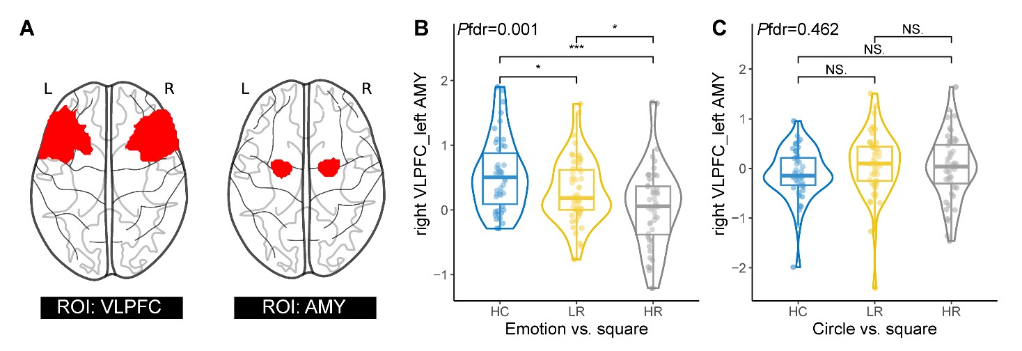
Figure 1. Localization of the bilateral ventrolateral prefrontal cortex (VLPFC) and amygdala (AMY) regions of interests (ROIs) (A). Between-group analysis results of functional connectivity between right VLPFC and left AMY during the emotion vs. square contrast (B) and the circle vs. square contrast (C). Abbreviations: HC, typically developing healthy controls; LR, low-risk attention deficits/hyperactive disorder group; HR, high-risk attention deficits/hyperactive disorder group; *, Pfdr<0.05; NS., not significant; L, left; R, right.
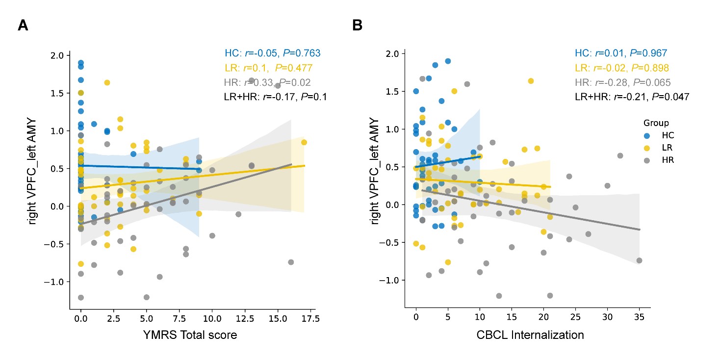
Figure 2. Correlations between functional connectivity between right ventrolateral prefrontal cortex (VLPFC) and left amygdala (AMY) during the emotion vs. square contrast and Young Mania Rating Scale (YMRS) total scores (A) and Child Behavior Checklist (CBCL) internalization subscale scores (B). Abbreviations: HC, typically developing healthy controls; LR, low-risk attention deficits/hyperactive disorder group; HR, high-risk attention deficits/hyperactive disorder group.