2544
Investigating the Effects of Antenatal Maternal Anaemia on Brain Structure in 6-Year-Old South African Children: A Neuroimaging Cohort Study1Department of Paediatrics and Child Health, University of Cape Town, Cape Town, South Africa, 2Neuroscience Institute, University of Cape Town, Cape Town, South Africa, 3Department of Neuroimaging, Kings College London, London, United Kingdom, 4Department of Psychiatry and Mental Health, University of Cape Town, Cape Town, South Africa, 5Department of Neurology, Psychiatry and Biobehavioral Sciences, University of California, Los Angeles, CA, United States, 6Department of Bioengineering, University of California, Los Angeles, CA, United States
Synopsis
Keywords: Neuro, Pediatric, Maternal anaemia, haemoglobin, high-field MRI, neurodevelopment, neuroimaging, child brain structure
Motivation: It is unknown whether recent findings linking antenatal maternal anaemia with altered brain structure in toddlers persist to later childhood.
Goal(s): This study aimed to determine whether associations of antenatal maternal anaemia with smaller corpus callosum, putamen, and caudate nucleus volumes described in 2–3-year-olds remain at age 6-7 years in the same cohort.
Approach: Linear modeling was used to investigate associations between antenatal maternal anaemia status, maternal haemoglobin concentrations, and child brain volumes.
Results: Antenatal maternal anaemia was associated with smaller volumes of the corpus callosum and caudate nucleus in school-age children, with comparable adjusted volume differences and coefficients to findings in toddlers.
Impact: Evidence that associations of maternal anaemia with brain volumes are consistent and persist from age 2-3 years through to age 6-7 years supports the importance of optimizing antenatal maternal health and reinforces these brain regions as a future research focus.
Introduction
Anaemia, indicated by low serum haemoglobin, is a strikingly prevalent health concern affecting approximately 30% of women of reproductive age and 36% of pregnant women worldwide.1 Although anaemia is a global health priority, it is particularly prevalent in Low- and Middle-Income Countries (LMICs), with 30-50% of women in Africa estimated to have anaemia (Figure 1).1 Given that progress in reducing anaemia has stagnated between 2000 and 2019, it has been identified by the World Health Organization (WHO) as a Sustainable Development Goal (SDG) for accelerated action.2While antenatal maternal anaemia has consistently been associated with poor developmental and neurocognitive outcomes in multiple settings including South Africa,3-6 neuroimaging research findings7-9 are limited. In addressing this research gap, a neuroimaging sub-study from a South African birth cohort (Drakenstein Child Health Study; DCHS) found that antenatal maternal anaemia was associated with smaller volumes of the bilateral caudate, left putamen, and total corpus callosum at 2-3 years of age.10 Furthermore, continuous maternal haemoglobin concentrations directly predicted regional child brain volumes. These findings raised the question of whether such associations would persist through childhood.
Methods
This neuroimaging sub-study is nested within the DCHS, a population-based cohort in a peri-urban district of South Africa.11 Pregnant women were enrolled between 2012 and 2015, and mother-child dyads were followed prospectively. Maternal haemoglobin was measured during pregnancy and a subgroup of children had haemoglobin measurements postnatally. Based on WHO guidelines,12 maternal anaemia was classified as haemoglobin levels <11g/dL in pregnancy with age-specific thresholds for child anaemia. A sub-group of DCHS children had MRI at 6-7 years on a 3T Siemens Skyra. T1-weighted structural data was processed with FreeSurfer 7.1.113 using automated volume segmentation pipelines. Given previous findings,10 the corpus callosum, caudate nucleus, and putamen volumes were of particular interest. Alongside this hypothesis-driven approach, exploratory analyses were conducted across all other subcortical structures. Associations between child brain volumes for regions of interest with maternal anaemia status and continuous maternal haemoglobin concentrations were conducted using (M)ANOVAs and multivariable linear regression models, respectively. Maternal anaemia models included relevant covariates to account for potential confounding and were conducted with and without the inclusion of child anaemia to assess the role of antenatal versus postnatal anaemia.Results
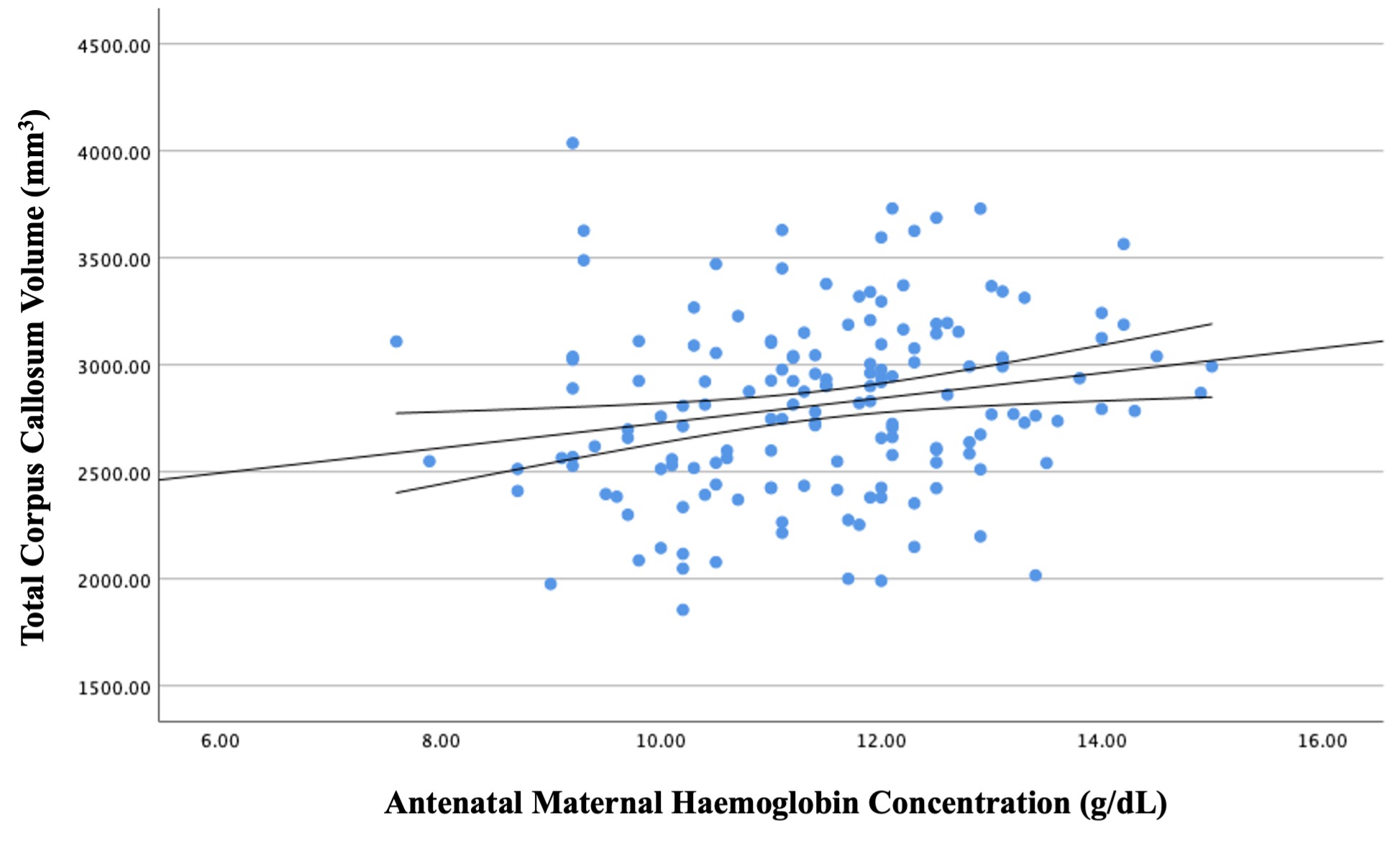
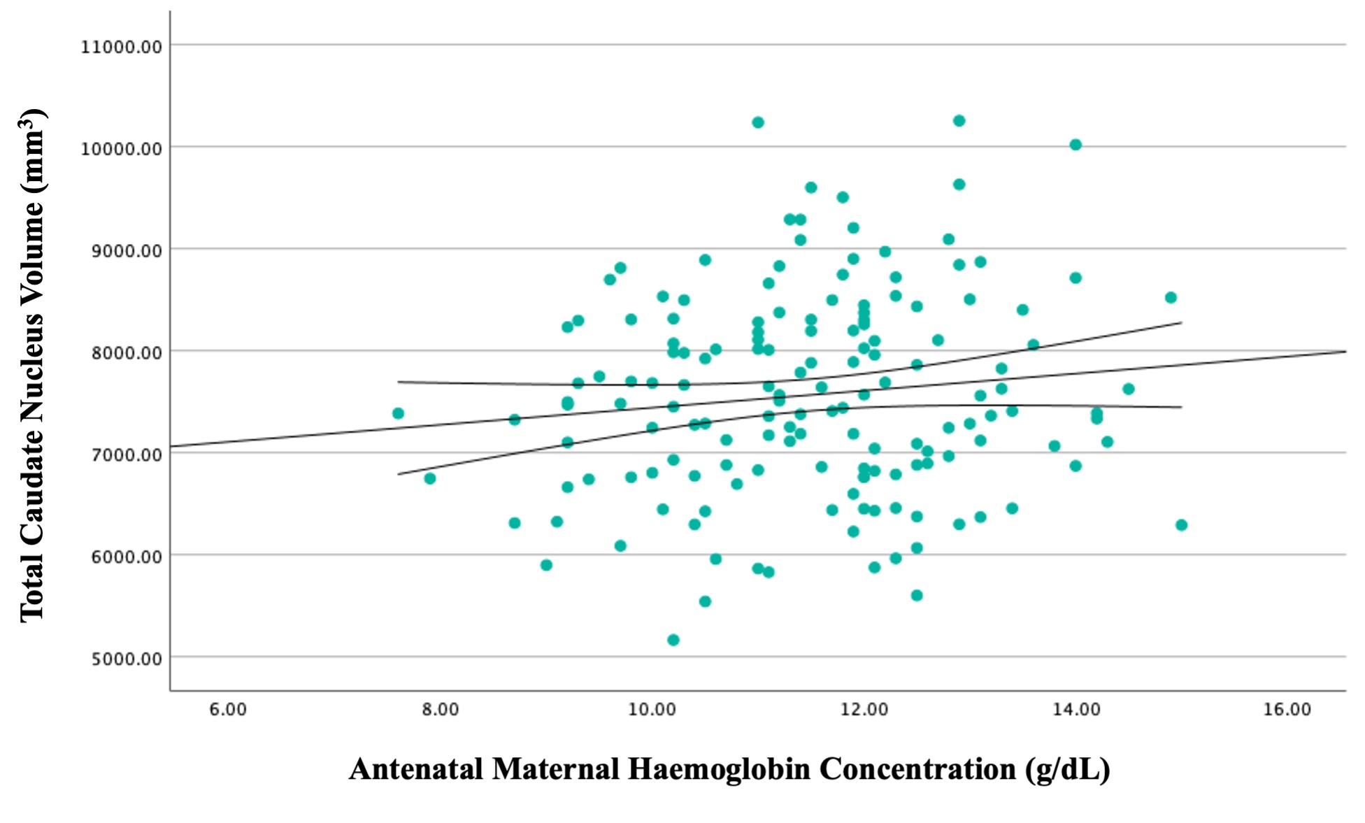
In this neuroimaging sub-study, 157 children (Mean [SD] age of 75.54[4.77] months; 84 [53.50%] male) were born to mothers with antenatal haemoglobin data. Overall, 50 (31.85%) of the mothers were anaemic during pregnancy. In fully adjusted models, antenatal maternal anaemia was associated with smaller volumes (Figure 2) of the total corpus callosum (adjusted percentage difference, -6.77%; p=0.003), left caudate nucleus (adjusted percentage difference, -5.98%; p=0.005), and right caudate nucleus (adjusted percentage difference, -6.12%; p=0.003). Similarly, continuous maternal haemoglobin in pregnancy was associated with total corpus callosum (β=0.239 [CI: 0.10 to 0.38]; p<0.001; Figure 3) and caudate nucleus (β=0.165 [CI: 0.02 to 0.31]; p=0.027; Figure 4) volumes. No other subcortical brain volume differences emerged in exploratory maternal anaemia analyses. In a sub-group of 89 children with child haemoglobin data (Mean [SD] age of 76.06[4.84] months; 49 [55%] male), the prevalence of maternal anaemia and child anaemia was 34 (38.20%) and 42 (47.19%), respectively. There was no association between maternal anaemia and child anaemia (χ2 = 0.799; p=0.372), and postnatal child anaemia did not contribute to regional brain volume differences associated with maternal anaemia.Discussion
Associations of antenatal maternal anaemia with brain volumes in children at 2-3 years of age persisted and were regionally consistent at 6-7 years of age. The adjusted volume differences for regions of interest were comparable between timepoints with smaller volumes of the corpus callosum (7% at 6-7 years versus 8% at 2-3 years) and caudate nucleus (6% at 6-7 years versus 5% at 2-3 years). Furthermore, the nature and the strength of the relationship between maternal haemoglobin concentrations and child volumes for the corpus callosum (standardised coefficient of 0.24 at both timepoints) and caudate nucleus (standardised coefficient of 0.17 at 6-7 years versus 0.15 at 2-3 years) remained similar.Given the magnitude of the volume difference in affected regions, the known role of the corpus callosum and caudate nucleus in neuropsychological functioning,14-16 and the previously demonstrated link between anaemia and poor cognitive outcomes in this cohort,6 these findings are likely to be clinically important.
Conclusion
The persistent associations of antenatal maternal anaemia with structural brain findings in childhood emphasize the need for optimised anaemia interventions in women of reproductive age before and during pregnancy. Additionally, the consistency of findings in key brain structures across early childhood reinforces the importance of these regions as a focus for future research.Acknowledgements
We would like to thank all the mothers and children who participated in the Drakenstein Child Health Study and made this neuroimaging research possible. Our sincere gratitude is also owed to the dedicated study staff members and leadership across sites, the radiographers at the Cape Universities Body Imaging Centre (CUBIC), and our international collaborators on the project. The DCHS is funded by the Bill & Melinda Gates Foundation (OPP 1017641), the National Research Foundation (NRF), and the National Institutes of Health. Additional funding for high-field MRI acquisition was provided by the National Institute on Alcohol Abuse and Alcoholism (NIAAA; R01AA026834-01) for K.A. Donald in support of the neuroimaging arm of the cohort. The first author, J.E. Ringshaw, is supported by a Wellcome Trust International Training Fellowship (224287/Z/21/Z).References
- Stevens GA, Paciorek CJ, Flores-Urrutia MC, et al. National, regional, and global estimates of anaemia by severity in women and children for 2000–19: a pooled analysis of population-representative data. The Lancet Global Health 2022; 10(5): e627-e39. doi:10.1016/S2214-109X(22)00084-5
- WHO. Accelerating anaemia reduction: a comprehensive framework for action. 2023.
- Radlowski EC, Johnson RW. Perinatal iron deficiency and neurocognitive development. Frontiers in Human Neuroscience 2013; 7: 585. doi:10.3389/fnhum.2013.00585
- Janbek J, Sarki M, Specht IO, Heitmann BL. A systematic literature review of the relation between iron status/anemia in pregnancy and offspring neurodevelopment. European Journal of Clinical Nutrition 2019; 73(12): 1561-78. doi:10.1038/s41430-019-0400-6
- Quezada-Pinedo HG, Cassel F, Duijts L, et al. Maternal iron status in pregnancy and child health outcomes after birth: a systematic review and meta-analysis. Nutrients 2021; 13(7): 2221. doi:10.3390/nu13072221
- Donald KA, Wedderburn CJ, Barnett W, et al. Risk and protective factors for child development: An observational South African birth cohort. PLoS Medicine 2019; 16(9): e1002920. doi:10.1371/journal.pmed.1002920
- Basu S, Kumar D, Anupurba S, Verma A, Kumar A. Effect of maternal iron deficiency anemia on fetal neural development. Journal of Perinatology 2018; 38(3): 233-9. doi:10.1038/s41372-017-0023-5
- Monk C, Georgieff MK, Xu D, et al. Maternal prenatal iron status and tissue organization in the neonatal brain. Pediatric Research 2016; 79(3): 482-8. doi:10.1038/pr.2015.248
- Loureiro B, Martinez-Biarge M, Foti F, Papadaki M, Cowan FM, Wusthoff CJ. MRI Patterns of brain injury and neurodevelopmental outcomes in neonates with severe anaemia at birth. Early Human Development 2017; 105: 17-22. doi:10.1016/j.earlhumdev.2017.01.001
- Wedderburn CJ, Ringshaw JE, Donald KA, et al. Association of Maternal and Child Anemia With Brain Structure in Early Life in South Africa. JAMA Network Open 2022; 5(12): e2244772-e. doi:10.1001/jamanetworkopen.2022.44772
- Zar H, Barnett W, Myer L, Stein D, Nicol M. Investigating the early-life determinants of illness in Africa: the Drakenstein Child Health Study. Thorax 2015; 70(6): 592-4. doi:10.1136/thoraxjnl-2014-206242
- World Health Organisation. Haemoglobin concentrations for the diagnosis of anaemia and assessment of severity: World Health Organization, 2011.
- Fischl B, Salat, D.H., Busa, E., et al. Whole brain segmentation: automated labeling of neuroanatomical structures in the human brain. Neuron 2002; 33(341-355). doi:10.1016/S0896-6273(02)00569-X
- Burgaleta M, MacDonald PA, Martínez K, et al. Subcortical regional morphology correlates with fluid and spatial intelligence. Human Brain Mapping 2014; 35(5): 1957-68. doi:10.1002/hbm.22305
- MacDonald PA, Ganjavi H, Collins DL, Evans AC, Karama S. Investigating the relation between striatal volume and IQ. Brain Imaging and Behavior 2014; 8(1): 52-9. doi:10.1007/s11682-013-9242-3
- Deoni SC, O’Muircheartaigh J, Elison JT, et al. White matter maturation profiles through early childhood predict general cognitive ability. Brain Structure and Function 2016; 221: 1189-203. doi:10.1007/s00429-014-0947-x
Figures
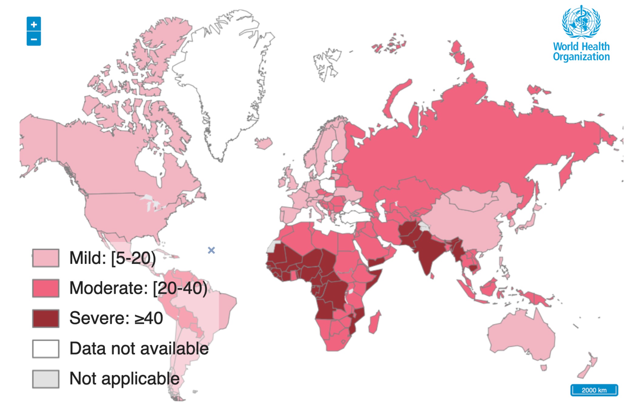
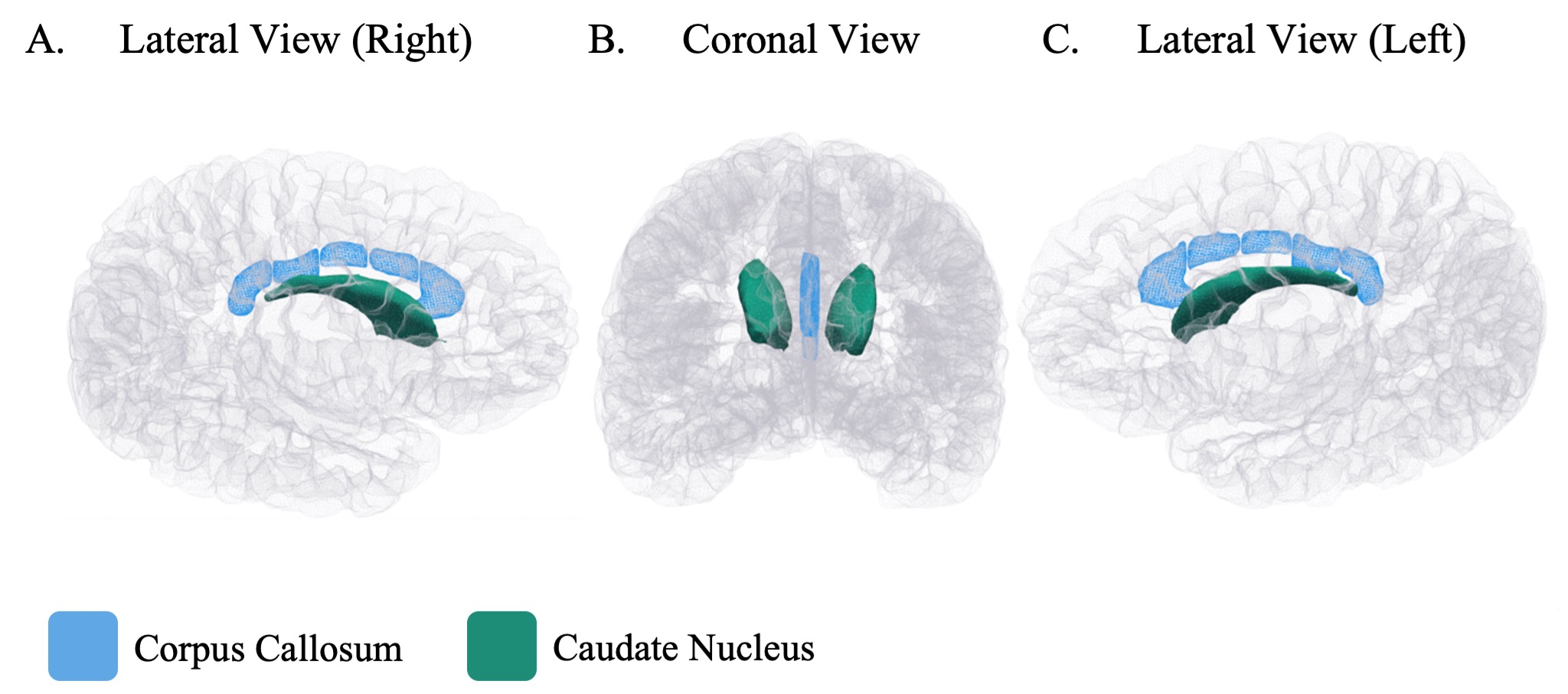
Figure 2. Corpus Callosum and Caudate Nucleus Volumes Associated With Antenatal Maternal Anaemia Visualised on a Cortical Surface. Images Were Created Using FreeSurfer.