2542
Clustering of local diffusion features reveals altered thalamic topography in newborns with congenital heart defects1MR-Research Center, University Children's Hospital Zurich, Zurich, Switzerland, 2Department of Information Technology and Electrical Engineering, ETH Zurich, Zurich, Switzerland, 3Child Development Center, University Children’s Hospital Zurich, Zurich, Switzerland, 4Department of Diagnostic Imaging, University Children’s Hospital Zurich, Zurich, Switzerland, 5Children’s Research Center, University Children’s Hospital Zurich, Zurich, Switzerland, 6Division of Pediatric Cardiology, Pediatric Heart Center, University Children's Hospital Zurich, Zurich, Switzerland
Synopsis
Keywords: Neonatal, Brain Connectivity, congenital heart defects
Motivation: Structural connectivity in the thalamus, essential for cortical-subcortical communication, is known to be disrupted in congenital heart defect (CHD) patients. Yet, assessing thalamic nuclei via local diffusion characteristics in vivo remains challenging.
Goal(s): We aimed to validate a diffusion-based clustering method to map the developing thalamus and explore topological differences between CHD infants and healthy controls
Approach: By refining a segmentation method and employing k-means and GMM clustering, we characterized thalamic nuclei
Results: Notable volume reductions in six thalamic clusters were identified in CHD infants, with significant mediodorsal nucleus group alterations, pertinent to prefrontal connectivity
Impact: Our validated segmentation technique enables robust delineation of thalamic nuclei in vivo, unveiling developmental alterations in CHD infants. This advancement paves the way for targeted clinical interventions and improves neurodevelopmental outcomes prediction
Introduction
The thalamus serves as crucial hub in the network of connections between the cortex and subcortical regions. It has been shown that structural connectivity topography in congenital heart defects is altered1, however, local diffusion features might be more reliable for thalamus clustering2. Therefore, our study aimed to reproduce these findings by applying a different clustering methodology based on local diffusion properties to the developing human thalamus, and further, to determine whether thalamic topology differs between infants with congenital heart defects and normally developing controls.Method
Our study refines a previously reported method1 for thalamic nuclei segmentation by estimating fiber orientation distribution functions (FOD) in MRtrix3 using constrained spherical deconvolution3,4. The mapping of thalamus topography relied on the spherical harmonic coefficients as feature vectors carrying biological information and spatial coordinates using an in-house implementation of clustering in the scikit-learn library5. To capture the developmental trajectories of local connectivity features within the thalamus, we employed two clustering methods: k-means clustering for voxel categorization based on proximity to the nearest cluster centroid, and Gaussian Mixture Models (GMM) for probabilistic voxel assignments, reflecting the complex nature of thalamic structures during early brain development.Results
To first optimize the clustering parameters and benchmark its within-subject reproducibility, we applied the k-means clustering to 30 open-source adult diffusion MRI datasets6, achieving successful and reproducible segmentation of the thalamus into seven distinct nuclei. Building on these results, we applied the technique to clinical MRI data acquired prior to corrective heart surgery in 49 newborns with congenital heart defects (CHD, birth age: 39.39 (±1.28) weeks), age at MRI: 40.23 (±1.30) weeks) and 42 healthy controls (birth age: 39.58 (±1.22) weeks), with MRI scans acquired on a 3.0T system, using both structural T2-weighted images (0.7 × 0.7 × 1.5 mm3 resolution) and diffusion tensor imaging with 35 gradient directions. The adult and infant data were preprocessed using in-house based pipeline, which incorporated denoising6, motion and distortion correction8,9, bias-field correction10 using MRtrix33, FSL11, ANTs12. The labeling and naming of clusters was based on the Morel Thalamus atlas non-linearly aligned with the subject's structural images, and seven thalamic nuclei groups were defined. We tested differences in the absolute and relative volumes between CHD and control newborns with ANOVA, the model was adjusted for age at scan and sex and corrected for multiple comparisons via the Benjamini-Hochberg method.The average intra-subject Dice Similarity Coefficient (DSC) for all clusters across 30 test-retest subjects was 0.811, with individual clusters displaying DSCs from 0.798 to 0.826, as shown in Figure 1. Utilizing the optimized k-means settings, we carried out GMM clustering on the CHD data.
We found that the absolute volumes were smaller in six of the seven clusters in the CHD group, with the most pronounced reductions in Clusters 4 and 7 (adjusted p-values at 0.0004, 0.0004 respectively) (Figure 2). No notable volume change was observed in Cluster 5. The altered topography of CHD was characterized by reduced relative volume proportion of the Cluster 2 (equivalent to the mediodorsal nuclei group, p = 0.001, adjusted p = 0.007) (Figure 3).
Conclusion
These findings underscore a significant reduction in thalamic volumes and suggest alterations in thalamic topology in CHD infants, which implies that the overarching brain connectivity architecture may be distinctively different in this group, potentially influencing neural network function and development. Specifically, the topography was characterized by smaller mediodorsal nucleus group, which relays connections to the prefrontal cortices. In conclusion, leveraging local diffusion properties from diffusion MRI, our validated segmentation technique offers robust thalamic delineation with potential clinical applications.Acknowledgements
The authors want to first thank all families who participated in this research
References
1. Jaimes C, Cheng HH, Soul J, Ferradal S, Rathi Y, Gagoski B, Newburger JW, Grant PE, Zöllei L. Probabilistic tractography-based thalamic parcellation in healthy newborns and newborns with congenital heart disease. J Magn Reson Imaging. 2018
2. Battistella G, Najdenovska E, Maeder P, Ghazaleh N, Daducci A, Thiran JP, Jacquemont S, Tuleasca C, Levivier M, Bach Cuadra M, Fornari E. Robust thalamic nuclei segmentation method based on local diffusion magnetic resonance properties. Brain Struct Funct. 2017 Jul;222(5):2203-2216.
3. Tournier JD, Smith R, Raffelt D, Tabbara R, Dhollander T, Pietsch M, Christiaens D, Jeurissen B, Yeh CH, Connelly A. MRtrix3: A fast, flexible and open software framework for medical image processing and visualisation. Neuroimage. 2019 Nov 15;202:116137.
4. Tournier JD, Calamante F, Connelly A. Robust determination of the fibre orientation distribution in diffusion MRI: non-negativity constrained super-resolved spherical deconvolution. Neuroimage. 2007 May 1;35(4):1459-72.
5. Pedregosa, F., Varoquaux, G., Gramfort, A., Michel, V., Thirion, B., Grisel, O., ... & Duchesnay, E. (2011). Scikit-learn: Machine learning in Python. Journal of Machine Learning Research, 12(Oct), 2825-2830
6. Boekel W, Forstmann BU, Keuken MC. A test-retest reliability analysis of diffusion measures of white matter tracts relevant for cognitive control. Psychophysiology. 2017 Jan;54(1):24-33.
7. Veraart J, Fieremans E, Novikov DS. Diffusion MRI noise mapping using random matrix theory. Magn Reson Med 2016;76(5):1582-1593
8. Andersson JLR, Skare S, Ashburner J. How to correct susceptibility distortions in spin-echo echo-planar images: application to diffusion tensor imaging. NeuroImage 2003;20(2):870-888.
9. Andersson JLR, Xu J, Yacoub E, Auerbach E, Moeller S, Ugurbil K. A comprehensive Gaussian Process framework for correcting distortions and movements in diffusion images. Proc ISMRM 2012;20:2426.
10. Tustison NJ, Avants BB, Cook PA, Zheng Y, Egan A, Yushkevich PA, Gee JC. N4ITK: Improved N3 bias correction. IEEE TMI 2010;29(6):1310-1320.
11. Jenkinson M, Beckmann CF, Behrens TE, Woolrich MW, Smith SM. FSL. NeuroImage 2012;62(2):782-790.
Figures
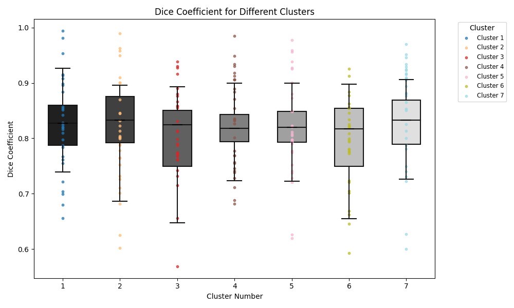
Figure1. Boxplot of DSC for of 7 clusters across 30 test-retest subjects
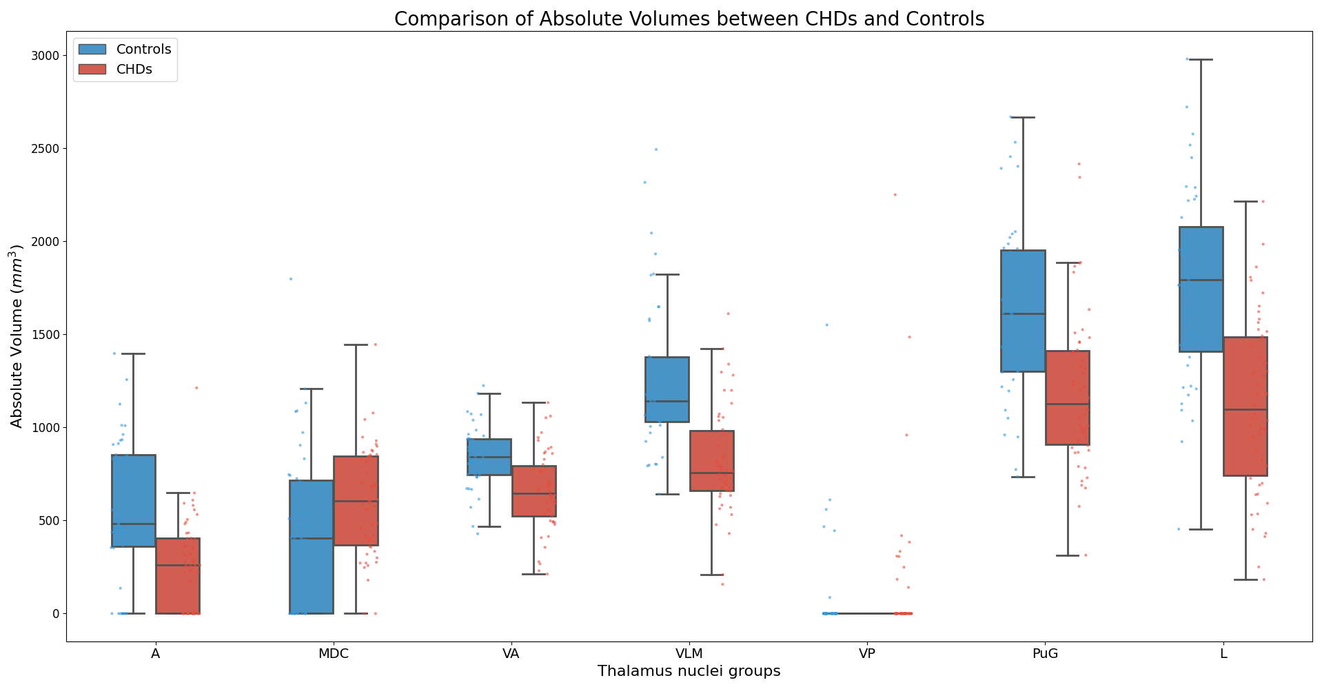
Figure 2. Absolute volume comparison between CHD and Controls for 7 thalamus clusters
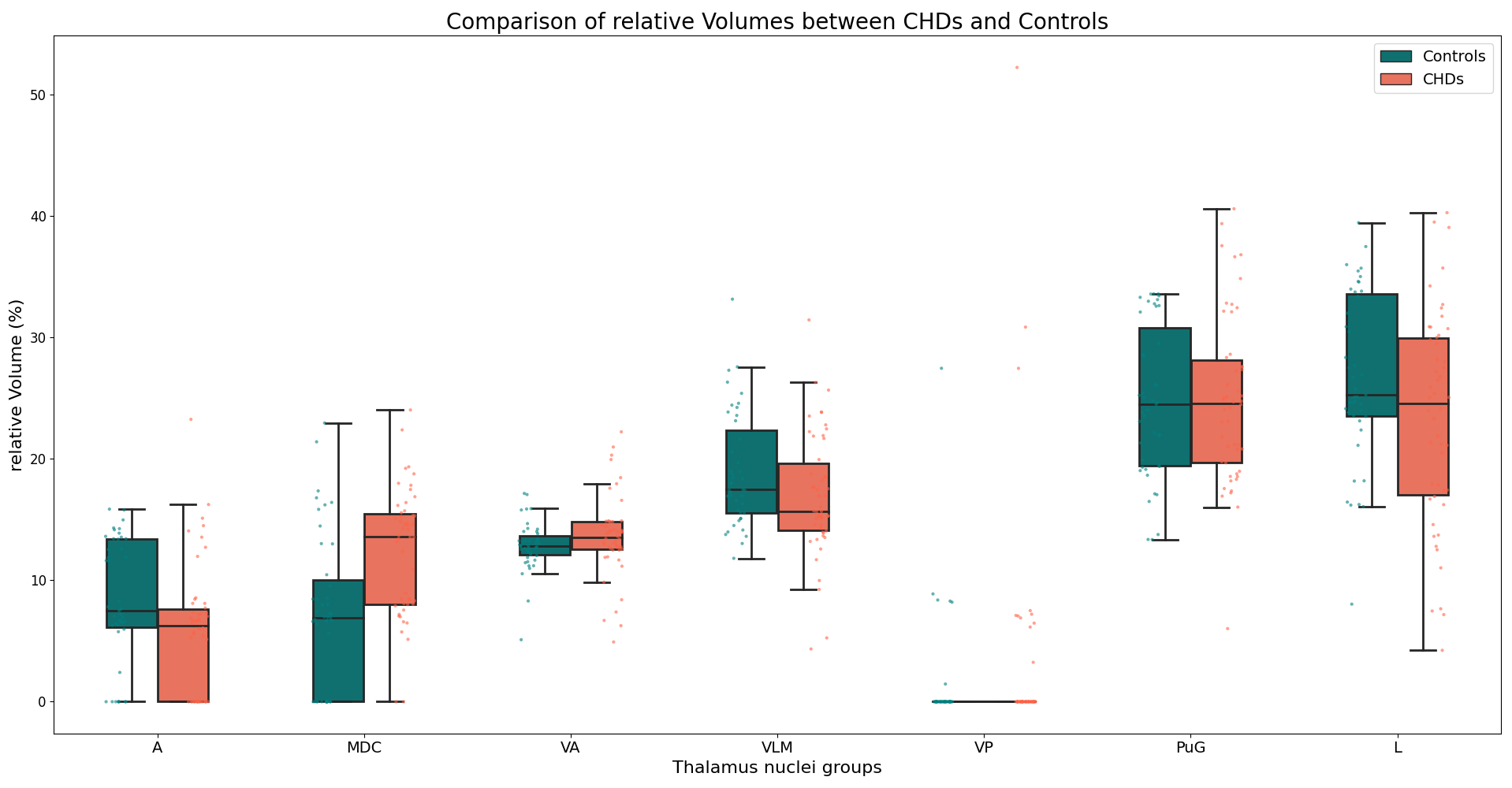
Figure 3. Relative volume comparison between CHD and Controls for 7 thalamus clusters